

Abstract
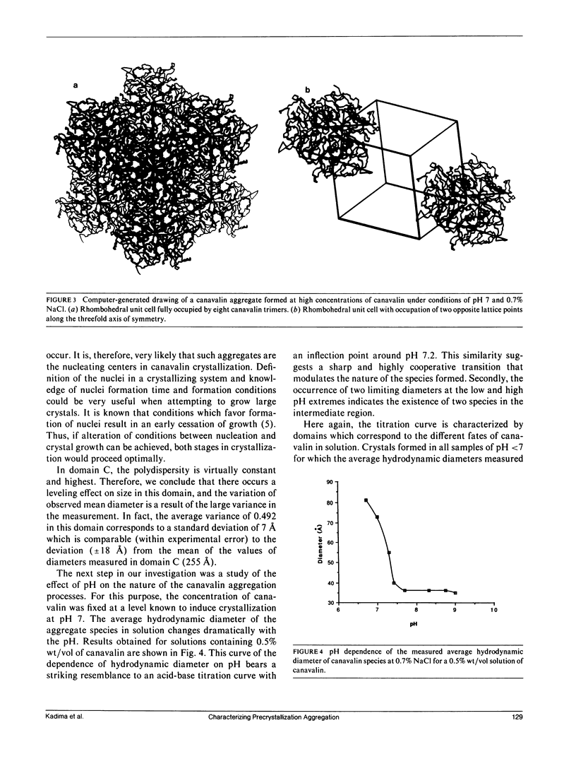
The aggregation processes leading to crystallization and precipitation of canavalin have been investigated by dynamic light scattering (DLS) in photon correlation spectroscopy (PCS) mode. The sizes of aggregates formed under various conditions of pH, salt concentration, and protein concentrations were deduced from the correlation functions generated by the fluctuating intensity of light scattered by the solutions of the protein. Results obtained indicate that the barrier to crystallization of canavalin is the formation of the trimer, a species that has been characterized by x-ray crystallographic studies (McPherson, A. 1980. J. Biol. Chem. 255:10472-10480). The dimensions of the trimer in solution are in good agreement with those obtained both from the crystal (McPherson, A. 1980. J. Biol. Chem. 255:10472-10480) and from a low angle x-ray scattering study in solution (Plietz, P., P. Damaschun, J. J. Müller, and B. Schlener. 1983. FEBS [Fed. Eur. Biochem. Soc.] Lett. 162:43-46). Furthermore, under conditions known to lead to the formation of rhombohedral crystals of canavalin, a limiting size is reached at high concentrations of canavalin. The size measured corresponds to an aggregate of trimers making a unit rhombohedral cell consistent with x-ray crystallographic data (McPherson, A. 1980. J. Biol. Chem. 255:10472-10480). Presumably, such aggregates are the nuclei from which crystal growth proceeds. The present study was undertaken primarily to test the potential of DLS (PCS) as a tool for rapid, routine screening to determine the ultimate fate of protein solutions (i.e., crystallization or amorphous precipitation) at an early stage, therefore eliminating the need for long-term visual observation. Achieving this goal would constitute amajor advance in the practive of protein crystallization. Delays imposed by visual observation would be considerably reduced, and a more systematic approach could be adopted to select experimental conditions.Our findings with canavalin demonstrate that DLS(PCS) is, indeed, a selective and sensitive probe of precrystallization conditions. Other advantages of this technique include the facts that it is noninvasive, nondestructive,universal, and does not require calibration.
Full text
PDF
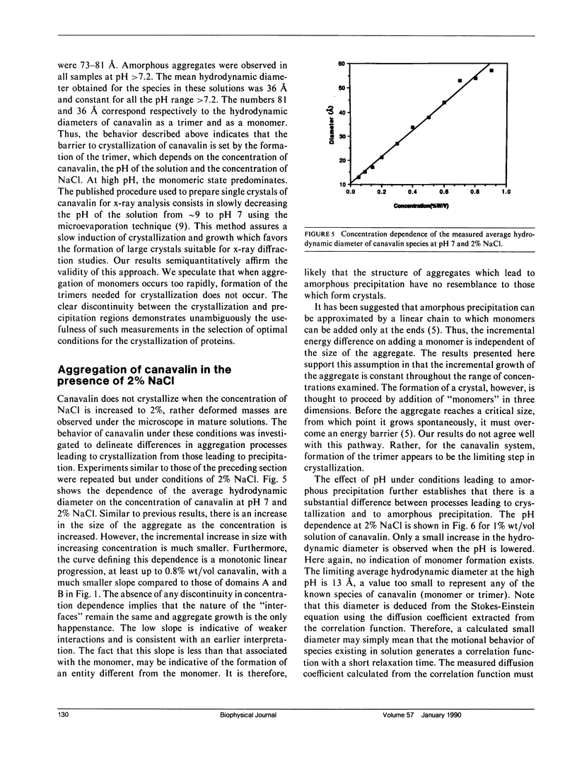
Selected References
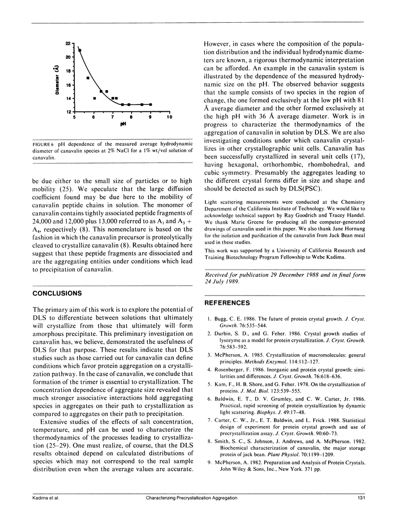
These references are in PubMed. This may not be the complete list of references from this article.
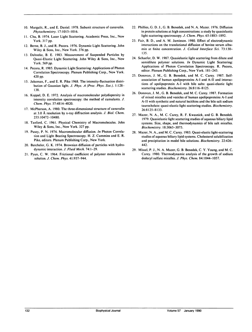
- Baldwin E. T., Crumley K. V., Carter C. W. Practical, rapid screening of protein crystallization conditions by dynamic light scattering. Biophys J. 1986 Jan;49(1):47–48. doi: 10.1016/s0006-3495(86)83587-1. [DOI] [PMC free article] [PubMed] [Google Scholar]
- Donovan J. M., Benedek G. B., Carey M. C. Self-association of human apolipoproteins A-I and A-II and interactions of apolipoprotein A-I with bile salts: quasi-elastic light scattering studies. Biochemistry. 1987 Dec 15;26(25):8116–8125. doi: 10.1021/bi00399a015. [DOI] [PubMed] [Google Scholar]
- Kam Z., Shore H. B., Feher G. On the crystallization of proteins. J Mol Biol. 1978 Aug 25;123(4):539–555. doi: 10.1016/0022-2836(78)90206-1. [DOI] [PubMed] [Google Scholar]
- Mazer N. A., Carey M. C., Kwasnick R. F., Benedek G. B. Quasielastic light scattering studies of aqueous biliary lipid systems. Size, shape, and thermodynamics of bile salt micelles. Biochemistry. 1979 Jul 10;18(14):3064–3075. doi: 10.1021/bi00581a024. [DOI] [PubMed] [Google Scholar]
- Mazer N. A., Carey M. C. Quasi-elastic light-scattering studies of aqueous biliary lipid systems. Cholesterol solubilization and precipitation in model bile solutions. Biochemistry. 1983 Jan 18;22(2):426–442. doi: 10.1021/bi00271a029. [DOI] [PubMed] [Google Scholar]
- McPherson A. Crystallization of macromolecules: general principles. Methods Enzymol. 1985;114:112–120. doi: 10.1016/0076-6879(85)14007-3. [DOI] [PubMed] [Google Scholar]
- McPherson A. The three-dimensional structure of canavalin at 3.0 A resolution by X-ray diffraction analysis. J Biol Chem. 1980 Nov 10;255(21):10472–10480. [PubMed] [Google Scholar]
- Smith S. C., Johnson S., Andrews J., McPherson A. Biochemical characterization of canavalin, the major storage protein of jack bean. Plant Physiol. 1982 Oct;70(4):1199–1209. doi: 10.1104/pp.70.4.1199. [DOI] [PMC free article] [PubMed] [Google Scholar]