

Abstract
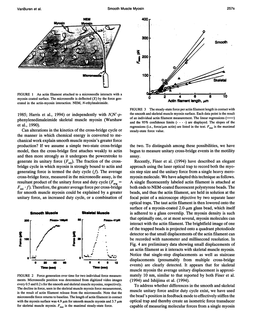
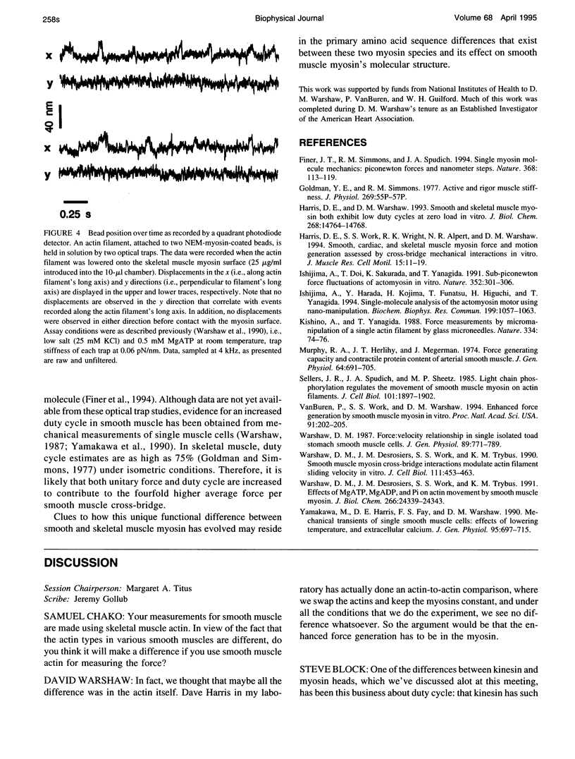
Smooth muscle generates as much force per cross sectional area of muscle as skeletal muscle with only one-fifth the myosin content. Although this apparent difference could be explained at the tissue or cellular level, it is possible that at the molecular level smooth muscle cross-bridges generate greater average force than skeletal muscle cross-bridges. To test this hypothesis, we used an in vitro motility assay (VanBuren et al., 1994) in which either chicken thiophosphorylated gizzard smooth or pectoralis skeletal muscle monomeric myosin is adhered to a nitrocellulose surface. A fluorescently labeled actin filament, attached to an ultracompliant (50-200 nm/pN) glass microneedle, is brought in contact with the myosin surface. Isometric force, being generated by myosin cross-bridges pulling on the attached actin filament, is calculated from the extent to which the calibrated microneedle is deflected. By measuring the density of myosin adhered to the surface, we estimated the number of myosin cross-bridges that are able to interact with a length of actin filament in contact with the myosin surface. In a direct comparison between smooth and skeletal muscle myosin, the average force per cross-bridge was 0.8 and 0.2 pN, respectively. Surprisingly, smooth muscle myosin generates approximately 4 times greater average force per cross-bridge head than skeletal muscle myosin. Because average isometric force is the product of the cross-bridge unitary force and duty cycle, we are presently using a laser optical trap in an attempt to measure unitary events from single myosin molecules. This approach should allow us to determine whether an increase in unitary force, duty cycle, or both contribute to smooth muscle myosin's enhanced force-generating capacity compared with skeletal muscle myosin.
Full text
PDF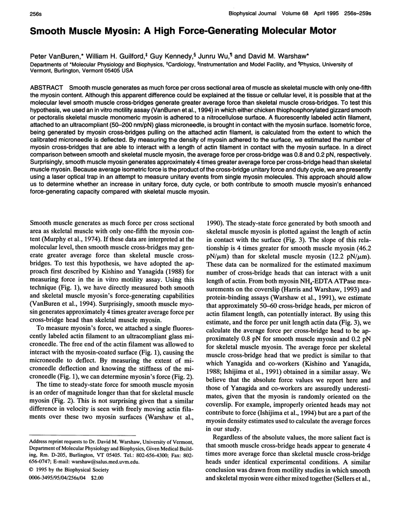
Images in this article
Selected References
These references are in PubMed. This may not be the complete list of references from this article.
- Finer J. T., Simmons R. M., Spudich J. A. Single myosin molecule mechanics: piconewton forces and nanometre steps. Nature. 1994 Mar 10;368(6467):113–119. doi: 10.1038/368113a0. [DOI] [PubMed] [Google Scholar]
- Goldman Y. E., Simmons R. M. Active and rigor muscle stiffness [proceedings]. J Physiol. 1977 Jul;269(1):55P–57P. [PubMed] [Google Scholar]
- Harris D. E., Warshaw D. M. Smooth and skeletal muscle myosin both exhibit low duty cycles at zero load in vitro. J Biol Chem. 1993 Jul 15;268(20):14764–14768. [PubMed] [Google Scholar]
- Harris D. E., Work S. S., Wright R. K., Alpert N. R., Warshaw D. M. Smooth, cardiac and skeletal muscle myosin force and motion generation assessed by cross-bridge mechanical interactions in vitro. J Muscle Res Cell Motil. 1994 Feb;15(1):11–19. doi: 10.1007/BF00123828. [DOI] [PubMed] [Google Scholar]
- Ishijima A., Doi T., Sakurada K., Yanagida T. Sub-piconewton force fluctuations of actomyosin in vitro. Nature. 1991 Jul 25;352(6333):301–306. doi: 10.1038/352301a0. [DOI] [PubMed] [Google Scholar]
- Ishijima A., Harada Y., Kojima H., Funatsu T., Higuchi H., Yanagida T. Single-molecule analysis of the actomyosin motor using nano-manipulation. Biochem Biophys Res Commun. 1994 Mar 15;199(2):1057–1063. doi: 10.1006/bbrc.1994.1336. [DOI] [PubMed] [Google Scholar]
- Kishino A., Yanagida T. Force measurements by micromanipulation of a single actin filament by glass needles. Nature. 1988 Jul 7;334(6177):74–76. doi: 10.1038/334074a0. [DOI] [PubMed] [Google Scholar]
- Murphy R. A., Herlihy J. T., Megerman J. Force-generating capacity and contractile protein content of arterial smooth muscle. J Gen Physiol. 1974 Dec;64(6):691–705. doi: 10.1085/jgp.64.6.691. [DOI] [PMC free article] [PubMed] [Google Scholar]
- Sellers J. R., Spudich J. A., Sheetz M. P. Light chain phosphorylation regulates the movement of smooth muscle myosin on actin filaments. J Cell Biol. 1985 Nov;101(5 Pt 1):1897–1902. doi: 10.1083/jcb.101.5.1897. [DOI] [PMC free article] [PubMed] [Google Scholar]
- VanBuren P., Work S. S., Warshaw D. M. Enhanced force generation by smooth muscle myosin in vitro. Proc Natl Acad Sci U S A. 1994 Jan 4;91(1):202–205. doi: 10.1073/pnas.91.1.202. [DOI] [PMC free article] [PubMed] [Google Scholar]
- Warshaw D. M., Desrosiers J. M., Work S. S., Trybus K. M. Effects of MgATP, MgADP, and Pi on actin movement by smooth muscle myosin. J Biol Chem. 1991 Dec 25;266(36):24339–24343. [PubMed] [Google Scholar]
- Warshaw D. M., Desrosiers J. M., Work S. S., Trybus K. M. Smooth muscle myosin cross-bridge interactions modulate actin filament sliding velocity in vitro. J Cell Biol. 1990 Aug;111(2):453–463. doi: 10.1083/jcb.111.2.453. [DOI] [PMC free article] [PubMed] [Google Scholar]
- Warshaw D. M. Force: velocity relationship in single isolated toad stomach smooth muscle cells. J Gen Physiol. 1987 May;89(5):771–789. doi: 10.1085/jgp.89.5.771. [DOI] [PMC free article] [PubMed] [Google Scholar]
- Yamakawa M., Harris D. E., Fay F. S., Warshaw D. M. Mechanical transients of single toad stomach smooth muscle cells. Effects of lowering temperature and extracellular calcium. J Gen Physiol. 1990 Apr;95(4):697–715. doi: 10.1085/jgp.95.4.697. [DOI] [PMC free article] [PubMed] [Google Scholar]