Abstract
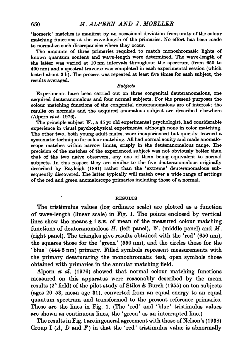
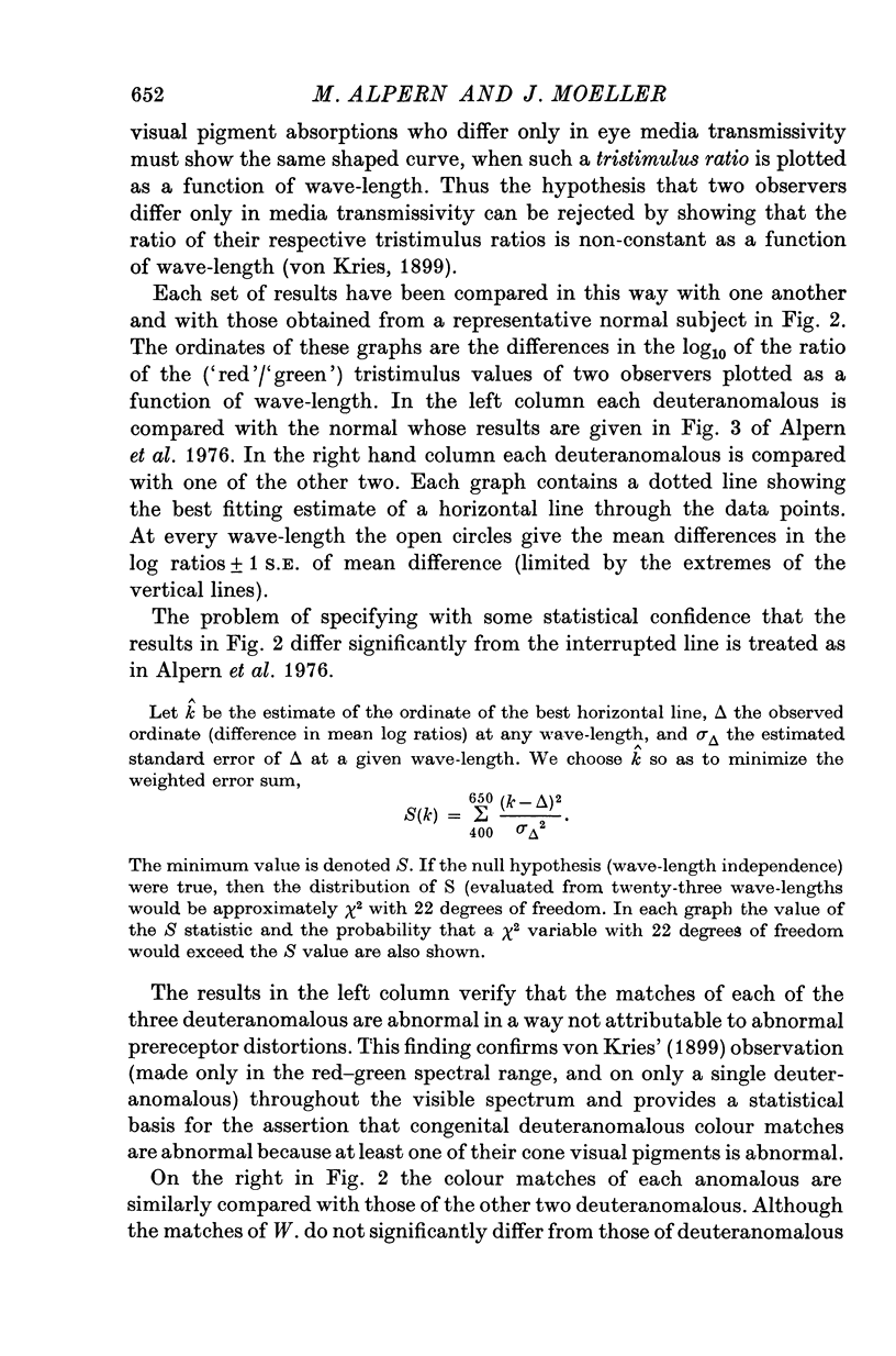
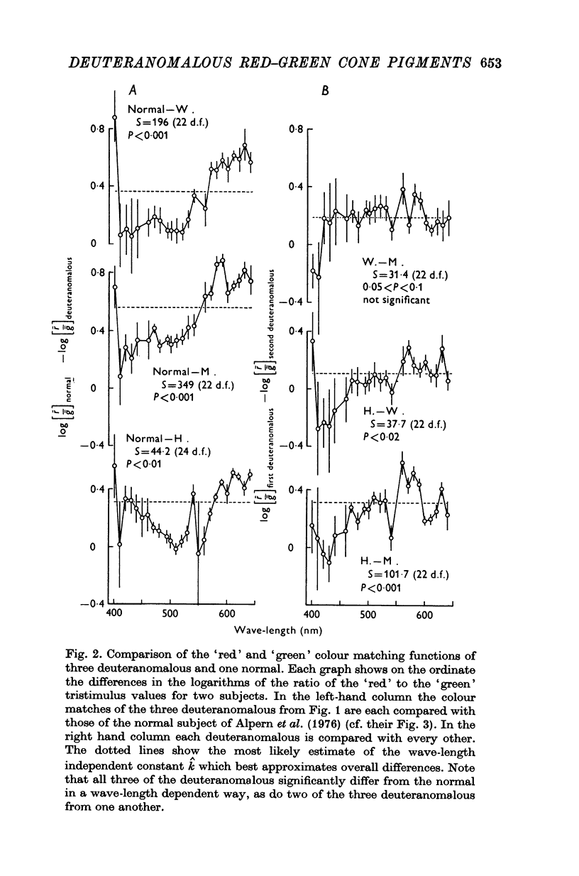
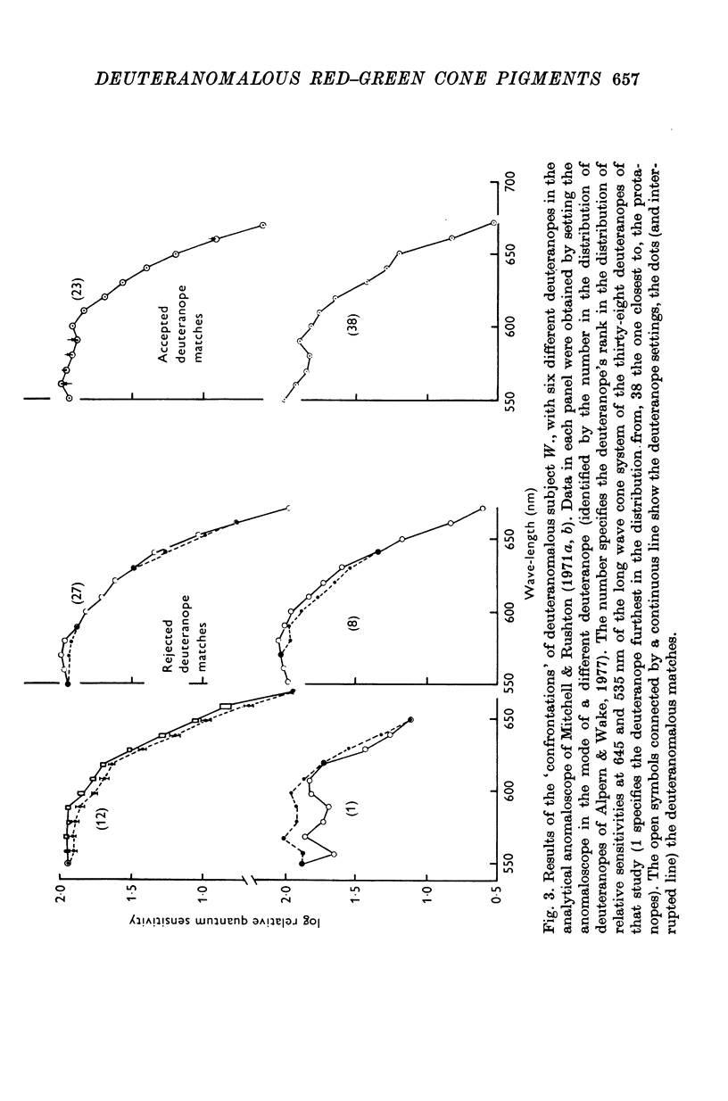
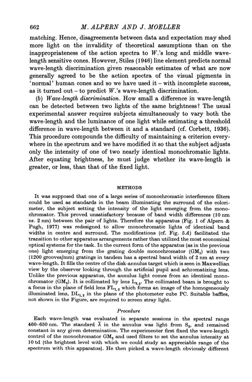
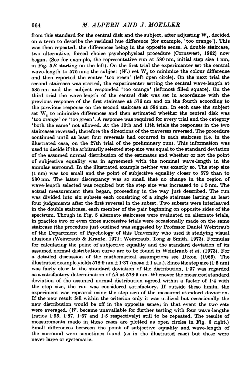
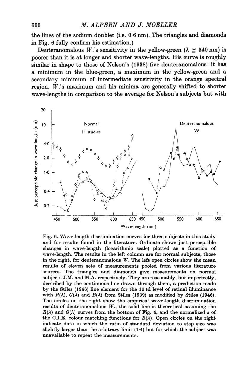
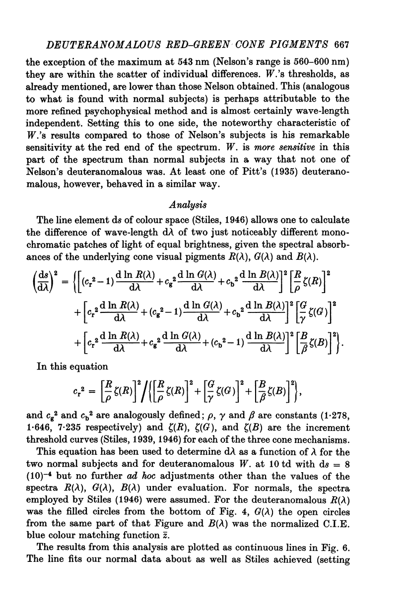
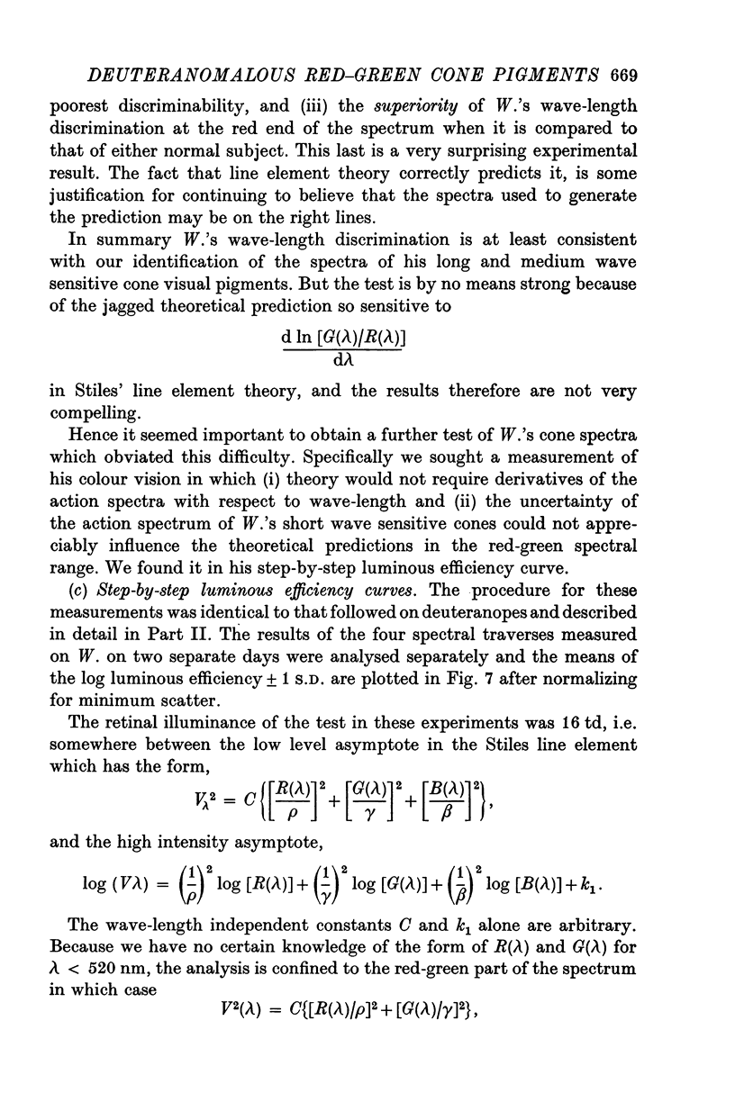
1. Three "simple" deuteranomalous trichromats match with abnormally low "red" tristimulus values throughout the spectrum and abnormally high "green" tristimulus values in the long wave end of the spectrum which become normal (and then low) in the yellow-green. The spectrum locus of this transition differs from one anomalous to the other. Differences in the matches of two of these cannot be due to differences in eye media transmissivities alone. Therefore these two deuteranomalous have different cone visual pigments. 2. The analytical anomaloscope was used in the confrontation of one deuteranomalous with six deuteranopes in turn. In each confrontation the deuteranope set the anomaloscope in his mode and adjusted the intensity of the monochromatic light for a match. Deuteranomalous matches were rejected by four of these six deuteranopes. 3. They were accepted by two of the six. These two rejected each other's matches in a way not attributable to differences in eye media transmissivity. 4. Three different psychophysical techniques were used to measure the action spectra of the long wave cones of these two deuternopes. All three methods reveal small but systematic differences in lambdamax and shape of the curve for the one deuteranope compared with that of the other. 5. In red-green spectral range, these spectra are accurately described by different linear combinations of the color matching functions of the same deuteranomalous whose matches the two deuteranopes accept. Linear combinations of those of a second deuteranomalous, with at least one different kind of cone, fit less well. 6. The wave length discrimination curve of the former deuteranomalous was measured with a new method. The curves of two normals were also obtained for comparison. Wave-length discrimination predictions from the Stiles (1946) line element theory were compared to the anomalous curve. The deuteranopic action spectra were used in the line element to compute this deuteranomalous' discrimination. There is reasonable first order correspondence between prediction and observation, but the prediction is sensitive to small changes in the derivatives of the logarithms of the action spectra. 7. Line element prediction of the deuteranomalous step-by-step luminous efficiency curve is insensitive to such uncertainties. The agreement with expectation from the above assumptions and the measured step-by-step deuteranomalous luminous efficiency curve in the red-green part of the spectrum is therefore good. 8. It is concluded that the erythrolabe in one deuternope's long wave cones has the action spectrum of this deuteranomalous' long and the erythrolabe in the other deuternope's long wave sensitive cones has that of this deuternomalous' medium wave cones. This leads to a general hypothesis about the nature of all forms of red-green colour vision defects transmitted recessively on the X chromosome.
Full text
PDF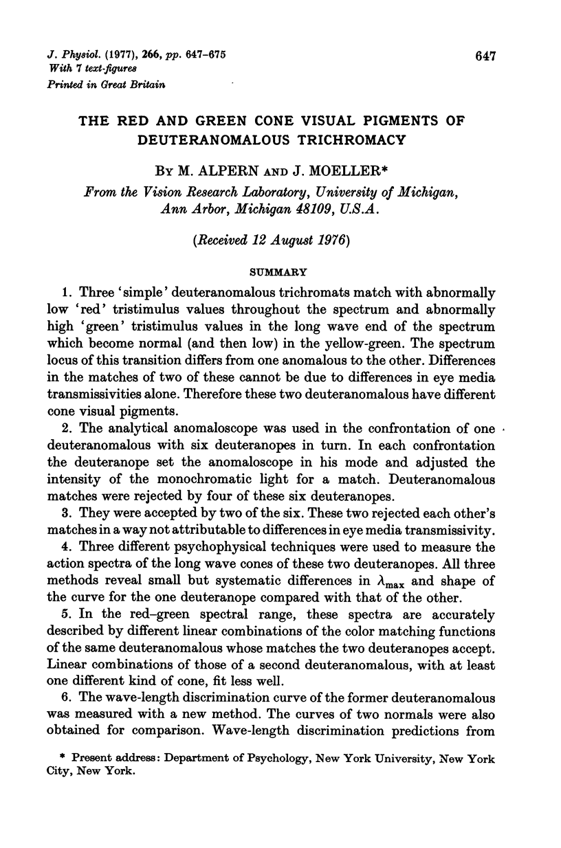



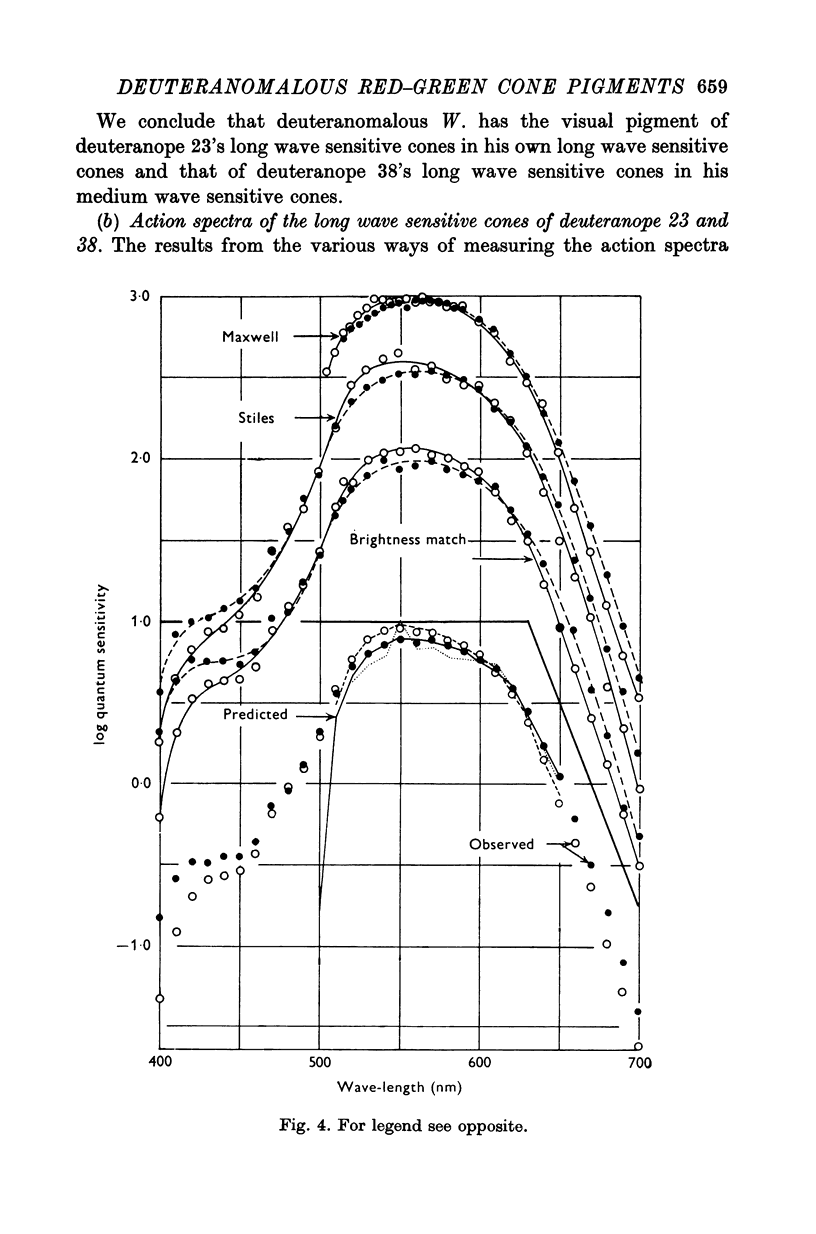





Selected References
These references are in PubMed. This may not be the complete list of references from this article.
- Alpern M., Bastian B., Pugh E. N., Jr, Gras W. Altered ocular pigments, photostable and labile: two causes of deuteranomalous trichromacy. Mod Probl Ophthalmol. 1976;17:273–291. [PubMed] [Google Scholar]
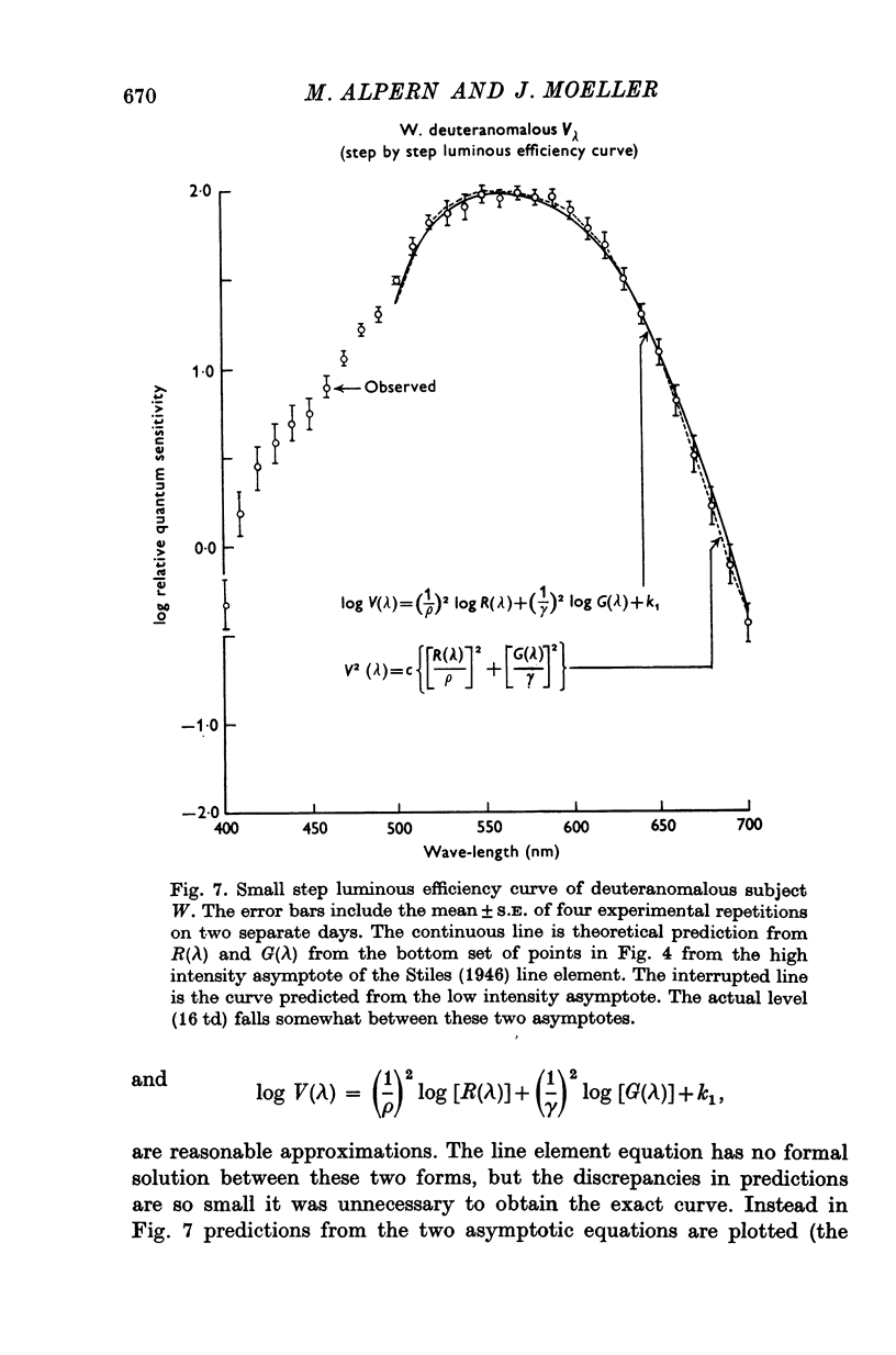
- Alpern M., Pugh E. N., Jr Variation in the action spectrum of erythrolabe among deuteranopes. J Physiol. 1977 Apr;266(3):613–646. doi: 10.1113/jphysiol.1977.sp011785. [DOI] [PMC free article] [PubMed] [Google Scholar]
- Alpern M., Torii S. The luminosity curve of the deuteranomalous fovea. J Gen Physiol. 1968 Nov;52(5):738–749. doi: 10.1085/jgp.52.5.738. [DOI] [PMC free article] [PubMed] [Google Scholar]
- Alpern M., Torii S. The luminosity curve of the protanomalous fovea. J Gen Physiol. 1968 Nov;52(5):717–737. doi: 10.1085/jgp.52.5.717. [DOI] [PMC free article] [PubMed] [Google Scholar]
- Alpern M. Tritanopia. Am J Optom Physiol Opt. 1976 Jul;53(7):340–349. doi: 10.1097/00006324-197607000-00003. [DOI] [PubMed] [Google Scholar]
- Alpern M., Wake T. Cone pigments in human deutan colour vision defects. J Physiol. 1977 Apr;266(3):595–612. doi: 10.1113/jphysiol.1977.sp011784. [DOI] [PMC free article] [PubMed] [Google Scholar]
- BEDFORD R. E., WYSZECKI G. W. Wavelength discrimination for point sources. J Opt Soc Am. 1958 Feb;48(2):129–135. doi: 10.1364/josa.48.000129. [DOI] [PubMed] [Google Scholar]
- BEDFORD R. E., WYSZECKI G. Axial chromatic aberration of the human eye. J Opt Soc Am. 1957 Jun;47(6):564–565. doi: 10.1364/josa.47.0564_1. [DOI] [PubMed] [Google Scholar]
- CORNSWEET T. N. The staircrase-method in psychophysics. Am J Psychol. 1962 Sep;75:485–491. [PubMed] [Google Scholar]
- Corbett H. V. Hue discrimination in normal and abnormal colour vision. J Physiol. 1936 Nov 6;88(2):176–190. doi: 10.1113/jphysiol.1936.sp003430. [DOI] [PMC free article] [PubMed] [Google Scholar]
- HEATH G. G. Luminosity curves of normal and dichromatic observers. Science. 1958 Oct 3;128(3327):775–776. doi: 10.1126/science.128.3327.775. [DOI] [PubMed] [Google Scholar]
- HECHT S., HSIA Y. Colorblind vision; luminosity losses in the spectrum for dichromats. J Gen Physiol. 1947 Nov 20;31(2):141–152. doi: 10.1085/jgp.31.2.141. [DOI] [PMC free article] [PubMed] [Google Scholar]
- HURVICH L. M., JAMESON D. Theoretical analysis of anomalous trichromatic color vision. J Opt Soc Am. 1956 Dec;46(12):1075–1089. doi: 10.1364/josa.46.001075. [DOI] [PubMed] [Google Scholar]
- King-Smith P. E. The optical density of erythrolabe determined by a new method. J Physiol. 1973 May;230(3):551–560. doi: 10.1113/jphysiol.1973.sp010203. [DOI] [PMC free article] [PubMed] [Google Scholar]
- King-Smith P. E. The optical density of erythrolabe determined by retinal densitometry using the self-screening method. J Physiol. 1973 May;230(3):535–549. doi: 10.1113/jphysiol.1973.sp010202. [DOI] [PMC free article] [PubMed] [Google Scholar]
- Mitchell D. E., Rushton W. A. The red-green pigments of normal vision. Vision Res. 1971 Oct;11(10):1045–1056. doi: 10.1016/0042-6989(71)90111-8. [DOI] [PubMed] [Google Scholar]
- Mitchell D. E., Rushton W. A. Visual pigments in dichromats. Vision Res. 1971 Oct;11(10):1033–1043. doi: 10.1016/0042-6989(71)90110-6. [DOI] [PubMed] [Google Scholar]
- Piantanida T. P., Sperling H. G. Isolation of a third chromatic mechanism in the deuteranomalous observer. Vision Res. 1973 Nov;13(11):2049–2058. doi: 10.1016/0042-6989(73)90181-8. [DOI] [PubMed] [Google Scholar]
- Pokorny J., Smith V. C., Katz I. Derivation of the photopigment absorption spectra in anomalous trichromats. J Opt Soc Am. 1973 Feb;63(2):232–237. doi: 10.1364/josa.63.000232. [DOI] [PubMed] [Google Scholar]
- RUSHTON W. A. A FOVEAL PIGMENT IN THE DEUTERANOPE. J Physiol. 1965 Jan;176:24–37. doi: 10.1113/jphysiol.1965.sp007532. [DOI] [PMC free article] [PubMed] [Google Scholar]
- Rushton W. A., Powell D. S., White K. D. Exchange thresholds in dichromats. Vision Res. 1973 Nov;13(11):1993–2002. doi: 10.1016/0042-6989(73)90177-6. [DOI] [PubMed] [Google Scholar]
- Rushton W. A., Powell D. S., White K. D. Pigments in anomalous trichromats. Vision Res. 1973 Nov;13(11):2017–2031. doi: 10.1016/0042-6989(73)90179-x. [DOI] [PubMed] [Google Scholar]
- Rushton W. A. Visual pigments and color blindness. Sci Am. 1975 Mar;232(3):64–74. doi: 10.1038/scientificamerican0375-64. [DOI] [PubMed] [Google Scholar]
- WALLS G. L. Graham's theory of color blindness. Am J Optom Arch Am Acad Optom. 1958 Sep;35(9):449–460. doi: 10.1097/00006324-195809000-00001. [DOI] [PubMed] [Google Scholar]


