Abstract
The Cdk1 inhibitor Wee1 is inactivated during mitotic entry by proteolysis, translational regulation, and transcriptional regulation. Wee1 is also regulated by posttranslational modifications, and here we have identified five phosphorylation sites in the N-terminal domain of embryonic Xenopus Wee1A through a combination of mutagenesis studies and matrix-assisted laser desorption ionization-time of flight mass spectrometry. All five sites conform to the Ser-Pro/Thr-Pro consensus for proline-directed kinases like Cdks. Three of the sites (Ser 38, Thr 53, and Ser 62) are required for the mitotic gel shift, and at least two of these sites (Ser 38 and Thr 53) regulate the proteolysis of Wee1A during interphase. The other two sites (Thr 104 and Thr 150) are primarily responsible for the mitotic inactivation of Wee1A. Alanine mutants of Thr 150 or Thr 104 had an increased capacity to inhibit mitotic entry in cyclin B-treated interphase extracts, and Thr 150 was found to be transiently phosphorylated just prior to nuclear envelope breakdown in cycling egg extracts. These findings establish the phosphorylation-dependent direct inactivation of Wee1A as a critical mechanism for the promotion of M-phase entry. These results also show that multisite phosphorylation cooperatively inactivates Wee1A and cooperatively promotes Wee1A proteolysis.
Wee1 was first identified through genetic studies of cell size control and cell cycle progression in Schizosaccharomyces pombe (30, 31, 38). Subsequent work established Wee1 as a critical regulator of the G2/M transition in diverse organisms and cell types (1, 3, 7). Wee1 functions by phosphorylating cyclin-Cdk complexes at a conserved tyrosine residue (Tyr 15 in Cdc2/Cdk1), thereby inactivating the kinase (8, 17, 20, 33). Wee1's activity is opposed by Cdc25A, -B, and -C, a small family of conserved phosphatases that dephosphorylate two inhibitory sites (Thr 14 and Tyr 15) in Cdk1 (1, 3, 4, 6, 7, 9, 13). In late G2 phase, the balance between Wee1-mediated Cdk1 phosphorylation and Cdc25-mediated dephosphorylation shifts in favor of dephosphorylation, bringing about activation of cyclin-Cdk1 complexes and entry into mitosis. Fungi and invertebrates possess a single Wee1 gene; vertebrates possess two, an embryonic Wee1 gene expressed predominantly in mature oocytes, testes, and early embryos, and a somatic gene expressed later in development (16, 28, 32, 45). (The nomenclature of the somatic and embryonic Wee1 genes is described in Materials and Methods.)
Although the biochemical function of Wee1 is relatively simple—its only known substrates are itself and the cyclin-Cdk complexes—Wee1's regulation is complicated and incompletely understood. The levels, activity, and localization of Wee1 are all regulated, and multiple mechanisms contribute to each of these aspects of Wee1 control. Wee1 levels are regulated transcriptionally (46), translationally (5, 25, 27), and by ubiquitylation and proteolysis (21). The degradation of Wee1 in M phase or late G2 phase has been reported to be triggered by three SCF-type ubiquitin ligases: SCFTome-1, SCFβ-TrCP1, and SCFβ-TrCP2 (2, 45). Recognition of Xenopus embryonic Wee1 by Tome-1 depends upon the phosphorylation of Ser 38 (2), an SP site that is present in Xenopus embryonic Wee1 (Wee1A) but not in human or mouse embryonic Wee1 (Fig. 1A). Recognition of somatic Wee1 by β-TrCPs depends upon the phosphorylation of two residues conserved in somatic Wee1 proteins but absent from embryonic Wee1 proteins: Ser 53 (a putative Plk1 phosphorylation site) and Ser 123 (a putative Cdk1 phosphorylation site) (45).
FIG. 1.
OP11-Wee1A is resistant to mitotic inactivation. (A) Sequence alignment of the Xenopus, mouse, and human embryonic Wee1 proteins. The 11 SP/TP motifs present in Xenopus Wee1A and mutated in OP11-Wee1A are shown. (B) In vitro kinase activity of wild-type (WT) Wee1A and OP11-Wee1A. Bead-bound Wee1A proteins were incubated with interphase Xenopus egg extracts or extracts treated with 200 nM Δ90-cyclin B (M-phase extracts). The Wee1A beads were then washed and incubated with a complex of kinase-minus T161A-Cdk1 and Δ65-cyclin B. The phosphorylation of Cdk1 was assessed by antiphosphotyrosine immunoblotting. Equal loading of substrate was verified by Cdk1 immunoblotting. (C) Autophosphorylation of wild-type Wee1A (WT), OP11-Wee1A (OP), and kinase-minus Wee1A (KM). Bead-bound Wee1A proteins were treated with interphase extracts or M-phase extracts as in panel B. The electrophoretic mobility of Wee1A was assessed by blotting with Flag antibody and the autophosphorylation of Wee1A assessed by antiphosphotyrosine immunoblotting. (D) Interphase extracts cause a shift in both wild-type Wee1A and OP11-Wee1A. Wee1A proteins were detected by Coomassie staining.
In addition, the specific activity of the Wee1 protein is regulated posttranslationally. During mitosis, the N-terminal noncatalytic domain of Wee1 becomes hyperphosphorylated, causing Wee1 to shift to a higher apparent molecular weight on sodium dodecyl sulfate (SDS) gels (19, 22, 34, 41). This hyperphosphorylation is accompanied by a marked decrease in the activity of the Wee1 protein (19, 22, 34, 41). Wee1 inactivation could be an important means of negatively regulating Wee1 function during M phase, particularly in systems like Xenopus embryos and extracts, where the overall abundance of Wee1A varies little between interphase and M phase (25).
Wee1 is positively regulated by phosphorylation as well. Xenopus Wee1A autophosphorylates on Tyr 90, Tyr 103, and Tyr 110, three poorly conserved residues in its N-terminal regulatory domain (23). Mutation of these sites to phenylalanines blocks Wee1A autophosphorylation and decreases Wee1A's ability to inhibit Xenopus oocyte maturation but, curiously, does not appear to decrease Wee1A's in vitro kinase activity toward cyclin B-Cdk1 (23). Autophosphorylation may decrease Wee1's susceptibility to negative regulatory factors or increase its access to positive regulatory factors that are present in cells but absent from in vitro kinase reaction mixtures. Two additional possible autophosphorylation sites have been identified in human somatic Wee1, Tyr 295 and Tyr 362, conserved sites that correspond to Tyr 206 and Tyr 273 in Xenopus Wee1A (14).
Wee1 is probably also regulated by the phosphorylation of Ser 549, a conserved residue in the C-terminal tail of the protein. Phosphorylation of this site allows the association of Wee1 with 14-3-3 proteins, and both Chk1 and Akt have been put forward as likely Ser 549 kinases (14, 15, 37, 44). The consequences of this phosphorylation and the consequent 14-3-3 binding are controversial. One group has reported that Ser 549 phosphorylation decreases Wee1 function and promotes mitosis (14), while three others have found that it increases Wee1 function and inhibits mitosis (15, 37, 44).
Here we have set out to identify the sites of mitotic phosphorylation in Wee1 and to assess their importance in Wee1 regulation. We chose to use Xenopus Wee1A and Xenopus egg extracts for these studies because extracts can be put in a permanent interphase state or a permanent mitotic state, allowing the phosphorylation of endogenous or added recombinant Wee1A to be driven to near-steady-state levels. Through a combination of matrix-assisted laser desorption ionization-time of flight (MALDI-TOF) mass spectrometry (MS) and mutagenesis, we have identified five mitotic phosphorylation sites. Three poorly conserved sites (Ser 38, Thr 53, and Ser 62) were found to be required for the mitotic gel shift and were largely dispensable for mitotic inactivation of Wee1A. Two better-conserved sites (Thr 104 and Thr 150) were required for mitotic inactivation of Wee1 but were dispensable for the gel shift. Finally, we found that Wee1A mutants that are resistant to mitotic inactivation are potent inhibitors of mitotic entry, establishing phosphorylation-dependent changes in the specific activity of Wee1A as critical for normal mitotic progression.
MATERIALS AND METHODS
Wee1 nomenclature.
Humans, mice, chickens, and frogs all possess two Wee1 genes. In Xenopus, the gene expressed in spermatocytes, mature oocytes, and early embryos is designated Wee1 or Wee1A and the gene expressed in later embryos and somatic cells is designated Wee2 or Wee1B (22, 32). The form of Wee1 being studied here is Xenopus embryonic Wee1, which by convention we designate Wee1A. In human cells, the somatic gene is termed Wee1A and the embryonic gene is termed Wee1 or Wee1B (28, 45, 46). Thus, Xenopus Wee1A corresponds to human Wee1B and Xenopus Wee1B corresponds to human Wee1A.
Mutagenesis and expression of recombinant Xenopus Wee1A proteins.
The cDNA of wild-type Xenopus Wee1A was provided by Bill Dunphy (California Institute of Technology, Pasadena). Kinase-minus K239I-Wee1A was provided by Sarah Walter (Stanford University, Stanford, CA). Wild-type and kinase-minus Xenopus K239I-Wee1A were subcloned into the BamHI and XhoI sites of a modified pFastBacHTc-His6 vector (pFastBacHTc-His6-Flag; J. Pomerening, Stanford University). Throughout the text, we have numbered amino acid residues according to the Xenopus Wee1A protein sequence described in reference 22. Eleven SP/TP sites—Ser 38, Thr 53, Ser 62, Ser 68, Ser 74, Thr 86, Thr 101, Thr 104, Ser 115, Thr 150, and Thr 450—were individually or combinatorially mutated to alanine with the QuikChange site-directed mutagenesis kit (Stratagene, La Jolla, CA). The oligonucleotides used for construction of OP11-Wee1A were described previously (35). The oligonucleotides used to produce the individual alanine mutations were as follows: S38A, 5′ATTAATGAAGGTCCCCAGAAGGGGGCTCCCGTGAGTTCCTGGAGGACCA3′; T53A, 5′CCAATAACTGCCCCTTCCCCATCGCCCCCCAGAGGAACGAGAGGGAAC TT3′; S62A, 5′CCCCAGAGGAACGAGAGGGAACTTGCTCCTACTCAA GAGCTGAGCCCAA3′; S68A, 5′GAACTTTCTCCTACTCAAGAGCTGGCCCCAAGTAGCGACTACTCGCCCG3′; S74A, 5′GAGCTGAGCCCAAGT AGCGACTACGCGCCCGACCCAAGTGTGGGGGCTG3′; T86A, 5′AGTGTGGGGGCTGAATGCCCTGGTGCCCCCCTTCATTACAGCACATGGA3′; T101A, 5′GGAAGAAGCTCAAGCTCTGTGACGCCCCTTATACCCCAA AGAGCC3′; T104A, 5′CTCAAGCTCTGTGACACCCCTTATGCCCCAAAGAGCCTTTTGTACAAAACGC3′; S115A, 5′AGCCTTTTGTACAAAACGCTTCCCGCTCCGGGGTCCCGCGTTCACTGCA3′ T150A, 5′CTCTGGTCAATATCAACCCCTTCGCCCCTGAATCCTACCGACAAACCC3′; T450A, 5′TTGCCCCACGTTCCCCAGCTGCTGGCTCCCGTCTTCCTTGCCCTG CTCA3′. The bases changed are in bold italics; the codons changed are underlined. Mutations were verified by DNA sequencing. Baculoviruses encoding Wee1A mutants were generated with the Bac-to-Bac system (Invitrogen, Carlsbad, CA) by the manufacturer's protocol. N-terminally His6- and Flag-tagged Wee1A proteins were expressed in Sf9 cells by infection with baculovirus encoding Wee1A for 72 h.
Preparation of Xenopus egg extracts.
Interphase egg extracts and cycling egg extracts were prepared as described previously (26). Unfertilized eggs were dejellied in 2% cysteine, activated with 0.4 μg/ml calcium ionophore A23187 (Sigma, St Louis, MO), and released to interphase in the absence (cycling extracts) or in the presence (interphase extracts) of 10 μg/ml cycloheximide. To drive interphase extracts into a stable mitotic state, nondegradable sea urchin Δ90-cyclin B was added to interphase extracts at a concentration of 150 to 200 nM. Demembranated sperm chromatin (40) was routinely added to the extracts at a concentration of 500 sperm cells/μl and stained with 4′,6′-diamidino-2-phenylindole (DAPI) to monitor progression into mitosis.
Wee1A autophosphorylation.
Sf9 cells (5 × 10⁶) overexpressing Wee1A protein were lysed in a buffer containing 50 mM Tris-Cl (pH 7.4), 150 mM NaCl, 1 mM EDTA, 10 mM β-mercaptoethanol, 1% Nonidet P-40, and protease inhibitors (10 μg/ml leupeptin, 10 μg/ml chymostatin, and 10 μg/ml pepstatin). Lysates were clarified by centrifugation at 14,000 rpm and incubated with 20 μl of packed anti-Flag beads for 2 h at 4°C. Protein-bound beads were washed and rotated in an interphase- or Δ90-cyclin B-treated extract for 40 min at room temperature. The reactions were stopped by washing the beads once with 1 ml of washing buffer (80 mM β-glycerophosphate, 20 mM EGTA, 15 mM MgCl2, 1 mM Na3VO4, 50 mM NaF, 5 mM Tris-Cl [pH 7.4], 15 mM NaCl, 0.1 mM EDTA, 1 mM β-mercaptoethanol, 0.1% Nonidet P-40) and twice with 1 ml of extract buffer (80 mM β-glycerophosphate, 20 mM EGTA, 15 mM MgCl2, 1 mM Na3VO4, 50 mM NaF), and the proteins were eluted with SDS sample buffer. Autophosphorylation was assessed by immunoblotting with antiphosphotyrosine antibody (PY99; Santa Cruz Biotechnology, Santa Cruz, CA). Chemiluminescence was quantified by densitometry.
Tyrosine kinase activity of Wee1A.
Flag bead-bound Wee1A protein was incubated with an interphase- or Δ90-cyclin B-treated extract for 40 min, washed, and supplemented with a complex of kinase-minus T161A-Cdk1 and nondegradable Δ65-cyclin B1 as a substrate in 30 μl of kinase buffer (20 mM HEPES, 150 mM NaCl, 5 mM MgCl2, 0.1 mM ATP, 10% sucrose, pH 7.7). The reaction progressed for 30 min at room temperature and was stopped by adding SDS sample buffer. T161A-Cdk1 (W. Dunphy, California Institute of Technology) and Δ65-cyclin B1 (J. Pomerening, Stanford University) were expressed by baculovirus infection of Sf9 cells, and T161A-Cdk1-Δ65-cyclin B1 complexes were prepared as previously described (22).
In-gel digestion and MS analysis.
The gel bands were excised with a scalpel, crushed, and destained by washing with 25 mM ammonium bicarbonate-50% acetonitrile. The gels were dehydrated by adding acetonitrile, rehydrated by adding 10 to 20 μl of 25 mM ammonium bicarbonate with 10 ng/μl of sequencing grade trypsin (Promega), and incubated at 37°C for 12 to 15 h. Peptides were extracted by adding 30 μl of a solution containing 60% acetonitrile-0.1% trifluoroacetic acid. The extraction was repeated three times and completed by adding 20 μl of acetonitrile. The resulting extracts were pooled and evaporated to dryness in a vacuum centrifuge. Peptides were mixed with saturated matrix solution, α-cyano-4-hydroxycinnamic acid in 60% acetonitrile-0.1% trifluoroacetic acid, and analyzed by MALDI-TOF MS (Voyager-DE STR; Applied Biosystems, Inc.). Mass accuracy was within 50 ppm. For interpretation of the mass spectra, we used the UCSF MS-Fit program available on the web (http://prospector.ucsf.edu/).
Western blot analysis.
Wee1A protein samples were separated on a 10% SDS-polyacrylamide gel (acrylamide/bisacrylamide ratio, 100:1), transferred to Immobilon P blotting membranes (Millipore, Bedford, MA), and probed with anti-Flag (M2; Sigma), anti-Wee1A (Zymed, San Francisco, CA), or antiphosphotyrosine (PY99; Santa Cruz Biotechnology) antibodies. Wee1A kinase assay samples were separated on a 12% SDS-polyacrylamide gel (acrylamide/bisacrylamide ratio, 29:1) and probed with anti-Cdc2/Cdk1 (C-19; Santa Cruz Biotechnology) or antiphosphotyrosine antibodies. Anti-phosphothreonine 150 (pT150) Wee1A serum was raised in rabbits against the synthetic peptide corresponding to VNINPF(pT150)PESY in Xenopus Wee1A and affinity purified over an unphosphorylated peptide column and a phosphopeptide column (BioSource International, Camarillo, CA). Mitotic phosphorylation of p42 mitogen-activated protein (MAPK) was analyzed with phospho-MAPK antibody (E10; Cell Signaling Technology, Beverly, MA). All blots were washed with Tris-buffered saline containing 0.05% Tween 20 and probed with a horseradish peroxidase-conjugated secondary antibody for detection with an Immun-Star horseradish peroxidase detection kit (Bio-Rad, Hercules, CA) or an alkaline phosphatase-conjugated antibody for detection with CDP-Star (Perkin-Elmer, Norwalk, CT).
Wee1A degradation.
Wee1A degradation assays were performed as previously described (2). [35S]methionine-labeled Wee1A proteins were produced by in vitro translation with the TNT coupled reticulocyte lysate system (Promega, Madison, WI). Cytostatic factor (CSF)-arrested extracts were prepared in the presence of cycloheximide (10 μg/ml), demembranated sperm chromatin (2,000/μl) was added, and the extracts were released into interphase with 0.8 mM CaCl2. Once nuclei had formed (generally 30 to 40 min), 25-μl aliquots of extract were incubated with 4 μl of in vitro-translated Wee1A. Samples were taken every 30 min, and the remaining 35S-labeled Wee1A was assessed by phosphoimaging and compared with the 35S-labeled Wee1A input.
RESULTS
OP11-Wee1A is active in Δ90-cyclin B-treated extracts.
Since activation of Cdk1 causes Wee1A to become hyperphosphorylated and inactivated and Cdks phosphorylate serine and threonine residues that are followed by prolines, we addressed whether any of the 11 SP and TP motifs present in Wee1A (Ser 38, Thr 53, Ser 62, Ser 68, Ser 74, Thr 86, Thr 101, Thr 104, Ser 115, Thr 150, and Thr 450) are required for mitotic hyperphosphorylation and inactivation of Wee1A. To this end, we generated OP11-Wee1A, which has all 11 potential SP/TP phosphorylation sites changed to alanines (Fig. 1A). N-terminally His6/Flag-tagged wild-type Wee1A and OP11-Wee1A were overexpressed in Sf9 cells and affinity purified with anti-Flag beads. Bead-bound Wee1A was incubated for 40 min in interphase Xenopus egg extracts or extracts pretreated (for 20 to 30 min) with Δ90-cyclin B (200 nM). Control experiments established that this concentration of Δ90-cyclin B was sufficient to cause Cdk1 activation and M-phase entry (as assessed by H1 kinase assay and the morphology of sperm chromatin) even in the presence of the immobilized wild-type Wee1A or OP11-Wee1A. The extract-treated beads were washed and mixed with a complex of kinase-minus T161A-Cdk1, nondegradable Δ65-cyclin B1, and MgATP. The resulting tyrosine phosphorylation of Cdk1 was assessed by anti-phosphotyrosine immunoblotting (Fig. 1B).
In interphase extracts, both wild-type Wee1A (Fig. 1B, lanes 2 to 5) and OP11-Wee1A (Fig. 1B, lanes 6 to 9) phosphorylated Cdk1 on tyrosine in a dose-dependent manner, with the activities of the two Wee1A proteins being similar. Incubation of wild-type Wee1A with Δ90-cyclin B-treated extracts (M-phase extracts) resulted in a marked inhibition of Wee1A kinase activity (lanes 11 to 14) without significantly decreasing the amount of Wee1A present (data not shown). In contrast, OP11-Wee1A was resistant to mitotic inactivation; its activity after incubation with M-phase extracts (Fig. 1B, lanes 15 to 18) was similar to its activity after incubation with interphase extracts (Fig. 1B, lanes 6 to 9). These data demonstrate that the 11 SP/TP motifs include sites that are required for mitotic inactivation of Wee1A.
Previous work has shown that Xenopus Wee1A can autophosphorylate on Tyr 90, Tyr 103, and Tyr 110 and that the level of Wee1A autophosphorylation is regulated during the first mitotic cell cycle and development (23, 24). As mentioned above, these studies also suggested that the extent of Wee1A autophosphorylation might be a more faithful indicator of Wee1A's biological activity than is its in vitro kinase activity (23). We therefore assessed the autophosphorylation of wild-type Wee1A and OP11-Wee1A in interphase extracts and M-phase extracts. As shown in Fig. 1C, addition of wild-type baculovirus-expressed Wee1A to interphase extracts caused a small increase in the apparent molecular weight of Wee1A (top blot, lanes 1 to 2) and caused Wee1A to become tyrosine phosphorylated (bottom blot, lanes 1 to 2). This small shift was also seen in OP11-Wee1A (Fig. 1D) but not in kinase-minus K239I-Wee1A (KM-Wee1A) (Fig. 1C, lane 7, and data not shown), suggesting that it might be directly due to autophosphorylation (which occurs in interphase wild-type Wee1A and OP11-Wee1A but not in KM-Wee1A; Fig. 1C, lanes 7 to 9). Note that if the tyrosine phosphorylation of Wee1A were actually due to some other kinase or occurred by intermolecular trans-autophosphorylation, KM-Wee1A would have been expected to exhibit strong tyrosine phosphorylation. Thus, the low levels of KM-Wee1A tyrosine phosphorylation seen in Fig. 1C, lanes 7 and 10, imply that, under these conditions (20 nM recombinant Wee1A added to an extract containing approximately 16 nM endogenous Wee1A (22), Wee1A tyrosine phosphorylation occurs primarily by intramolecular autophosphorylation.
Incubation with M-phase extracts caused Wee1A to shift to a still higher apparent molecular weight and caused a marked decrease in Wee1A tyrosine phosphorylation (Fig. 1C, lane 3). In contrast, OP11-Wee1A showed similar high levels of tyrosine phosphorylation and similar electrophoretic mobilities when treated with either interphase extracts or M-phase extracts (Fig. 1C, lanes 5 to 6). The difference in autophosphorylation between mitotic wild-type Wee1A and OP11-Wee1A was fivefold on average, which was similar to the difference in autophosphorylation between interphase- and M-phase-treated wild-type Wee1A (Fig. 1C, lanes 3, 6, 11, and 12; also see Fig. 3C). These findings support the conclusion that OP11-Wee1 has a normal, high protein kinase activity toward itself and toward Cdk1-cyclin B during interphase but is resistant to inactivation during M phase.
FIG. 3.
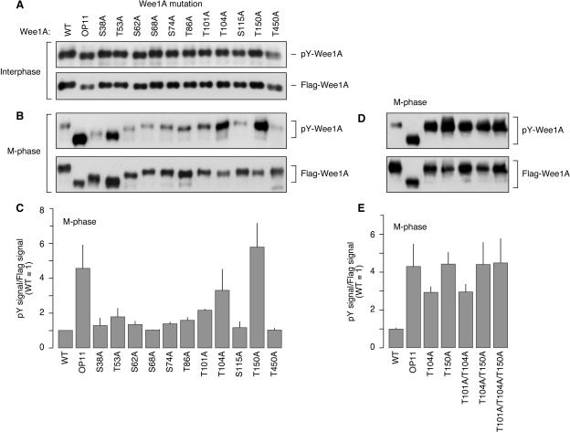
Mutating putative phosphorylation sites. (A and B) Effects of single alanine mutations on interphase (A) and M-phase (B) electrophoretic mobility and autophosphorylation of Wee1A. Wild-type (WT) Wee1A, OP11-Wee1A, and 11 individual alanine mutants were expressed, purified, and incubated with interphase extracts (A) or M-phase extracts (B). Wee1A protein levels and electrophoretic mobilities were assessed by Flag blotting. Wee1A autophosphorylation was assessed by antiphosphotyrosine immunoblotting. (C) M-phase autophosphorylation was quantified by densitometry and normalized to the amount of Wee1A protein used. Bars represent means ± standard errors (n = 3). (D and E) Thr 101, Thr 104, and Thr 150 were mutated combinatorially. The resulting autophosphorylation was assessed by phosphotyrosine immunoblotting, and the resulting gel shift was assessed by Flag blotting. Bars represent means ± standard errors (n = 2).
MALDI-TOF analysis of interphase and M-phase Wee1A.
Next we attempted to map the mitotic phosphorylation sites in wild-type Wee1A by MALDI-TOF MS. Flag bead-bound recombinant Wee1A was treated with interphase extracts, M-phase extracts, or no agent; separated on an SDS-polyacrylamide gel; visualized by Coomassie staining (Fig. 2A); excised from the gel; digested with trypsin; and subjected to MS. The peptides identified covered 44 to 60% of the total Wee1A sequence and included 5 of the 11 SP/TP residues (Fig. 2B). We found evidence for three phosphorylations in mitotic Wee1A (Fig. 2C and D). The first was on a peptide that comprised amino acids 45 to 56 and included one TP site, Thr 53 (TNNCPFPIT53PQR). The interphase Wee1A sample yielded a peak corresponding to the mass of nonphosphorylated TNNCPFPITPQR (1,458.7 Da), whereas in the M-phase sample the 1,458.7-Da peak disappeared and new peaks corresponding to phosphorylated TNNCPFPITPQR (1,538.7 Da) and phosphorylated TNNCPFPITPQRNER (1,937.9 Da) appeared (Fig. 2C and D). The second phosphorylation was on a peptide comprising amino acids 130 to 155 and again including one TP site, Thr 150 (FVAGTGAELDDPSLVNINPFT150PESYR). A peak corresponding to the nonphosphorylated peptide (2,809.4 Da) was detected in interphase Wee1A and was replaced by a peak corresponding to the phosphorylated peptide (2,889.4 Da) in M-phase Wee1A (Fig. 2C and D). The third possible phosphorylation was on a peptide that included amino acids 37 to 44, with one SP site, Ser 38 (GS38PVSSWR). A peak corresponding to nonphosphorylated GSPVSSWR (875.4 Da) was detected in interphase Wee1A and was not detected in M-phase Wee1A. No peptide corresponding to phosphorylated GSPVSSWR (955.4 Da) was detected in either interphase Wee1A or M-phase Wee1A; however, as described below, mutational analysis supported the conclusion that this peptide was in fact phosphorylated at Ser 38 in M phase.
FIG. 2.
Identification of mitotic phosphorylation sites in Wee1A. (A) Coomassie-stained gels of recombinant Wee1A incubated with buffer or extracts. (B) Summary of the peptides identified by MALDI-TOF MS. Coverage ranged from 44 to 60%. Peptides containing 5 of the 11 SP/TP sites were detected. WT, wild type. (C) MALDI-TOF MS spectrum of trypsin-digested interphase Wee1A. Three nonphosphorylated peptide peaks are indicated. Their observed m/z ratios are 875.4405 (identified as GSPVSSWR; theoretical m/z = 875.4375), 1,458.7101 (identified as TNNCPFPITPQR; theoretical m/z = 1,458.7164), and 2,809.3623 (identified as FVAGTGAELDDPSLVNINPFTPESYR; theoretical m/z = 2,809.3685). (D) MALDI-TOF MS spectrum of trypsin-digested M-phase Wee1A. Three phosphorylated peptide peaks are indicated. Their observed m/z ratios are 1,538.6667 [identified as TNNCPFPITPQR(-PO4); theoretical m/z = 1,538.6827], 1,937.8879 [identified as TNNCPFPITPQRNER(-PO4); theoretical m/z = 1,937.8693], and 2,889.3784 [identified as FVAGTGAELDDPSLVNINPFTPESYR(-PO4); theoretical m/z = 2,889.3348].
Mutational analysis of the SP/TP motifs.
Each of the three phosphorylated tryptic peptides identified in M-phase Wee1A contained one SP or TP motif, but each contained other potential phosphorylation sites as well. To determine whether the SP/TP motifs in these three peptides were actual phosphorylation sites, and to look for additional SP/TP phosphorylation sites in the regions of Wee1A from which tryptic peptides were not recovered, we constructed a series of Wee1A mutants in which each of the 11 SP/TP sites was individually replaced with alanine (S38A, T53A, S62A, S68A, S74A, T86A, T101A, T104A, S115A, T150A, and T450A). Each of these proteins was expressed and purified, incubated with interphase or M-phase extract, and subjected to anti-Flag immunoblotting to determine protein levels and electrophoretic mobilities and anti-phosphotyrosine immunoblotting to determine autophosphorylation.
In interphase, the electrophoretic mobilities and autophosphorylation of all of the Wee1A alanine mutants were similar to those of wild-type Wee1A and OP11-Wee1A (Fig. 3A). However, in M phase there were clear differences among the various mutants. First, the S38A-, T53A-, and S62A-Wee1A bands were only partially shifted in M phase (Fig. 3B). This finding established Ser 38 and Thr 53 as the phosphorylation sites in GS38PVSSWR and TNNCPFPIT53PQR, peptides whose phosphorylation had been established by the MALDI-TOF data in Fig. 2C and D, and added Ser 62 as an additional mitotic phosphorylation site. Although their mitotic gel shifts were incomplete, all of these mutants exhibited normal low levels of autophosphorylation in M phase (Fig. 3B and C). Note that the high T53A-Wee1A tyrosine phosphorylation signal in the top part of Fig. 3B reflects uneven protein loading; when normalized to the Flag-Wee1A signal and averaged, the T53A-Wee1A tyrosine phosphorylation was no higher than that of wild-type Wee1A (Fig. 3C).
Two additional mutants, T104A-Wee1A and T150A-Wee1A, were found to be normal with respect to the mitotic gel shift (Fig. 3B) but resistant to mitotic inactivation (Fig. 3C). The T104A and T150A mutants were approximately three- to sixfold higher in mitotic autophosphorylation than was wild-type Wee1A (Fig. 3C). In addition, their mitotic autophosphorylation was similar to the interphase autophosphorylation of all of the Wee1A proteins (Fig. 1C and 3A to C). This evidence established Thr 150 as the phosphorylation site in FVAGTGAELDDPSLVNINPFT150PESYR, consistent with the MALDI-TOF data (Fig. 2C and D), and added a fifth SP/TP site (Thr 104) to the list of mitotic phosphorylation sites.
Taken together, the MALDI-TOF MS data and mutagenesis data established five SP/TP sites as being phosphorylated in M-phase Wee1A. Three of the sites, Ser 38, Thr 53, and Ser 62, were required for the mitotic gel shift of Wee1A but were largely dispensable for mitotic inactivation of Wee1A, whereas two other sites, Thr 104 and Thr 150, were required for mitotic inactivation of Wee1A but were dispensable for the gel shift.
Multisite mutations.
We next examined how combinations of phosphorylation site mutations affected the mitotic inactivation and mitotic gel shift of Wee1A. In the first group, mutations at Ser 38, Thr 53, and Ser 62 were combined (S38A/T53A, T53A/S62, S38A/S62A, and S38A/T53A/S62A). All of these mutations decreased the mitotic gel shift of Wee1A, with the decrease being most pronounced in the S38A/T53A/S62A triple mutant. None of these mutations compromised the mitotic inactivation of Wee1A (data not shown).
Next we combined mutations of the two sites implicated in mitotic inactivation of Wee1A, Thr 104 and Thr 150. As shown in Fig. 3C, mutation of Thr 150 fully blocked mitotic inactivation of Wee1A whereas mutation of Thr 104 partially blocked it. The T104A/T150A double mutant was no more resistant to inactivation than the T150A single mutant (Fig. 3D and E). We also examined combinations of T101A with T104A and T150A. T101A/T104A-Wee1A had no greater activity than the T104A single mutant, and T101A/T104A/T150A-Wee1A had no greater activity than T104A/T150A (Fig. 3D and E). None of these mutations compromised the mitotic gel shift of Wee1A (Fig. 3D).
Finally, mutations at Ser 38, Thr 53, and Ser 62 were combined with a mutation at Thr 104 or Thr 150 (data not shown). In all cases, the status of Ser 38/Thr 53/Ser 62 determined the mitotic gel shift and the status of Thr 104/Thr 150 determined whether the protein could be inactivated in M phase.
Taken together, these findings indicate that the activity of Wee1A and the electrophoretic mobility of Wee1A are independently regulated by distinct sets of phosphorylations. The phosphorylations of three N-terminal sites, Ser 38, Thr 53, and Ser 62, combined to produce the gel shift seen in mitotic Wee1A. Moreover, they combined in a graded, additive fashion: OP11-Wee1A and S38A/T53A/S62A-Wee1A showed no mitotic gel shift, whereas the Wee1A mutants with one or two of the N-terminal sites showed partial shifts. Two other phosphorylation sites, Thr 104 and Thr 150, are required for mitotic inactivation of Wee1A. Mutating Thr 150 tended to have a stronger effect than mutation of Thr 104, but both Thr 104 and Thr 150 were required for full mitotic inactivation of Wee1A.
Wee1 stability.
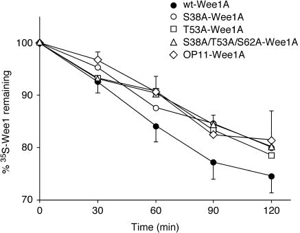
Previous work has demonstrated that Wee1A is relatively unstable in postreplication interphase extracts (21). Ser 38 phosphorylation has been implicated in the control of Wee1A degradation (2). We therefore asked whether any of our alanine mutations resulted in a change in Wee1A stability. Wild-type and mutant 35S-labeled Wee1A proteins were translated in reticulocyte lysates and added to chromatin-supplemented interphase Xenopus egg extracts. The amount of 35S-labeled Wee1A remaining was then assessed as a function of time. As shown in Fig. 4, wild-type Wee1A was degraded with a half-life of 260 ± 20 min. The S38A mutant had a significantly longer half-life, 364 ± 49 min, in qualitative (but not quantitative) agreement with the trend seen by Ayad et al. (2). The T53A mutant had a similarly long half-life, 353 ± 48 min, suggesting that Thr 53 phosphorylation is also required for Wee1A degradation. The half-lives of the S38A/T53A/S62A triple mutant and OP11-Wee1A were similar (373 ± 27 min and 380 ± 49 min, respectively) to those of the S38A and T53A single mutants (Fig. 4). These data suggest that the phosphorylation of multiple poorly conserved phosphorylation sites in the N terminus of Wee1A contributes to the regulation of Wee1A degradation.
FIG. 4.
Degradation of Wee1A mutants in interphase Xenopus egg extracts. Wild-type (wt) Wee1A and four phosphorylation site mutants (S38A-Wee1A, T53A-Wee1, S38A/T53A/S62A-Wee1A, and OP11-Wee1A) were translated in vitro in reticulocyte lysates in the presence of [35S]methionine. Wee1A proteins were mixed with chromatin-supplemented interphase extracts, and the amount of 35S-labeled Wee1A remaining was assessed as a function of time by phosphorimaging. The data points represent the averages of five to eight determinations per point from three independent experiments. Half-error (standard error) bars are shown for the wild-type Wee1A and OP11-Wee1 data; all other standard errors ranged between 1% and 6%. Half-times were calculated by nonlinear least-squares curve fitting to the equation Wee1Aremaining = 100 × (0.5)t/τ, where t represents time and τ represents the half-life. The calculated half-lives were as follows: wild-type Wee1A, 261 ± 20 min (standard error); S38A-Wee1A, 364 ± 49 min; T53A-Wee1A, 353 ± 48 min; S38A/T53A/S62A-Wee1A, 373 ± 27 min; OP11-Wee1A, 380 ± 49 min.
Generation of a phospho-Thr 150 antibody and phosphorylation of Thr 150 in vivo.
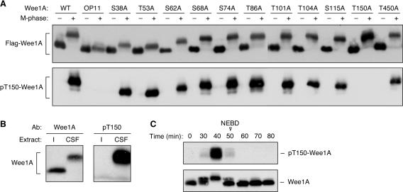
To see whether and when the Thr 150 residue is phosphorylated in vivo, we generated an affinity-purified antibody to phospho-Thr 150 (pT150). The specificity of the antibody was verified by incubating wild-type Wee1A, OP11-Wee1A, and each of the 11 single alanine mutations with buffer or M-phase extract and then subjecting the Wee1A proteins to immunoblotting with Flag antibody and pT150 antibody. As shown in Fig. 5A, the pT150 antibody recognized M-phase wild-type Wee1A but not buffer-treated Wee1A. Moreover, the pT150 antibody recognized the M-phase forms of all of the Wee1A mutants except OP11-Wee1A and T150A-Wee1A (Fig. 5A), the two mutants that lacked the Thr 150 phosphorylation site. These findings established the specificity of the antibody for Thr 150-phosphorylated forms of Wee1A and furthermore demonstrated that Thr 150 phosphorylation does not depend upon prior phosphorylation of Thr 104 (the other site whose phosphorylation was required for mitotic inactivation of Wee1A) or any of the other SP/TP sites. However, one Wee1 multisite alanine mutant—OP9-Wee1A, which has all of the SP/TP sites mutated except Thr 150 and Thr 450—was found not to undergo Thr 150 phosphorylation (data not shown). This could mean that Thr 150 phosphorylation depends upon the prior phosphorylation of at least one of the N-terminal SP/TP sites, even though no single individual site is required. It is also possible that the regulatory domain of OP9-Wee1A is misfolded, rendering Thr 150 unphosphorylatable.
FIG. 5.
Phosphorylation of Thr 150 in vitro and in cycling extracts. (A) Recognition of T150-phosphorylated forms of recombinant Wee1A by an affinity-purified pT150 antibody (Ab). (B) Endogenous Wee1A is phosphorylated at Thr 150 in CSF-arrested extracts but not in interphase extracts. (C) Time course of the Wee1A gel shift and Thr 150 phosphorylation in cycling extracts. Extracts underwent nuclear envelope breakdown (NEBD) at 50 min. WT, wild type.
Next we immunoblotted endogenous Wee1A from interphase extracts or CSF-arrested M-phase extracts. As shown in Fig. 5B, Wee1A antibodies detected Wee1A bands in both interphase and M phase. In contrast, the pT150 antibody detected only the hyperphosphorylated M-phase Wee1A protein.
Finally, we subjected cycling Xenopus egg extracts to immunoblot analysis with a Wee1A antibody and the pT150 antibody. As shown in Fig. 5C, Thr 150 was transiently phosphorylated just prior to nuclear envelope breakdown. The mitotic gel shift of Wee1A also preceded nuclear envelope breakdown, consistent with previous findings (43). However, the gel shift was half maximal by 30 min, at which time Thr 150 phosphorylation was barely detectable (Fig. 5C). Assuming that the shift was due to phosphorylation of Ser 38, Thr 53, and Ser 62, this finding implies that those phosphorylations generally precede the phosphorylation of Thr 150, even though they are not strictly required for Thr 150 phosphorylation (Fig. 5A). These results also imply that the “mitotic” phosphorylation of Wee1A may actually occur during late interphase.
Functional significance of the mitotic inactivation of Wee1A.
Having identified two sites (Thr 104 and Thr 150) required for mitotic inactivation of Wee1A, we can assess whether the inactivation of Wee1A is important for the mitotic activation of Cdk1 or, alternatively, is a backup mechanism that is normally unimportant because of the various additional ways of inactivating Wee1A (decreased transcription, decreased translation, and proteolysis). To this end, we compared the efficacies of wild-type Wee1A, T104A-Wee1A, T150A-Wee1, and OP11-Wee1A at blocking cyclin-driven Cdk1 activation. As shown in Fig. 6, both T104A-Wee1A and T150A-Wee1A inhibited mitotic entry more strongly than did wild-type Wee1A. This could be seen as a subtle shift to the right in the cyclin-versus-Cdk1 activity dose-response curves (Fig. 6A) and as a more dramatic decrease in the mitotic phosphorylation of p42 MAPK (Fig. 6B) (see reference 36 for other examples of relatively small changes in Cdk1 activity resulting in more pronounced changes in p42 MAPK phosphorylation). These findings suggest that the phosphorylation-mediated inactivation of Wee1A is important for mitotic entry.
FIG. 6.
Effectiveness of wild-type (WT) Wee1A, T104A-Wee1A, T150A-Wee1A, and OP11-Wee1A at inhibiting mitotic entry. Interphase extracts were supplemented with 40 nM Wee1A and various concentrations of nondegradable Δ90-cyclin B. Extracts were incubated for 120 min, and then aliquots were taken for H1 kinase assays (A and B, top) and phospho-MAPK blot assays (B, bottom).
OP11-Wee1A was somewhat more effective at inhibiting mitotic entry than either the T104A- or T150A-Wee1A mutant, as shown by both H1 kinase assays (Fig. 6A) and phospho-MAPK blots (Fig. 6B). This finding was somewhat surprising given that M-phase OP11-Wee1A was not detectably higher in its autophosphorylation than M-phase T150A-Wee1A (Fig. 3C and E). This suggests that the phosphorylation of Ser 38, Thr 53, and/or Ser 62, or possibly some additional unidentified sites, has some sort of negative effect on Wee1A function.
DISCUSSION
We have identified two sets of regulatory phosphorylation sites in mitotic Xenopus Wee1A. The first set consists of Ser 38, Thr 53, and Ser 62. These sites are all poorly conserved; none of the three SP/TP motifs is present in mouse embryonic Wee1, and only one of the three is present in human embryonic Wee1 (Fig. 1A). Alanine substitution at any of these sites decreases the mitotic gel shift of Wee1A (Fig. 3B), and mutating all three sites abolishes the gel shift (Fig. 1, 3, and 5 and data not shown). In addition, alanine substitution at Ser 38 and Thr 53 resulted in a modest stabilization of the Wee1A protein. This suggests that at least two of the sites that regulate Wee1's mitotic gel shift also regulate Wee1A stability. Perhaps the phosphorylation of Ser 38, Thr 53, and Ser 62 results in some gross conformational change that produces both an electrophoretic mobility shift and a change in the interaction of Wee1A with ubiquitin ligases like Tome-1 (2).
The second set of phosphorylation sites consists of Thr 104 and Thr 150. These sites are perfectly conserved in the other species of embryonic Wee1 (Fig. 1A) and are conserved in the somatic Wee1 proteins and the single Drosophila Dwee1 protein as well (Fig. 7). The sequences around the two sites are similar to each other as well, with the notable exception of the +2 position: at the proximal site (Thr 104 in Xenopus Wee1A) this residue is always a lysine, whereas at the distal site (Thr 150 in Xenopus Wee1A) it is an aspartic acid or a glutamic acid (Fig. 7). Neither Thr 104 nor Thr 150 is important for the mitotic gel shift of Wee1A, but they are crucial for mitotic inactivation of Wee1A (Fig. 3B to E). Both sites must be present for mitotic inactivation of Wee1A to proceed normally, although the resistance to inactivation is somewhat more pronounced in the T150A mutants than in the T104A mutants (Fig. 3C and E). Thr 104 is required for inactivation but is not required for Thr 150 phosphorylation (Fig. 5A). It is possible that Thr 150 phosphorylation is required for Thr 104 phosphorylation, but given that the T150A phenotype is somewhat stronger than the T104A phenotype, it seems likely that Thr 150 phosphorylation has an effect on Wee1A activity above and beyond any possible effect on Thr 104 phosphorylation. Overall, the logic of Wee1A inactivation appears to be analogous to that of MAPK activation, with the phosphorylation of two well-conserved sites cooperating to produce an effect.
FIG. 7.
Conservation of the sequence around the Thr 104 and Thr 150 phosphorylation sites.
Thus, the mitotic gel shift and degradation of Wee1A, on the one hand, and the mitotic inactivation of Wee1A, on the other, appear to be strictly separable aspects of Wee1A regulation. The S38A and T53A mutants showed decreased gel shifts and proteolysis but no resistance to mitotic inactivation. Conversely, the T104A and T150A mutants were resistant to inactivation but exhibited normal gel shifts.
We also showed that Thr 150 is phosphorylated in vivo in M-phase Xenopus egg extracts and just prior to M phase in cycling Xenopus egg extracts (Fig. 5). These findings are consistent with a role for M-phase Wee1A inactivation in maintaining the positive feedback that helps maintain mitosis, as well as a role for late-interphase inactivation of Wee1A in triggering M-phase entry. It will be of particular interest to determine what kinase(s) is responsible for the phosphorylation of Thr 104 and Thr 150 during late interphase and mitosis.
The identification of Thr 104 and Thr 150 as sites that are required for the inactivation of Wee1A allowed us to assess whether or not mitotic inactivation of Wee1A is important for normal M-phase entry. On the basis of quantitative titration experiments comparing the biological activity of wild-type Wee1A with that of T104A-Wee1A and T150A-Wee1A, we conclude that it is (Fig. 6). This does not rule out the possibility that other regulatory mechanisms, such as proteolysis, translational regulation, and transcriptional regulation, also contribute to the control of Wee1A at the G2/M boundary. Indeed, the fact that OP11-Wee1A had a somewhat stronger effect on M-phase entry than T150A-Wee1A did, despite the two proteins' indistinguishable in vitro activities, suggests that the control of Wee1A is shared by multiple regulatory mechanisms.
Wee1A is an important component of a double-negative feedback loop: active Wee1A inhibits cyclin B-Cdk1, while active cyclin B-Cdk1 inhibits Wee1A. Loops of this sort have the potential to function as a bistable switch (29, 42), and indeed recent experimental work indicates that the Cdk1/Wee1A/Cdc25 system generates bistable steady-state responses (36, 39) and helps produce sustained cell cycle oscillations (35). For the Cdk1/Wee1A/Cdc25 system to produce a bistable response, it is helpful if some component of the intertwined loops exhibits a highly ultrasensitive response to its upstream activator or inhibitor (10, 11). The multisite phosphorylation of Wee1A could provide a mechanism for generating the required ultrasensitivity, either through the two-step phosphorylation of Thr 104 and Thr 150 (which could yield a Hill coefficient between 1 and 2, depending upon the kinetic constants for the two phosphorylation-dephosphorylation reactions) or through competition among all five mitotic phosphorylation sites for access to their regulatory kinase(s) and phosphatase(s), which potentially could yield higher Hill coefficients (18). It will be of interest to determine how quantitative aspects of the regulation of Wee1A contribute to the overall function of the cell cycle oscillator.
Since the original submission of the manuscript, Harvey and coworkers have published an important study of the mitotic phosphorylation of Swe1, the budding yeast homolog of Wee1 (12). Like Wee1, Swe1 possesses a C-terminal kinase domain and an N-terminal regulatory domain. There is no obvious sequence conservation between the N termini of Swe1 and Wee1A or -B, but the Swe1 regulatory domain possesses numerous potential Cdk phosphorylation sites, as do the Wee1A and -B regulatory domains. And as was true of Wee1A, the mitotic phosphorylation of Swe1 had not been previously examined in detail.
There are a number of interesting similarities in the findings of our two studies. Both Swe1 and Wee1A undergo multisite phosphorylation during mitosis, with Swe1 becoming phosphorylated at at least 28 sites in vivo (12), compared with the 5 identified here for Wee1A. Swe1 hyperphosphorylation was found to depend upon the continual presence of Cdc28 activity (12); we have found similar evidence for Wee1A (data not shown).
However, there are interesting differences as well. Many of the Swe1 phosphorylation sites, including 11/19 sites phosphorylated by Cdc28 in vitro, do not conform to the canonical S/TP Cdk consensus. In contrast, we have not yet identified any noncanonical Cdk phosphorylation sites in Wee1A. The possibility of noncanonical phosphorylation of Wee1A by Cdk1 (particularly in the 40% of the mitotic Wee1A sequence that was not covered in our MS analysis) certainly warrants further investigation.
Moreover, there were striking differences in the behaviors of alanine substitution mutants in the two studies. Harvey et al. found that replacement of 18 Swe1 phosphorylation sites with alanine residues resulted in a mutant that was almost completely resistant to the mitotic mobility shift, similar to the absence of a shift that we saw in OP11-Wee1A and the Ser 38/Thr 53/Ser 62-Wee1A proteins. However, whereas we found that OP11-Wee1A had a normal kinase activity in interphase and an elevated kinase activity in M phase and was more effective than wild-type Wee1A in delaying M-phase entry, Harvey et al. found that swe1-18A cells had low basal Swe1 activity and entered mitosis prematurely. This constitutes a major difference in behavior between the two Wee1 proteins: Swe1 can apparently be positively regulated by Cdc28, whereas we and others have thus far found evidence only for negative regulation of Xenopus Wee1A by mitotic phosphorylation.
Acknowledgments
We thank Joe Pomerening and Jianbo Yue for help with extract preparation and members of the Ferrell lab for advice and critical reading of the manuscript.
This work was supported by grants from the National Institutes of Health (R01 GM46383) and from KOSEF (FPR05AZ-480).
REFERENCES
- 1.Atherton-Fessler, S., G. Hannig, and H. Piwnica-Worms. 1993. Reversible tyrosine phosphorylation and cell cycle control. Semin. Cell Biol. 4:433-442. [DOI] [PubMed] [Google Scholar]
- 2.Ayad, N. G., S. Rankin, M. Murakami, J. Jebanathirajah, S. Gygi, and M. W. Kirschner. 2003. Tome-1, a trigger of mitotic entry, is degraded during G1 via the APC. Cell 113:101-113. [DOI] [PubMed] [Google Scholar]
- 3.Berry, L. D., and K. L. Gould. 1996. Regulation of Cdc2 activity by phosphorylation at T14/Y15. Prog. Cell Cycle Res. 2:99-105. [DOI] [PubMed] [Google Scholar]
- 4.Busino, L., M. Chiesa, G. F. Draetta, and M. Donzelli. 2004. Cdc25A phosphatase: combinatorial phosphorylation, ubiquitylation and proteolysis. Oncogene 23:2050-2056. [DOI] [PubMed] [Google Scholar]
- 5.Charlesworth, A., J. Welk, and A. M. MacNicol. 2000. The temporal control of Wee1 mRNA translation during Xenopus oocyte maturation is regulated by cytoplasmic polyadenylation elements within the 3′-untranslated region. Dev. Biol. 227:706-719. [DOI] [PubMed] [Google Scholar]
- 6.Chen, M. S., J. Hurov, L. S. White, T. Woodford-Thomas, and H. Piwnica-Worms. 2001. Absence of apparent phenotype in mice lacking Cdc25C protein phosphatase. Mol. Cell. Biol. 21:3853-3861. [DOI] [PMC free article] [PubMed] [Google Scholar]
- 7.Coleman, T. R., and W. G. Dunphy. 1994. Cdc2 regulatory factors. Curr. Opin. Cell Biol. 6:877-882. [DOI] [PubMed] [Google Scholar]
- 8.Featherstone, C., and P. Russell. 1991. Fission yeast p107wee1 mitotic inhibitor is a tyrosine/serine kinase. Nature 349:808-811. [DOI] [PubMed] [Google Scholar]
- 9.Ferguson, A. M., L. S. White, P. J. Donovan, and H. Piwnica-Worms. 2005. Normal cell cycle and checkpoint responses in mice and cells lacking Cdc25B and Cdc25C protein phosphatases. Mol. Cell. Biol. 25:2853-2860. [DOI] [PMC free article] [PubMed] [Google Scholar]
- 10.Ferrell, J. E. 2002. Self-perpetuating states in signal transduction: positive feedback, double-negative feedback and bistability. Curr. Opin. Cell Biol. 14:140-148. [DOI] [PubMed] [Google Scholar]
- 11.Ferrell, J. E., Jr., and W. Xiong. 2001. Bistability in cell signaling: how to make continuous processes discontinuous, and reversible processes irreversible. Chaos 11:227-236. [DOI] [PubMed] [Google Scholar]
- 12.Harvey, S. L., A. Charlet, W. Haas, S. P. Gygi, and D. R. Kellogg. 2005. Cdk1-dependent regulation of the mitotic inhibitor Wee1. Cell 122:407-420. [DOI] [PubMed] [Google Scholar]
- 13.Hutchins, J. R., and P. R. Clarke. 2004. Many fingers on the mitotic trigger: post-translational regulation of the Cdc25C phosphatase. Cell Cycle 3:41-45. [PubMed] [Google Scholar]
- 14.Katayama, K., N. Fujita, and T. Tsuruo. 2005. Akt/protein kinase B-dependent phosphorylation and inactivation of WEE1Hu promote cell cycle progression at G2/M transition. Mol. Cell. Biol. 25:5725-5737. [DOI] [PMC free article] [PubMed] [Google Scholar]
- 15.Lee, J., A. Kumagai, and W. G. Dunphy. 2001. Positive regulation of Wee1 by Chk1 and 14-3-3 proteins. Mol. Biol. Cell 12:551-563. [DOI] [PMC free article] [PubMed] [Google Scholar]
- 16.Leise, W., III, and P. R. Mueller. 2002. Multiple Cdk1 inhibitory kinases regulate the cell cycle during development. Dev. Biol. 249:156-173. [DOI] [PubMed] [Google Scholar]
- 17.Lundgren, K., N. Walworth, R. Booher, M. Dembski, M. Kirschner, and D. Beach. 1991. mik1 and wee1 cooperate in the inhibitory tyrosine phosphorylation of cdc2. Cell 64:1111-1122. [DOI] [PubMed] [Google Scholar]
- 18.Markevich, N. I., J. B. Hoek, and B. N. Kholodenko. 2004. Signaling switches and bistability arising from multisite phosphorylation in protein kinase cascades. J. Cell Biol. 164:353-359. [DOI] [PMC free article] [PubMed] [Google Scholar]
- 19.McGowan, C. H., and P. Russell. 1995. Cell cycle regulation of human WEE1. EMBO J. 14:2166-2175. [DOI] [PMC free article] [PubMed] [Google Scholar]
- 20.McGowan, C. H., and P. Russell. 1993. Human Wee1 kinase inhibits cell division by phosphorylating p34cdc2 exclusively on Tyr15. EMBO J. 12:75-85. [DOI] [PMC free article] [PubMed] [Google Scholar]
- 21.Michael, W. M., and J. Newport. 1998. Coupling of mitosis to the completion of S phase through Cdc34-mediated degradation of Wee1. Science 282:1886-1889. (Errata, 283: 35 and 2102, 1999.) [DOI] [PubMed] [Google Scholar]
- 22.Mueller, P. R., T. R. Coleman, and W. G. Dunphy. 1995. Cell cycle regulation of a Xenopus Wee1-like kinase. Mol. Biol. Cell 6:119-134. [DOI] [PMC free article] [PubMed] [Google Scholar]
- 23.Murakami, M. S., T. D. Copeland, and G. F. Vande Woude. 1999. Mos positively regulates Xe-Wee1 to lengthen the first mitotic cell cycle of Xenopus. Genes Dev. 13:620-631. [DOI] [PMC free article] [PubMed] [Google Scholar]
- 24.Murakami, M. S., S. A. Moody, I. O. Daar, and D. K. Morrison. 2004. Morphogenesis during Xenopus gastrulation requires Wee1-mediated inhibition of cell proliferation. Development 131:571-580. [DOI] [PubMed] [Google Scholar]
- 25.Murakami, M. S., and G. F. Vande Woude. 1998. Analysis of the early embryonic cell cycles of Xenopus; regulation of cell cycle length by Xe-wee1 and Mos. Development 125:237-248. [DOI] [PubMed] [Google Scholar]
- 26.Murray, A. W. 1991. Cell cycle extracts. Methods Cell Biol. 36:581-605. [PubMed] [Google Scholar]
- 27.Nakajo, N., S. Yoshitome, J. Iwashita, M. Iida, K. Uto, S. Ueno, K. Okamoto, and N. Sagata. 2000. Absence of Wee1 ensures the meiotic cell cycle in Xenopus oocytes. Genes Dev. 14:328-338. [PMC free article] [PubMed] [Google Scholar]
- 28.Nakanishi, M., H. Ando, N. Watanabe, K. Kitamura, K. Ito, H. Okayama, T. Miyamoto, T. Agui, and M. Sasaki. 2000. Identification and characterization of human Wee1B, a new member of the Wee1 family of Cdk-inhibitory kinases. Genes Cells 5:839-847. [DOI] [PubMed] [Google Scholar]
- 29.Novak, B., and J. J. Tyson. 1993. Numerical analysis of a comprehensive model of M-phase control in Xenopus oocyte extracts and intact embryos. J. Cell Sci. 106:1153-1168. [DOI] [PubMed] [Google Scholar]
- 30.Nurse, P. 1975. Genetic control of cell size at cell division in yeast. Nature 256:547-551. [DOI] [PubMed] [Google Scholar]
- 31.Nurse, P. 2004. Wee beasties. Nature 432:557. [DOI] [PubMed] [Google Scholar]
- 32.Okamoto, K., N. Nakajo, and N. Sagata. 2002. The existence of two distinct Wee1 isoforms in Xenopus: implications for the developmental regulation of the cell cycle. EMBO J. 21:2472-2484. [DOI] [PMC free article] [PubMed] [Google Scholar]
- 33.Parker, L. L., S. Atherton-Fessler, M. S. Lee, S. Ogg, J. L. Falk, K. I. Swenson, and H. Piwnica-Worms. 1991. Cyclin promotes the tyrosine phosphorylation of p34cdc2 in a wee1+ dependent manner. EMBO J. 10:1255-1263. [DOI] [PMC free article] [PubMed] [Google Scholar]
- 34.Parker, L. L., P. J. Sylvestre, M. J. Byrnes III, F. Liu, and H. Piwnica-Worms. 1995. Identification of a 95-kDa WEE1-like tyrosine kinase in HeLa cells. Proc. Natl. Acad. Sci. USA 92:9638-9642. [DOI] [PMC free article] [PubMed] [Google Scholar]
- 35.Pomerening, J. R., S. Y. Kim, and J. E. Ferrell, Jr. 2005. Systems-level dissection of the cell cycle oscillator: bypassing positive feedback produces damped oscillations. Cell 122:565-578. [DOI] [PubMed] [Google Scholar]
- 36.Pomerening, J. R., E. D. Sontag, and J. E. Ferrell, Jr. 2003. Building a cell cycle oscillator: hysteresis and bistability in the activation of Cdc2. Nat. Cell Biol. 5:346-351. [DOI] [PubMed] [Google Scholar]
- 37.Rothblum-Oviatt, C. J., C. E. Ryan, and H. Piwnica-Worms. 2001. 14-3-3 binding regulates catalytic activity of human Wee1 kinase. Cell Growth Differ. 12:581-589. [PubMed] [Google Scholar]
- 38.Russell, P., and P. Nurse. 1987. Negative regulation of mitosis by wee1+, a gene encoding a protein kinase homolog. Cell 49:559-567. [DOI] [PubMed] [Google Scholar]
- 39.Sha, W., J. Moore, K. Chen, A. D. Lassaletta, C. S. Yi, J. J. Tyson, and J. C. Sible. 2003. Hysteresis drives cell-cycle transitions in Xenopus laevis egg extracts. Proc. Natl. Acad. Sci. USA 100:975-980. [DOI] [PMC free article] [PubMed] [Google Scholar]
- 40.Smythe, C., and J. W. Newport. 1991. Systems for the study of nuclear assembly, DNA replication, and nuclear breakdown in Xenopus laevis egg extracts. Methods Cell Biol. 35:449-468. [DOI] [PubMed] [Google Scholar]
- 41.Tang, Z., T. R. Coleman, and W. G. Dunphy. 1993. Two distinct mechanisms for negative regulation of the Wee1 protein kinase. EMBO J. 12:3427-3436. [DOI] [PMC free article] [PubMed] [Google Scholar]
- 42.Thron, C. D. 1996. A model for a bistable biochemical trigger of mitosis. Biophys. Chem. 57:239-251. [DOI] [PubMed] [Google Scholar]
- 43.Walter, S. A., T. M. Guadagno, and J. E. Ferrell, Jr. 1997. Induction of a G2-phase arrest in Xenopus egg extracts by activation of p42 MAP kinase. Mol. Biol. Cell 8:2157-2169. [DOI] [PMC free article] [PubMed] [Google Scholar]
- 44.Wang, Y., C. Jacobs, K. E. Hook, H. Duan, R. N. Booher, and Y. Sun. 2000. Binding of 14-3-3β to the carboxyl terminus of Wee1 increases Wee1 stability, kinase activity, and G2-M cell population. Cell Growth Differ. 11:211-219. [PubMed] [Google Scholar]
- 45.Watanabe, N., H. Arai, Y. Nishihara, M. Taniguchi, T. Hunter, and H. Osada. 2004. M-phase kinases induce phospho-dependent ubiquitination of somatic Wee1 by SCFβ-TrCP. Proc. Natl. Acad. Sci. USA 101:4419-4424. [DOI] [PMC free article] [PubMed] [Google Scholar]
- 46.Watanabe, N., M. Broome, and T. Hunter. 1995. Regulation of the human WEE1Hu CDK tyrosine 15-kinase during the cell cycle. EMBO J. 14:1878-1891. [DOI] [PMC free article] [PubMed] [Google Scholar]


