Abstract
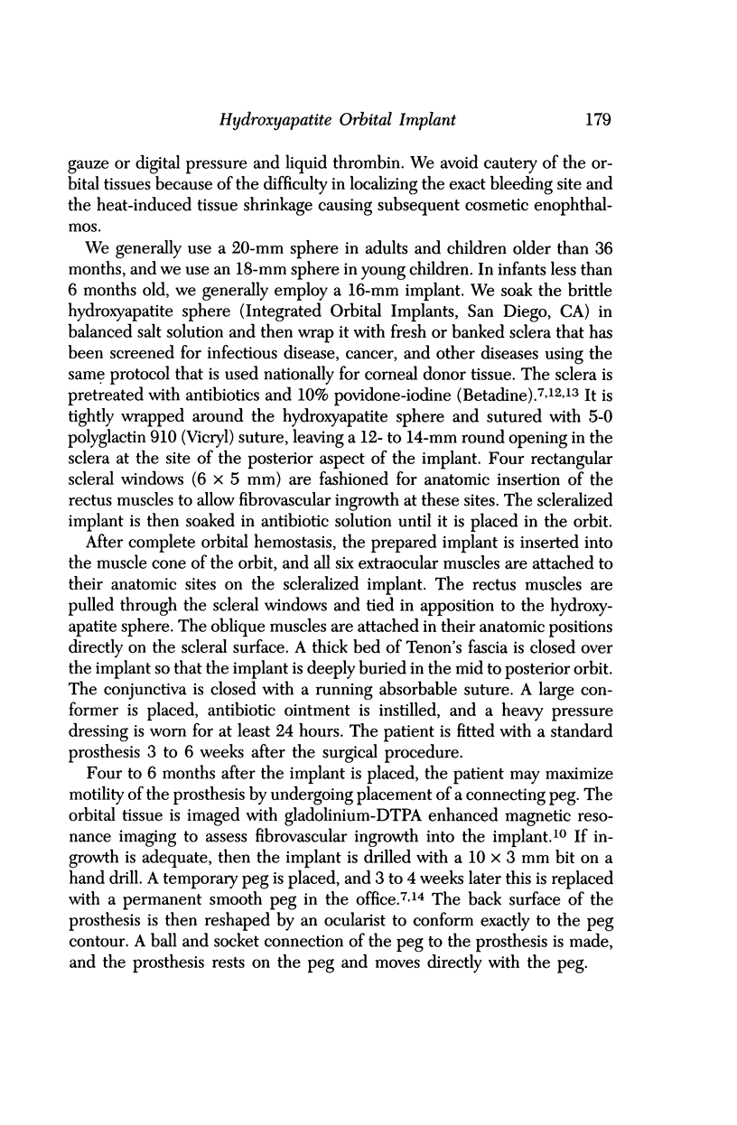
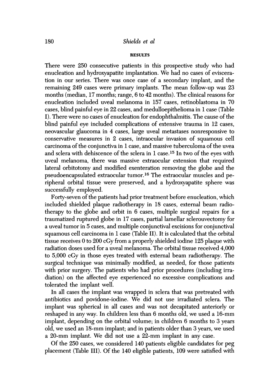
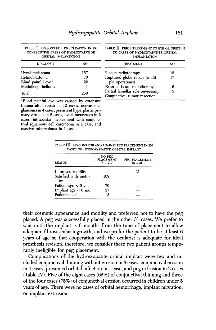
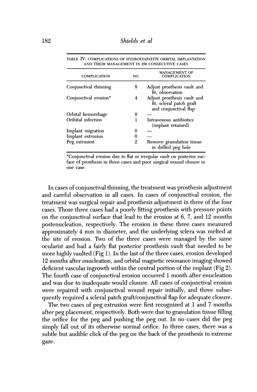
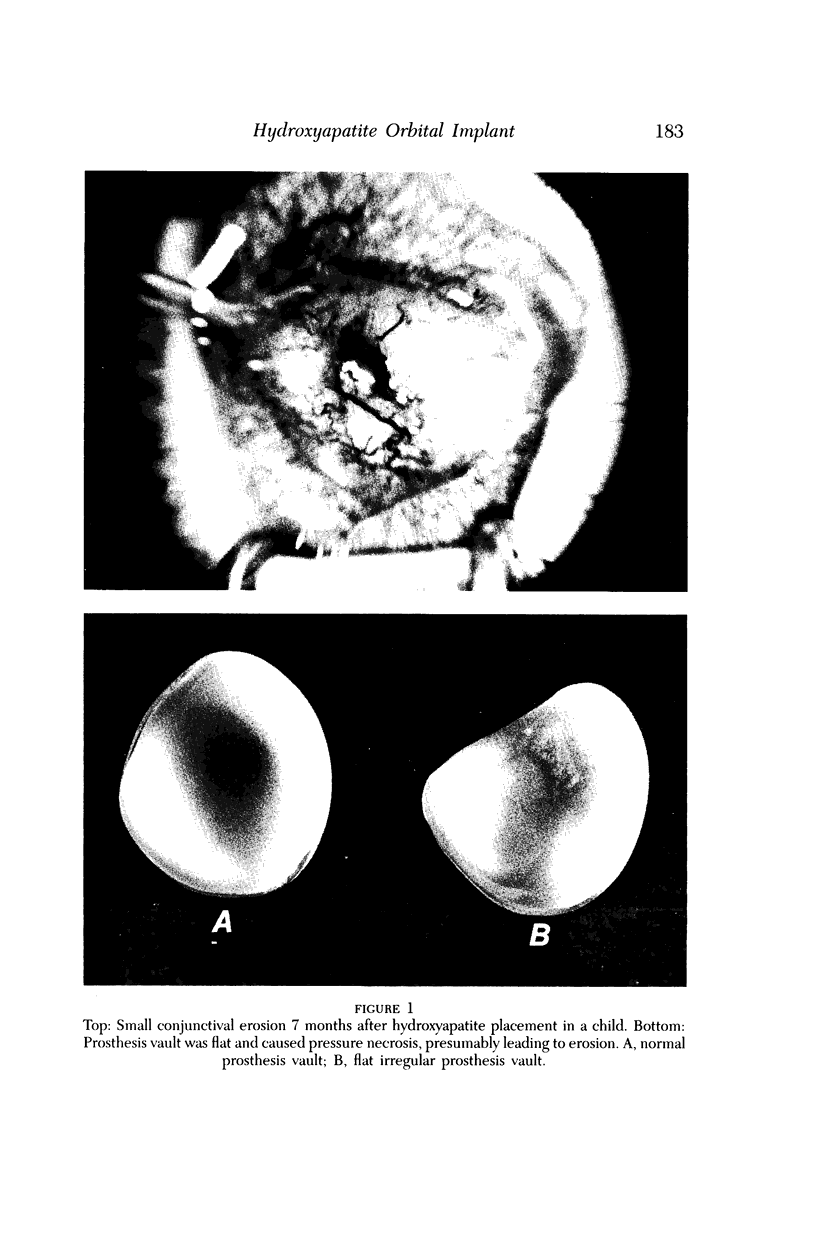
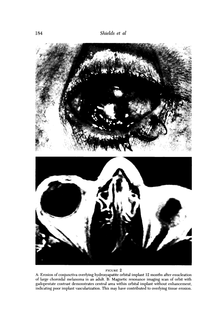
The coral-derived hydroxyapatite sphere is a popular, new integrated orbital implant designed to provide improved motility of the ocular prosthesis following enucleation. Although the implant has rapidly become widely used by ophthalmologists, there is little information available regarding the complications of this technique in a large series of cases. We report our results on our initial 250 consecutive cases of hydroxyapatite implantation for eyes enucleated primarily for intraocular neoplasms, with specific emphasis on the complication an their management. The reasons for enucleation included uveal melanoma (157 cases), retinoblastoma (70 cases), blind painful eye (22 cases), and intraocular medulloepithelioma (1 case). Prior treatment to the eye was performed before enucleation in 47 cases and included repair of ruptured globe (17 cases), plaque radiotherapy (18 cases), external beam radiotherapy (6 cases), and others (6 cases). During a mean of 23 months follow-up (range, 6 to 42 months), there have been no recognizable cases of orbital hemorrhage related to the implant and no cases of implant extrusion or implant migration. There was one case of presumed orbital infection (culture-negative) that resolved with intravenous antibiotics, and the implant was retained within the orbit. Other problems included conjunctival thinning in eight cases managed by observation and prosthesis adjustment and conjunctival erosion in four cases managed by combinations of scleral patch graft, conjunctival flap, and prosthesis adjustment. The conjunctival erosion was caused by a poorly fitting prosthesis in three cases and wound dehiscence in one case. The complication rate in eyes receiving prior radiotherapy or surgery was not increased. The hydroxyapatite integrated orbital implant is a well-tolerated motility implant without the high rate of extrusion and infection seen with other motility implants.
Full text
PDF
Images in this article
Selected References
These references are in PubMed. This may not be the complete list of references from this article.
- Benevento W. J., Murray P., Reed C. A., Pepose J. S. The sensitivity of Neisseria gonorrhoeae, Chlamydia trachomatis, and herpes simplex type II to disinfection with povidone-iodine. Am J Ophthalmol. 1990 Mar 15;109(3):329–333. doi: 10.1016/s0002-9394(14)74560-x. [DOI] [PubMed] [Google Scholar]
- Buettner H., Bartley G. B. Tissue breakdown and exposure associated with orbital hydroxyapatite implants. Am J Ophthalmol. 1992 Jun 15;113(6):669–673. doi: 10.1016/s0002-9394(14)74792-0. [DOI] [PubMed] [Google Scholar]
- De Potter P., Shields C. L., Shields J. A., Flanders A. E., Rao V. M. Role of magnetic resonance imaging in the evaluation of the hydroxyapatite orbital implant. Ophthalmology. 1992 May;99(5):824–830. doi: 10.1016/s0161-6420(92)31918-9. [DOI] [PubMed] [Google Scholar]
- De Potter P., Shields J. A., Shields C. L., Santos R. Modified enucleation via lateral orbitotomy for choroidal melanoma with orbital extension: a report of two cases. Ophthal Plast Reconstr Surg. 1992;8(2):109–113. doi: 10.1097/00002341-199206000-00004. [DOI] [PubMed] [Google Scholar]
- Dutton J. J. Coralline hydroxyapatite as an ocular implant. Ophthalmology. 1991 Mar;98(3):370–377. doi: 10.1016/s0161-6420(91)32304-2. [DOI] [PubMed] [Google Scholar]
- Dutton J. J. Coralline hydroxyapatite as an ocular implant. Ophthalmology. 1991 Mar;98(3):370–377. doi: 10.1016/s0161-6420(91)32304-2. [DOI] [PubMed] [Google Scholar]
- Dutton J. J. Coralline hydroxyapatite as an ocular implant. Ophthalmology. 1991 Mar;98(3):370–377. doi: 10.1016/s0161-6420(91)32304-2. [DOI] [PubMed] [Google Scholar]
- Goldberg R. A., Holds J. B., Ebrahimpour J. Exposed hydroxyapatite orbital implants. Report of six cases. Ophthalmology. 1992 May;99(5):831–836. doi: 10.1016/s0161-6420(92)31920-7. [DOI] [PubMed] [Google Scholar]
- Goldberg R. A., Holds J. B., Ebrahimpour J. Exposed hydroxyapatite orbital implants. Report of six cases. Ophthalmology. 1992 May;99(5):831–836. doi: 10.1016/s0161-6420(92)31920-7. [DOI] [PubMed] [Google Scholar]
- Harbison M. A., Hammer S. M. Inactivation of human immunodeficiency virus by Betadine products and chlorhexidine. J Acquir Immune Defic Syndr. 1989;2(1):16–20. [PubMed] [Google Scholar]
- Holmes R. E. Bone regeneration within a coralline hydroxyapatite implant. Plast Reconstr Surg. 1979 May;63(5):626–633. doi: 10.1097/00006534-197905000-00004. [DOI] [PubMed] [Google Scholar]
- Kenney E. B., Lekovic V., Sa Ferreira J. C., Han T., Dimitrijevic B., Carranza F. A., Jr Bone formation within porous hydroxylapatite implants in human periodontal defects. J Periodontol. 1986 Feb;57(2):76–83. doi: 10.1902/jop.1986.57.2.76. [DOI] [PubMed] [Google Scholar]
- Merriam J. C., Rosskothen H. D., Benedek E. Right-angled peg for hydroxyapatite orbital implant. Ophthalmic Surg. 1992 Aug;23(8):564–564. [PubMed] [Google Scholar]
- Nerad J. A., Hurtig R. R., Carter K. D., Bulgarelli D. M., Yeager D. C. A system for measurement of prosthetic eye movements using a magnetic search coil technique. Ophthal Plast Reconstr Surg. 1991;7(1):31–40. doi: 10.1097/00002341-199103000-00004. [DOI] [PubMed] [Google Scholar]
- Piecuch J. F. Extraskeletal implantation of a porous hydroxyapatite ceramic. J Dent Res. 1982 Dec;61(12):1458–1460. doi: 10.1177/00220345820610121801. [DOI] [PubMed] [Google Scholar]
- Piecuch J. F., Topazian R. G., Skoly S., Wolfe S. Experimental ridge augmentation with porous hydroxyapatite implants. J Dent Res. 1983 Feb;62(2):148–154. doi: 10.1177/00220345830620021301. [DOI] [PubMed] [Google Scholar]
- Regillo C. D., Shields C. L., Shields J. A., Eagle R. C., Jr, Lehr J. Ocular tuberculosis. JAMA. 1991 Sep 18;266(11):1490–1490. [PubMed] [Google Scholar]
- Rosen H. M. Surgical correction of the vertically deficient chin. Plast Reconstr Surg. 1988 Aug;82(2):247–256. doi: 10.1097/00006534-198808000-00006. [DOI] [PubMed] [Google Scholar]
- Sartoris D. J., Gershuni D. H., Akeson W. H., Holmes R. E., Resnick D. Coralline hydroxyapatite bone graft substitutes: preliminary report of radiographic evaluation. Radiology. 1986 Apr;159(1):133–137. doi: 10.1148/radiology.159.1.3513246. [DOI] [PubMed] [Google Scholar]
- Saunders P. P., Alvarez E., Kantarjian H. M. Determination of nicotinamide-adenine dinucleotide and thiazole-4-carboxamide-adenine dinucleotide in human leukocytes by reversed-phase high-performance liquid chromatography. J Chromatogr. 1992 May 20;577(1):37–41. doi: 10.1016/0378-4347(92)80596-i. [DOI] [PubMed] [Google Scholar]
- Shields C. L., Shields J. A., De Potter P. Hydroxyapatite orbital implant after enucleation. Experience with initial 100 consecutive cases. Arch Ophthalmol. 1992 Mar;110(3):333–338. doi: 10.1001/archopht.1992.01080150031022. [DOI] [PubMed] [Google Scholar]
- Shields C. L., Shields J. A., Eagle R. C., Jr, De Potter P. Histopathologic evidence of fibrovascular ingrowth four weeks after placement of the hydroxyapatite orbital implant. Am J Ophthalmol. 1991 Mar 15;111(3):363–366. doi: 10.1016/s0002-9394(14)72323-2. [DOI] [PubMed] [Google Scholar]
- Shields J. A., Shields C. L., De Potter P. Enucleation technique for children with retinoblastoma. J Pediatr Ophthalmol Strabismus. 1992 Jul-Aug;29(4):213–215. doi: 10.3928/0191-3913-19920701-06. [DOI] [PubMed] [Google Scholar]
- Soll D. B. The anophthalmic socket. Ophthalmology. 1982 May;89(5):407–423. doi: 10.1016/s0161-6420(82)34774-0. [DOI] [PubMed] [Google Scholar]
- White E., Shors E. C. Biomaterial aspects of Interpore-200 porous hydroxyapatite. Dent Clin North Am. 1986 Jan;30(1):49–67. [PubMed] [Google Scholar]


