Abstract
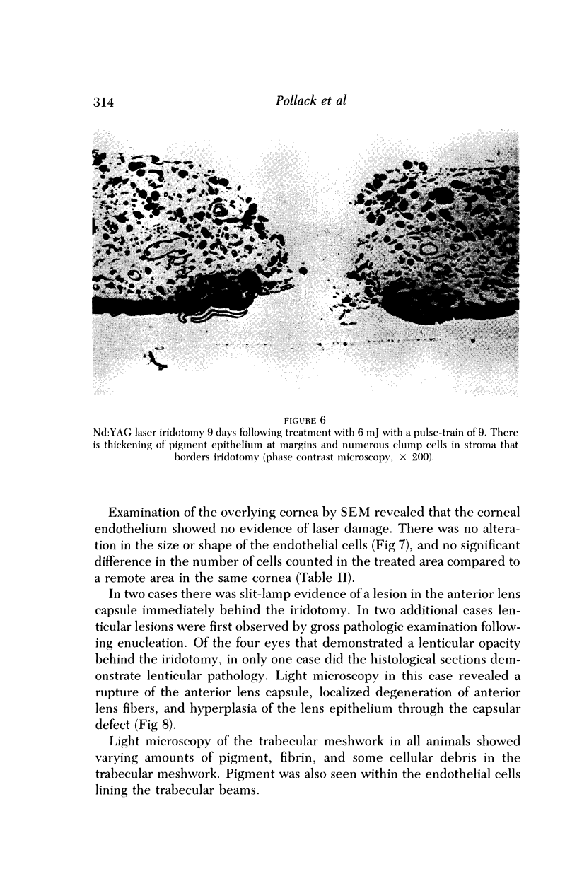
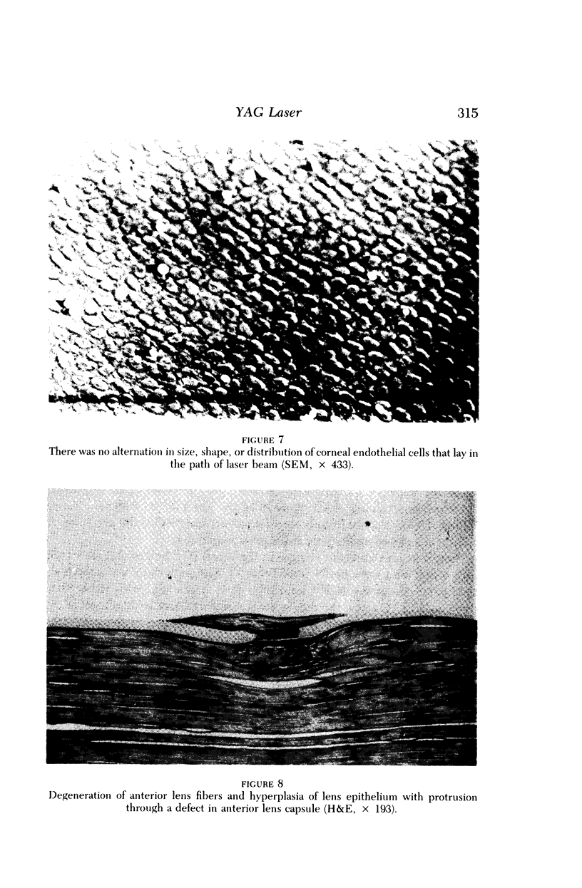
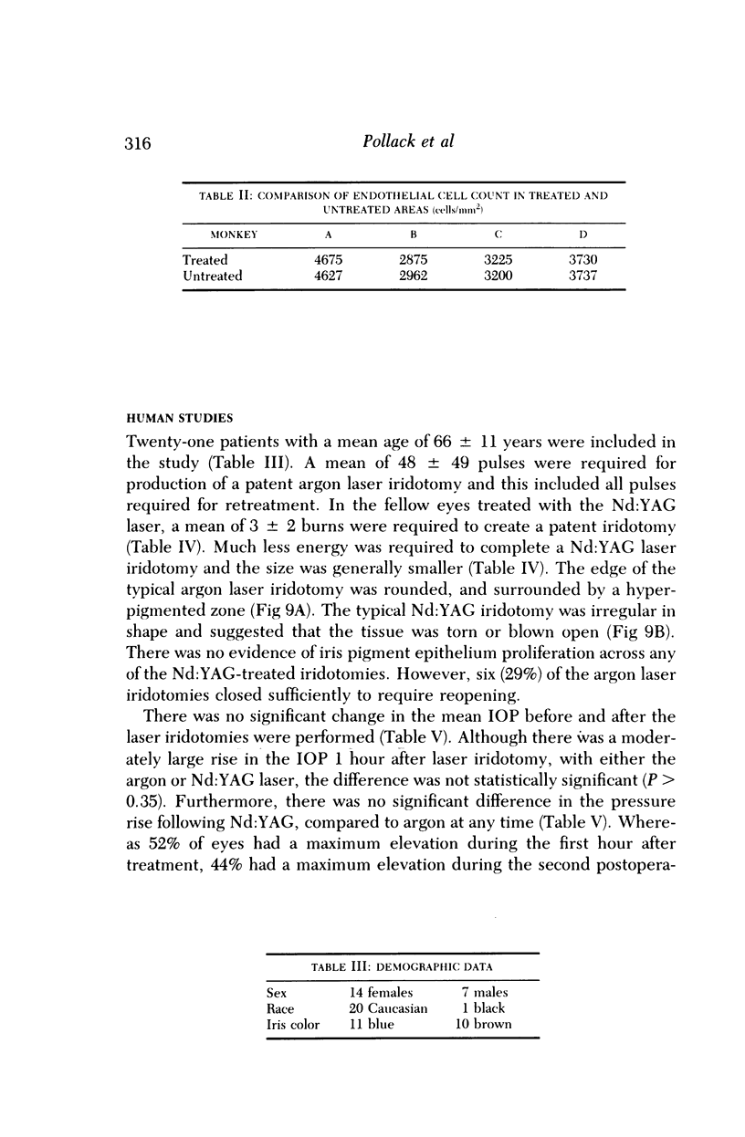
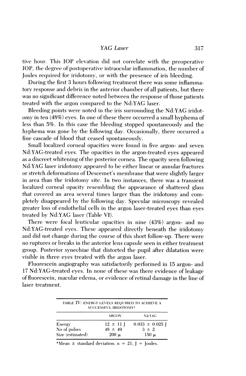
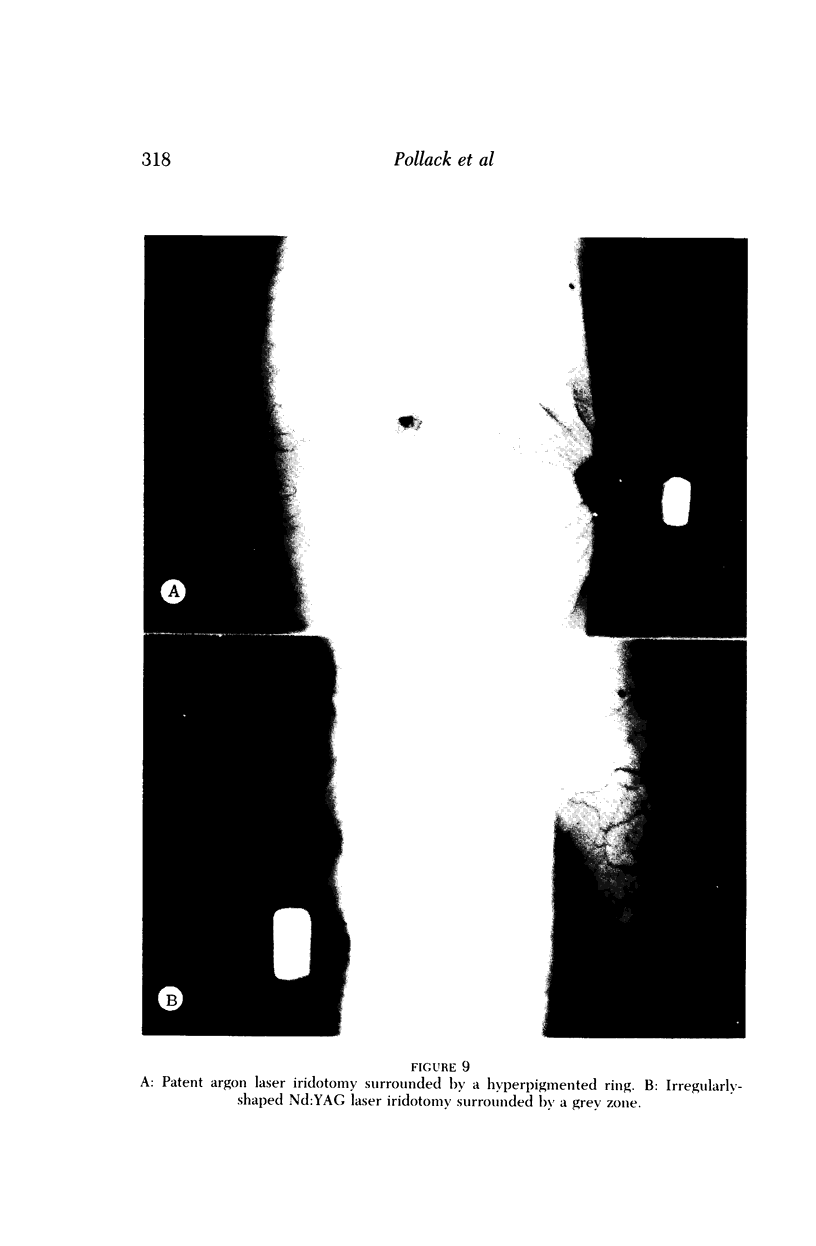
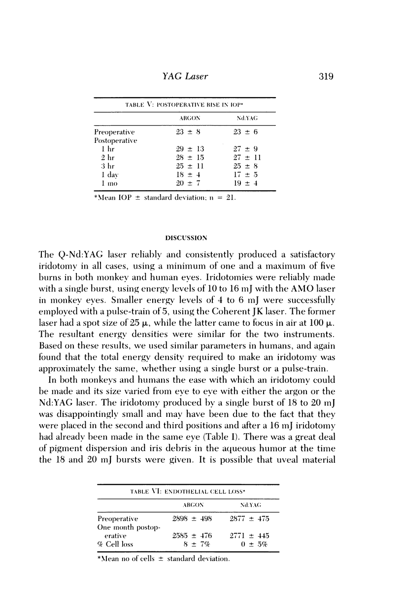
In a prospective study the Nd:YAG laser was used to create iridotomies in cynomolgus monkeys, using various levels of energy and pulse-trains of 1 to 9. Although no change occurred in the endothelial cell count of the cornea, opacities of the corneal endothelium and lens did occur. In addition, one eye showed rupture of the anterior lens capsule immediately behind the iridotomy. Most of the iridotomies in these animals closed within 3 to 4 weeks. Attenuated pigment epithelium bridged the gap within 9 days and in many sections it appeared that the iridotomies closed by fibrous contraction or early stromal regeneration. A prospective short-term clinical study evaluated argon and Q-Nd:YAG laser iridotomies in 42 eyes of 21 patients with primary chronic angle-closure glaucoma. In each patient one eye was randomly treated with an argon laser iridotomy and the fellow eye with a Nd:YAG laser iridotomy. In every case a patent iridotomy was created in one session. A mean of 12 +/- 11 and 0.033 +/- 0.025 Joules was required to complete an iridotomy with the argon and Nd:YAG lasers, respectively. Thirty percent of the argon iridotomies became sufficiently closed with pigment to require retreatment; whereas none of the Nd:YAG iridotomies closed. A postoperative rise in IOP greater than 10 mm Hg was seen in 38% argon- and 29% Nd:YAG-treated eyes. Although bleeding around the iridotomy occurred in 48% of eyes, in no case was this of significant consequence. No acute lens damage was observed in the Nd:YAG-treated human eyes, while 43% of lenses in the argon group had focal opacities. Thirty-three percent of Nd:YAG- and 24% of argon-treated eyes had focal, nonprogressive corneal opacities above the iridotomy. Specular microscopy showed a significant central corneal epithelial cell loss in argon laser eyes only. The potential of creating a laser iridotomy with a single burst of energy is extremely attractive and worthy of further investigation.
Full text
PDF
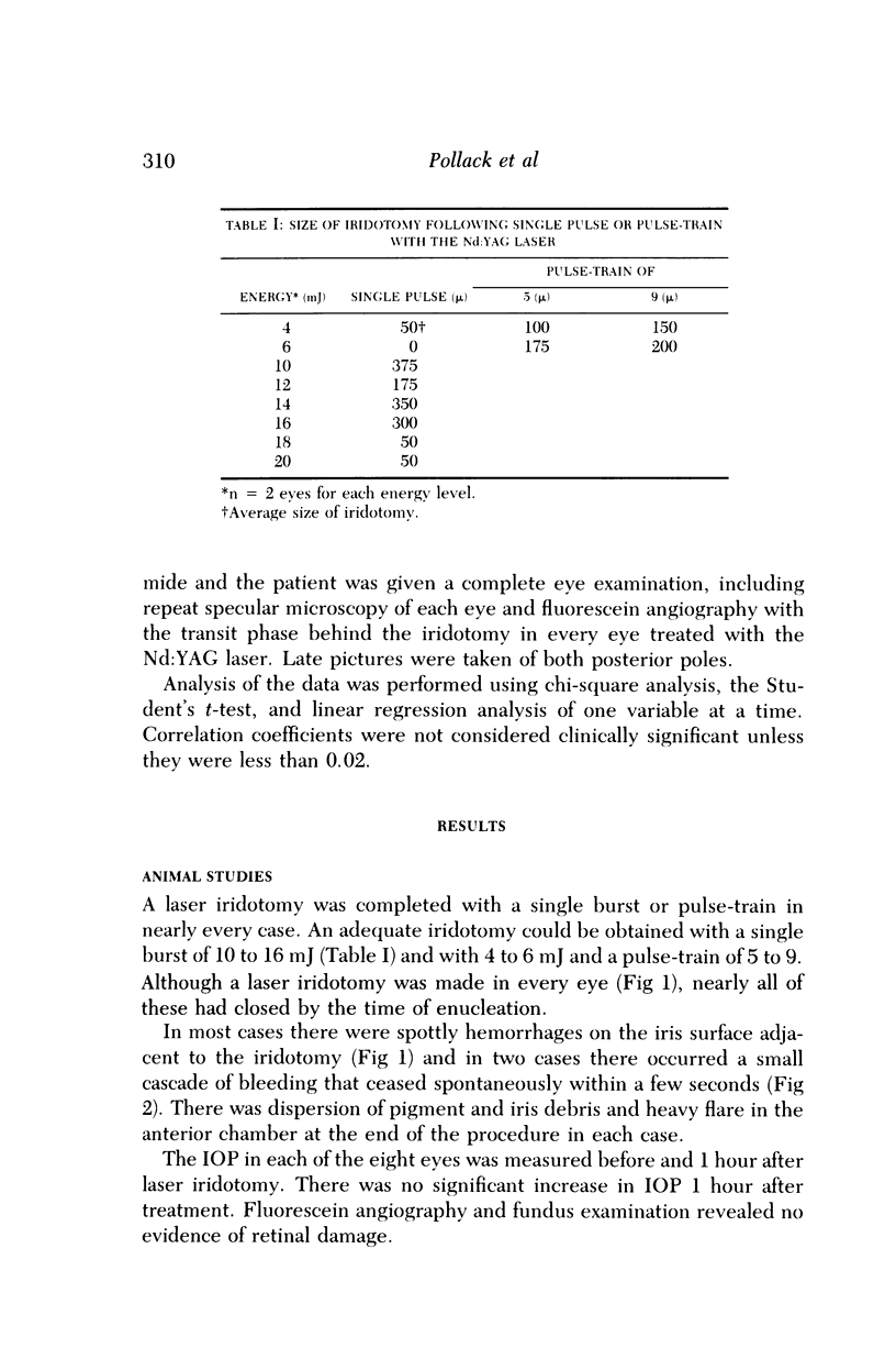
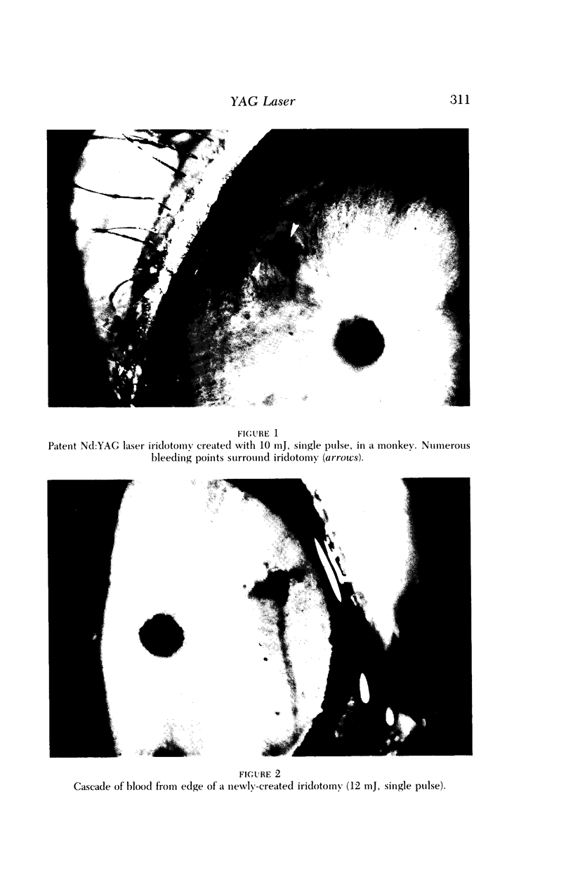
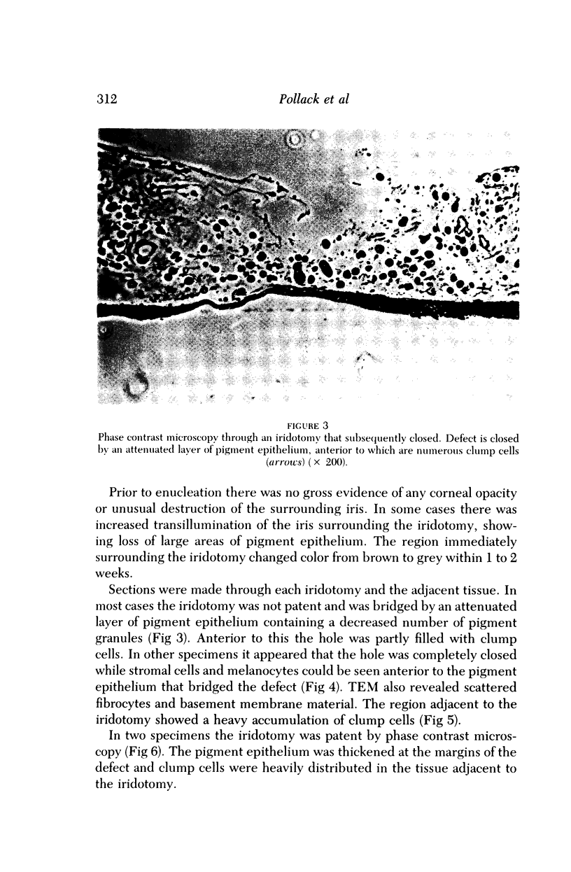
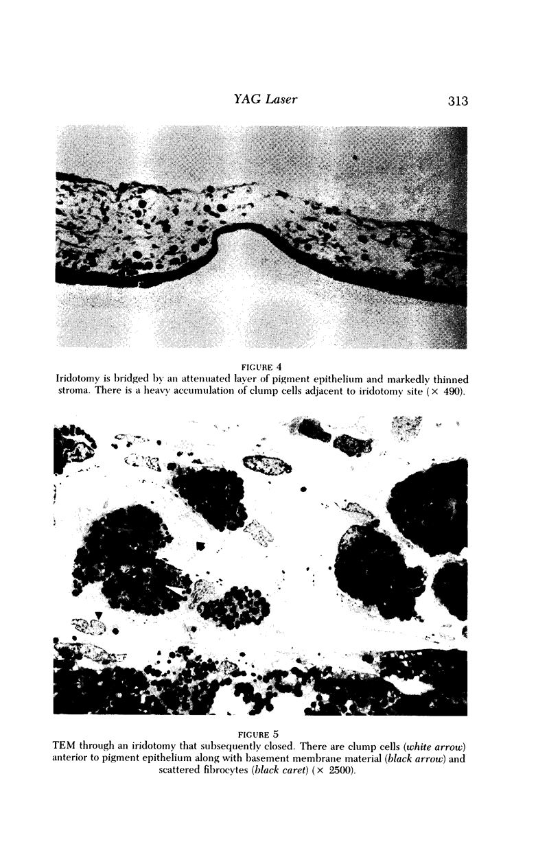
Images in this article
Selected References
These references are in PubMed. This may not be the complete list of references from this article.
- BLUM F. G., Jr, GATES L. K., JAMES B. R. How important are peripheral fields. AMA Arch Ophthalmol. 1959 Jan;61(1):1–8. doi: 10.1001/archopht.1959.00940090003001. [DOI] [PubMed] [Google Scholar]
- Bonney C. H., Gaasterland D. E. Low-energy, Q-switched ruby laser iridotomies in Macaca mulatta. Invest Ophthalmol Vis Sci. 1979 Mar;18(3):278–287. [PubMed] [Google Scholar]
- CHAMLIN M. Visual field changes in optic neuritis. AMA Arch Ophthalmol. 1953 Dec;50(6):699–713. doi: 10.1001/archopht.1953.00920030710005. [DOI] [PubMed] [Google Scholar]
- CHAMLIN M. Visual field defects due to optic nerve compression by mass lesions. AMA Arch Ophthalmol. 1957 Jul;58(1):37–58. doi: 10.1001/archopht.1957.00940010049005. [DOI] [PubMed] [Google Scholar]
- Corbett J. J., Savino P. J., Thompson H. S., Kansu T., Schatz N. J., Orr L. S., Hopson D. Visual loss in pseudotumor cerebri. Follow-up of 57 patients from five to 41 years and a profile of 14 patients with permanent severe visual loss. Arch Neurol. 1982 Aug;39(8):461–474. doi: 10.1001/archneur.1982.00510200003001. [DOI] [PubMed] [Google Scholar]
- Gilbert C. M., Robin A. L., Pollack I. P. Iridotomy using the Q-switched neodymium (Nd):YAG laser. Ophthalmology. 1984 Sep;91(9):1123–1123. [PubMed] [Google Scholar]
- Hanna C., Roy F. H. Iris wound healing. Arch Ophthalmol. 1972 Sep;88(3):296–304. doi: 10.1001/archopht.1972.01000030298015. [DOI] [PubMed] [Google Scholar]
- Hart W. M., Jr, Yablonski M., Kass M. A., Becker B. Quantitative visual field and optic disc correlates early in glaucoma. Arch Ophthalmol. 1978 Dec;96(12):2209–2211. doi: 10.1001/archopht.1978.03910060511007. [DOI] [PubMed] [Google Scholar]
- Hirst L. W., Robin A. L., Sherman S., Green W. R., D'Anna S., Dunkelberger G. Corneal endothelial changes after argon-laser iridotomy and panretinal photocoagulation. Am J Ophthalmol. 1982 Apr;93(4):473–481. doi: 10.1016/0002-9394(82)90137-4. [DOI] [PubMed] [Google Scholar]
- Keltner J. L., Johnson C. A. Automated and manual perimetry-a six-year overview. Special emphasis on neuro-ophthalmic problems. Ophthalmology. 1984 Jan;91(1):68–85. doi: 10.1016/s0161-6420(84)34328-7. [DOI] [PubMed] [Google Scholar]
- LeBlanc E. P., Becker B. Peripheral nasal field defects. Am J Ophthalmol. 1971 Aug;72(2):415–419. doi: 10.1016/0002-9394(71)91314-6. [DOI] [PubMed] [Google Scholar]
- Pollack I. P., Patz A. Argon laser iridotomy: an experimental and clinical study. Ophthalmic Surg. 1976 Spring;7(1):22–30. [PubMed] [Google Scholar]
- Pollack I. P., Robin A. L. Iridotomies in cynomolgus monkeys using a Q-switched ruby laser. Trans Am Ophthalmol Soc. 1980;78:88–106. [PMC free article] [PubMed] [Google Scholar]
- Pollack I. P. Use of argon laser energy to produce iridotomies. Trans Am Ophthalmol Soc. 1979;77:674–706. [PMC free article] [PubMed] [Google Scholar]
- Robin A. L., Pollack I. P. The Q-switched ruby laser in glaucoma. Ophthalmology. 1984 Apr;91(4):366–372. doi: 10.1016/s0161-6420(84)34278-6. [DOI] [PubMed] [Google Scholar]
- Safran A. B., Glaser J. S. Statokinetic dissociation in lesions of the anterior visual pathways. A reappraisal of the Riddoch phenomenon. Arch Ophthalmol. 1980 Feb;98(2):291–295. doi: 10.1001/archopht.1980.01020030287009. [DOI] [PubMed] [Google Scholar]
- Trautmann J. C., Laws E. R., Jr Visual status after transsphenoidal surgery at the Mayo Clinic, 1971-1982. Am J Ophthalmol. 1983 Aug;96(2):200–208. doi: 10.1016/s0002-9394(14)77788-8. [DOI] [PubMed] [Google Scholar]
- Trobe J. D., Krischer J. P. Cost-benefit analysis in screening. Unexplained visual loss. Surv Ophthalmol. 1983 Nov-Dec;28(3):189–193. doi: 10.1016/0039-6257(83)90096-6. [DOI] [PubMed] [Google Scholar]
- WALSH F. B. Syphilis of the optic nerve. Trans Am Acad Ophthalmol Otolaryngol. 1956 Jan-Feb;60(1):39–42. [PubMed] [Google Scholar]
- Wirtschafter J. D., Coffman S. M. Comparison of manual Goldmann and automated static visual fields using the Dicon 2000 perimeter in the detection of chiasmal tumors. Ann Ophthalmol. 1984 Aug;16(8):733–741. [PubMed] [Google Scholar]
- Younge B. R., Trautmann J. C. Computer-assisted perimetry in neuro-ophthalmic disease. Mayo Clin Proc. 1980 Apr;55(4):207–222. [PubMed] [Google Scholar]


