Abstract
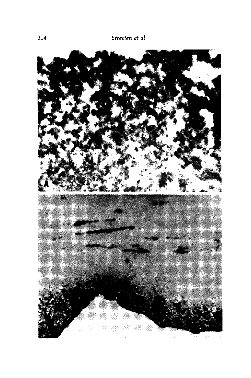
Because of histochemical similarities, there is reason for thinking that some component or components of PSX material are also present in zonular fibers. Since the zonule is a member of the elastic microfibrillar system, PSX material on the lens capsule was tested for immunologic affinity to elastic MFP from three widely divergent sources, using an indirect immunoperoxidase electron microscopic method. Positive staining was obtained with all three antibodies on all components of lens PSX material, including the superficial aggregates, deep fibrogranular zone, and capsular inclusions. The results support our hypothesis that PSX material derives from abnormal polymerization of glycoprotein associated with the zonular-elastic microfibrillar system. Similar staining of the abnormal material within the lens capsule indicates that the lens epithelial cell is involved in processing this protein. It might be suspected that other microfibrillar-secreting cells, even beyond the present range of suspected sources, could produce similar material.
Full text
PDF
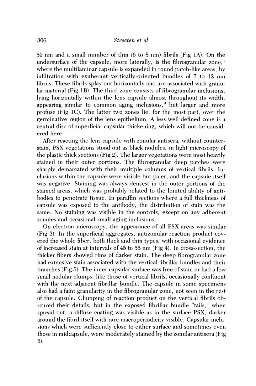
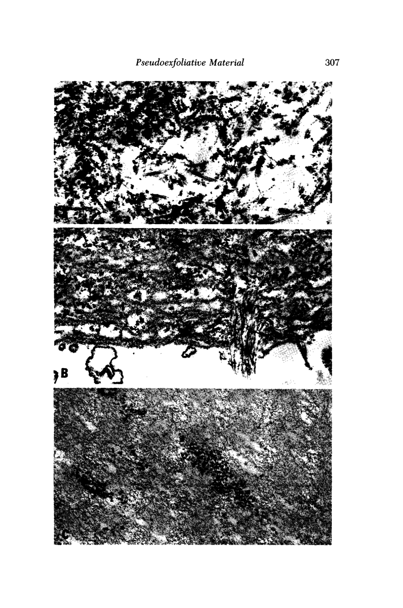
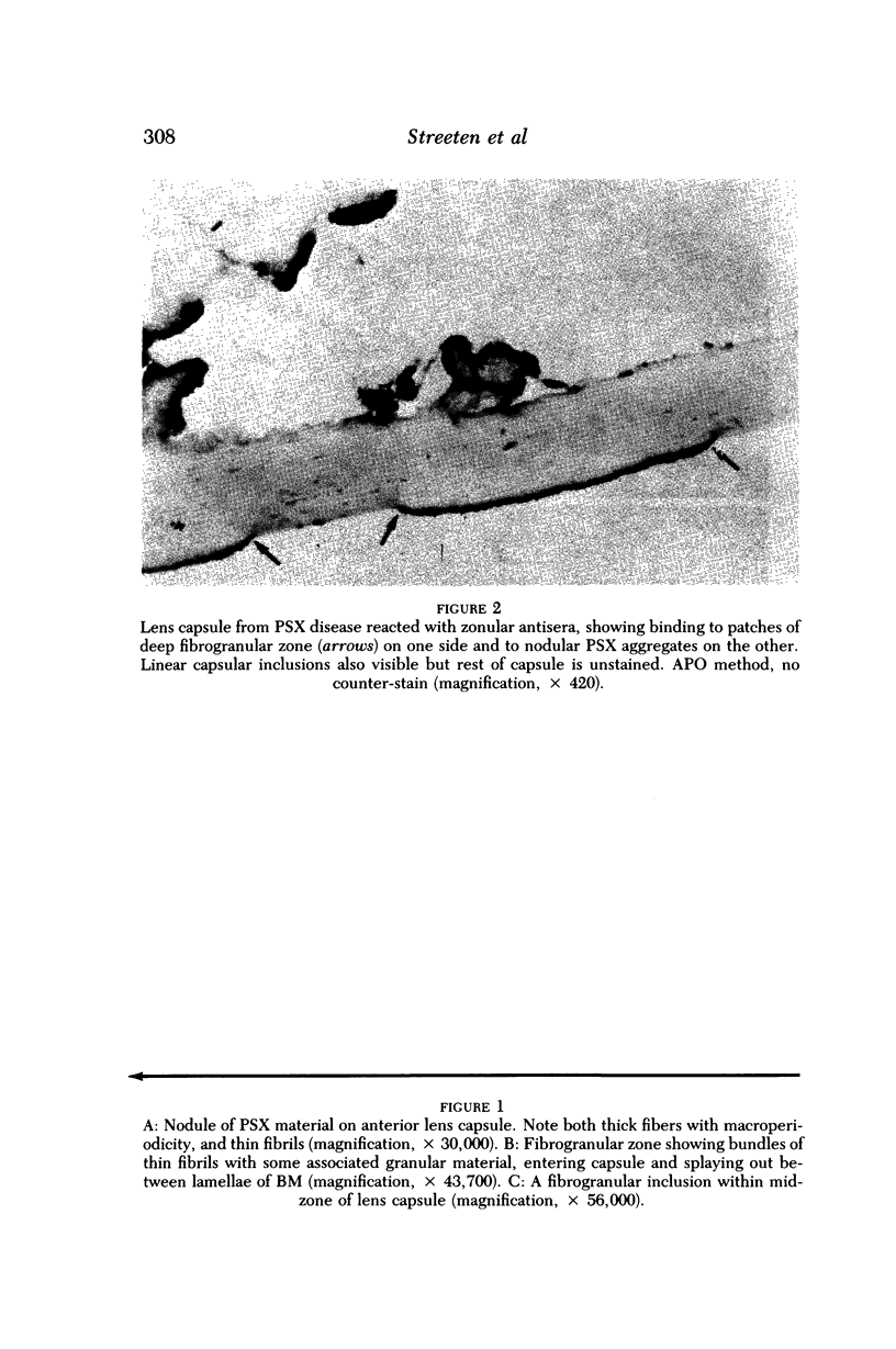
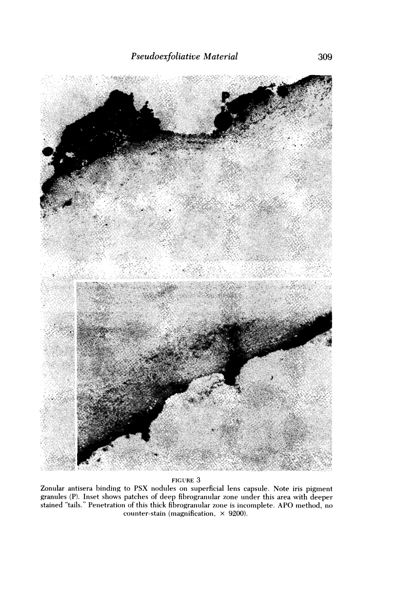
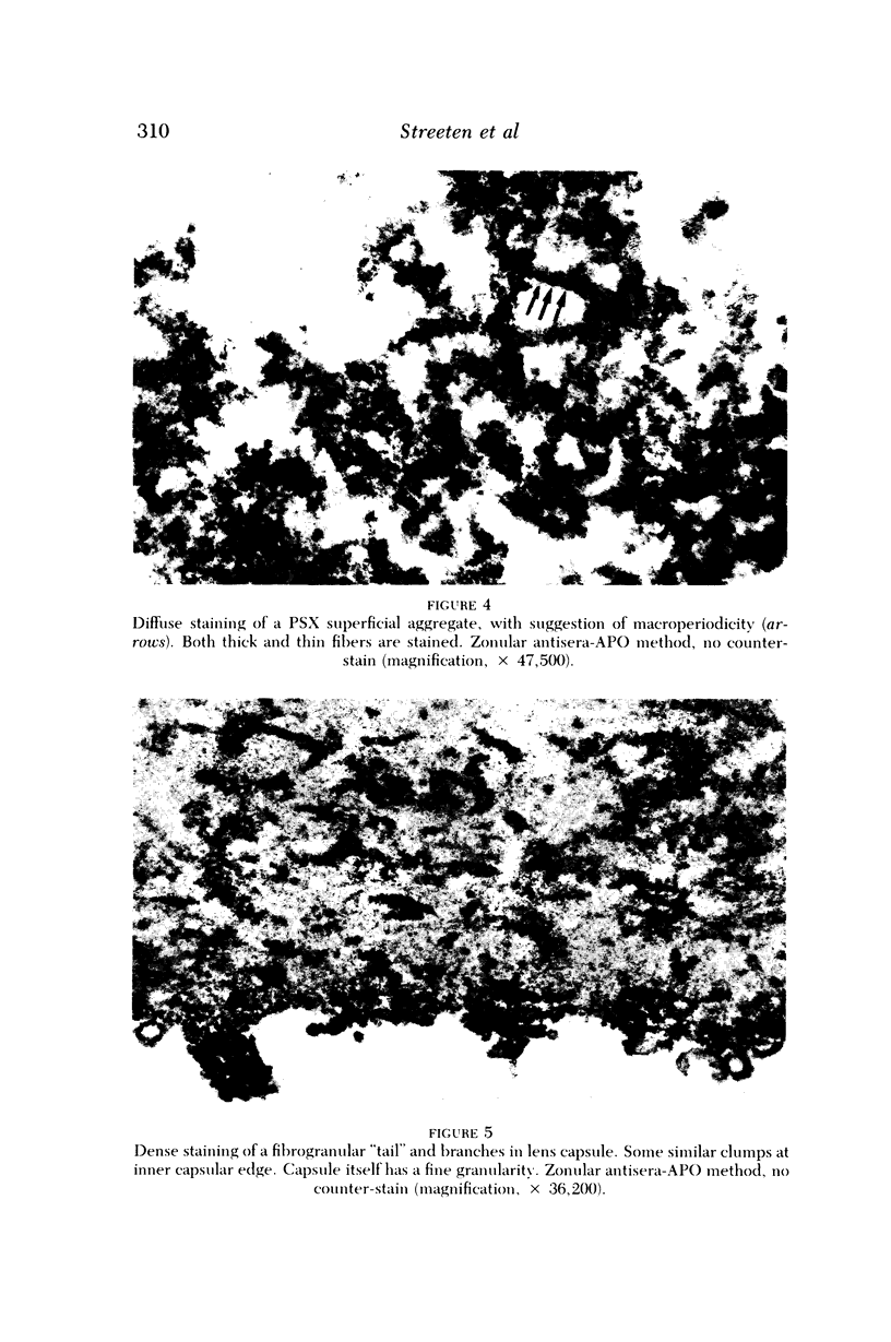
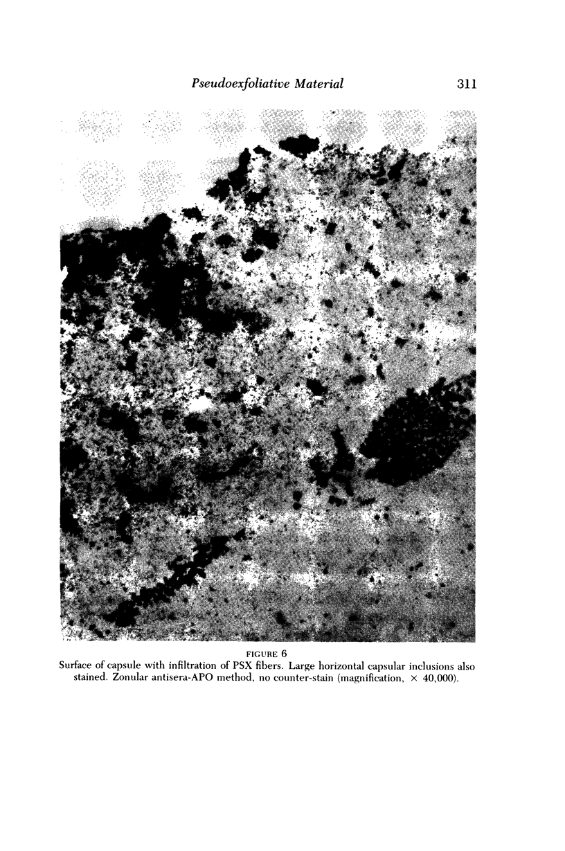
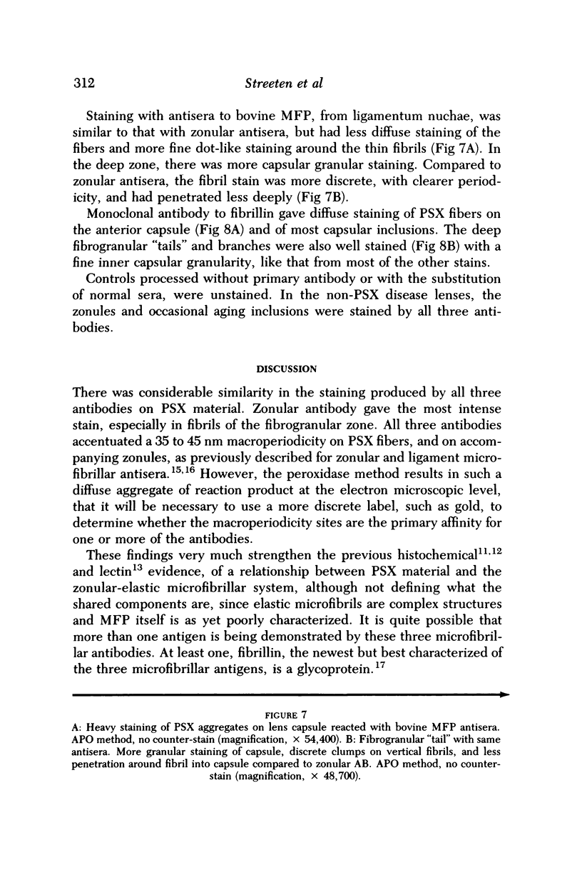
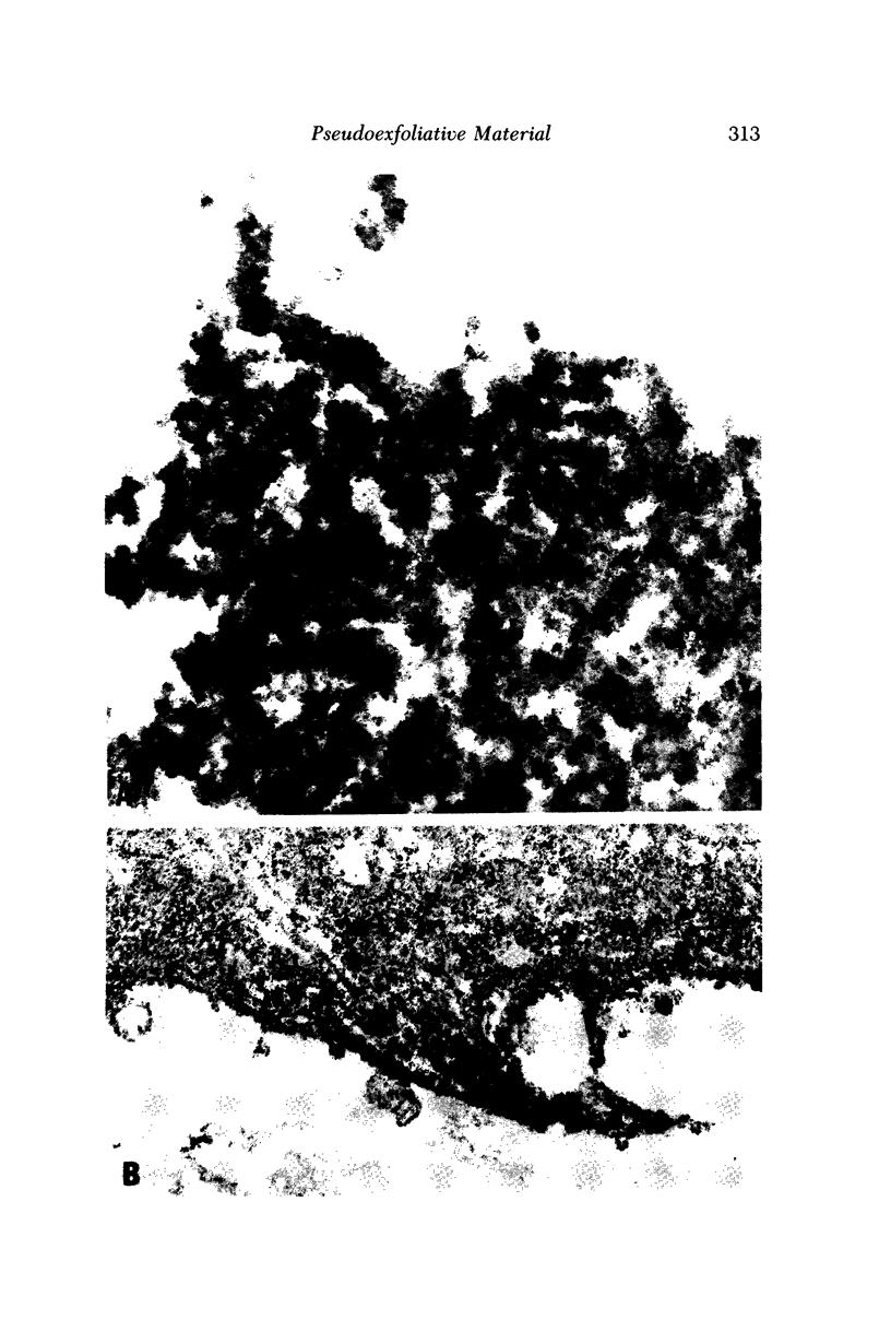
Images in this article
Selected References
These references are in PubMed. This may not be the complete list of references from this article.
- BERTELSEN T. I., DRABLOES P. A., FLOOD P. R. THE SOCALLED SENILE EXFOLIATION (PSEUDOEXFOLIATION) OF THE ANTERIOR LENS CAPSULE, A PRODUCT OF THE LENS EPITHELIUM. FIBRILLOPATHIA EPITHELIOCAPSULARIS. A MICROSCOPIC, HISTOCHEMIC AND ELECTRON MICROSCOPIC INVESTIGATION. Acta Ophthalmol (Copenh) 1964;42:1096–1113. doi: 10.1111/j.1755-3768.1964.tb03676.x. [DOI] [PubMed] [Google Scholar]
- Dark A. J., Streeten B. W., Cornwall C. C. Pseudoexfoliative disease of the lens: a study in electron microscopy and histochemistry. Br J Ophthalmol. 1977 Jul;61(7):462–472. doi: 10.1136/bjo.61.7.462. [DOI] [PMC free article] [PubMed] [Google Scholar]
- Dark A. J., Streeten B. W., Jones D. Accumulation of fibrillar protein in the aging human lens capsule, with special reference to the pathogenesis of pseudoexfoliative disease of the lens. Arch Ophthalmol. 1969 Dec;82(6):815–821. doi: 10.1001/archopht.1969.00990020807016. [DOI] [PubMed] [Google Scholar]
- Davanger M. Studies on the pseudo-exfoliation material: a review. Albrecht Von Graefes Arch Klin Exp Ophthalmol. 1978 Nov 8;208(1-3):65–68. doi: 10.1007/BF00406982. [DOI] [PubMed] [Google Scholar]
- Eagle R. C., Jr, Font R. L., Fine B. S. The basement membrane exfoliation syndrome. Arch Ophthalmol. 1979 Mar;97(3):510–515. doi: 10.1001/archopht.1979.01020010254014. [DOI] [PubMed] [Google Scholar]
- Garner A., Alexander R. A. Pseudoexfoliative disease: histochemical evidence of an affinity with zonular fibres. Br J Ophthalmol. 1984 Aug;68(8):574–580. doi: 10.1136/bjo.68.8.574. [DOI] [PMC free article] [PubMed] [Google Scholar]
- Ghosh M., Speakman J. S. The iris in senile exfoliation of the lens. Can J Ophthalmol. 1974 Jul;9(3):289–297. [PubMed] [Google Scholar]
- Harnisch J. P., Barrach H. J., Hassell J. R., Sinha P. K. Identification of a basement membrane proteoglycan in exfoliation material. Albrecht Von Graefes Arch Klin Exp Ophthalmol. 1981;215(4):273–278. doi: 10.1007/BF00407666. [DOI] [PubMed] [Google Scholar]
- Inoue S., Leblond C. P. The basement-membrane-like matrix of the mouse EHS tumor: I. Ultrastructure. Am J Anat. 1985 Dec;174(4):373–386. doi: 10.1002/aja.1001740402. [DOI] [PubMed] [Google Scholar]
- Ringvold A., Husby G. Pseudo-exfoliation material--an amyloid-like substance. Exp Eye Res. 1973 Nov 11;17(3):289–299. doi: 10.1016/0014-4835(73)90180-2. [DOI] [PubMed] [Google Scholar]
- Ringvold A. On the occurrence of pseudo-exfoliation material in extrabulbar tissue from patients with pseudo-exfoliation syndrome of the eye. Acta Ophthalmol (Copenh) 1973;51(3):411–418. doi: 10.1111/j.1755-3768.1973.tb06018.x. [DOI] [PubMed] [Google Scholar]
- SHAKIB M., ASHTON N., BLACH R. ELECTRON MICROSCOPIC STUDY OF PSEUDO-EXFOLIATION OF THE LENS CAPSULE. II. IRIS AND CILIARY BODY. Invest Ophthalmol. 1965 Apr;4:154–161. [PubMed] [Google Scholar]
- Speakman J. S., Ghosh M. The conjunctiva in senile lens exfoliation. Arch Ophthalmol. 1976 Oct;94(10):1757–1759. doi: 10.1001/archopht.1976.03910040531012. [DOI] [PubMed] [Google Scholar]
- Streeten B. W., Dark A. J., Barnes C. W. Pseudoexfoliative material and oxytalan fibers. Exp Eye Res. 1984 May;38(5):523–531. doi: 10.1016/0014-4835(84)90130-1. [DOI] [PubMed] [Google Scholar]
- Streeten B. W., Gibson S. A., Li Z. Y. Lectin binding to pseudoexfoliative material and the ocular zonules. Invest Ophthalmol Vis Sci. 1986 Oct;27(10):1516–1521. [PubMed] [Google Scholar]
- Streeten B. W., Licari P. A., Marucci A. A., Dougherty R. M. Immunohistochemical comparison of ocular zonules and the microfibrils of elastic tissue. Invest Ophthalmol Vis Sci. 1981 Jul;21(1 Pt 1):130–135. [PubMed] [Google Scholar]
- Streeten B. W., Licari P. A. The zonules and the elastic microfibrillar system in the ciliary body. Invest Ophthalmol Vis Sci. 1983 Jun;24(6):667–681. [PubMed] [Google Scholar]


