Abstract
Two virulence markers, cagA and babA2, were characterized by PCR in 101 Helicobacter pylori isolates from a population in Taiwan. cagA was detected in 99% of the isolates, while babA2 was present in all of the isolates. Base deletions and substitutions at the forward babA2 primer annealing sites were found. Given their high prevalence, cagA and babA2 cannot be useful markers for predicting the high-risk patients of H. pylori infection in Taiwan.
Helicobacter pylori is a gram-negative spiral bacterium that inhabits the gastric mucosa of the human stomach in approximately half of the world's population for a lifetime (9). Infection of this unique ecological niche by H. pylori induces gastric mucosal inflammation, which may progress into peptic ulcers. Persistent infection also increases an individual's risk for development of gastric adenocarcinoma and gastric mucosa-associated lymphoid tissue lymphoma (17). In 1994, H. pylori was declared a group I carcinogen (28) for gastric cancer, the fourth leading cause of cancer death in Taiwan.
Different degrees of bacterial virulence, environmental influences, and host factors are believed to contribute to the differential clinical sequelae of the infection. For the bacterium to succeed in its long-term colonization in the human stomach, a set of bacterial virulence determinants was developed for initial adhesion, for maintenance, and for the altering of gastric physiology. Of these factors, vacuolating toxin (VacA), cytotoxin-associated antigen (CagA), and the blood group antigen-binding adhesion molecule (BabA) have been shown to be related to more severe clinical outcomes (13). VacA is an 87-kDa exotoxin that can induce intracellular vacuolation in epithelial cells (18), leading to swelling and cell death. Alleles of the vacA gene vary among strains, particularly in the region encoding the signal sequence (typed s1 or s2) and in the 300-amino-acid midregion (typed m1 or m2) (2). Strains producing VacA or the vacAs1 genotype have been associated with increased gastric damage and peptic ulceration (2, 3). CagA is a 120-kDa immunodominant antigen and can be translocated by the type IV secretion system into the epithelial cells, where it is tyrosine phosphorylated, possibly for host-cell signaling (6, 7, 24, 25). The CagA-positive phenotype has been detected in a higher proportion of patients with peptic ulcer disease (7, 8), atrophic gastritis (17), and gastric adenocarcinoma (4). BabA is a 75-kDa adhesion molecule that mediates the attachment of H. pylori to Lewis b (α-1,3/4-difucosylated) blood group antigens on human gastric epithelial cells (5, 11, 12). Three bab alleles have been identified: babA1, babA2, and babB (14). babA1 and babA2 are identical alleles except that babA1 has a 10-bp deletion of the signal peptide sequence that leads to elimination of the translational initiation codon. The babA2 and babB alleles, which encode homologous proteins, have polymorphic midregion sequences but rather conserved sequences in the 5′ and 3′ regions (1, 14). Only the babA2 gene product is necessary for Lewis b binding activity (14). By use of a mismatch PCR method to characterize the presence of babA2, about 70% of H. pylori strains in Western countries were typed as babA2, which was associated with increased virulence (13). Moreover, the triple-positive phenotype (babA2, cagA, and vacAs1) was detected at a higher frequency in isolates from patients with ulcers and adenocarcinomas, which might serve as useful markers of high-risk patients in Western countries (13).
There is, on the other hand, a high prevalence of cagA- and vacAs1-positive bacteria in Asian countries (15, 19, 20, 22, 27, 29). In some regions, nearly all isolates are vacAs1 and cagA positive. In a recent report from Japan, a higher proportion (85%) of strains belonged to the babA2 genotype (21). Furthermore, no significant difference was found between the babA2 genotype and different clinical diseases, in contrast with that in found in Western countries. The aim of this study was to characterize the presence of babA2 and cagA in clinical isolates from a central Taiwan population. The correlation with various clinical outcomes was also investigated.
A total of 101 patients (54 male and 47 female) undergoing upper digestive endoscopy for the evaluation of dyspeptic symptoms at Taichung Veterans General Hospital, Taichung, Taiwan, between June 1996 and April 2001 were enrolled in this study. The subjects ranged in age from 32 to 83 years (mean ± standard deviation, 55.9 ± 13.0). Patients were classified at the time of endoscopy as having gastritis (n = 41), duodenal ulcers (n = 31), gastric ulcers (n = 15), or gastric cancer (n = 14). All these patients were H. pylori positive on the basis of bacterial culture performed on biopsy samples as previously described (16). H. pylori strains from patients were isolated, identified, stored, and recovered as previously described (23). For PCR genotyping, the oligonucleotide primer sequences were 5′-AATCCAAAAAGGAGAAAAAGTATGAAA-3′ (babA2-F), 5′-AATCGAAAAAGGAGAAAACATGAAAAA-3′ (nthu_babA2-F), 5′-TGTTAGTGATTTCGGTGTAGGACA-3′ (babA2-R), 5′-GATAACAGGCAAGCTTTTGAGG-3′ (Cf1), and 5′-CTGCAAAAGATTGTTTGCGAGA-3′ (Cr1). The babA2 genotype was determined by using a PCR method developed by Gerhard et al. (13) with babA2-F and babA2-R or with nthu_babA2-F and babA2-R. The babA2 gene was amplified directly from the genomic DNA of v254 by babA2-F and babA-stop (5′-TTAGTAAGCGAACACATAATTC-3′, nucleotide positions 2205 to 2226 of GenBank strain AF033654) and cloned into pGEM-T (Promega, Madison, Wis.) to generate pGEM-babA2. A babA1 fragment that had a 10-bp deletion in the signal sequence region (13) was prepared by PCR with pGEM-babA2 as a template and with two primers, babA1-F (5′-AATCGAAAAAGGAGAAAACACATCCTTTCATTAGC-3′, corresponding to positions −21 to 26 of AF033654) and babA-stop. This PCR product was cloned into pGEM-T to produce pGEM-babA1 as a negative control in babA2 typing. The presence of the cagA gene was detected by a PCR method with primers Cf1 and Cr1 to produce a 0.35-kb fragment (26, 29). A PCR-amplified 522-bp 16S rRNA fragment by Rf1 and Rr1 (10, 29) was used as an internal control in each assay. PCR amplification was carried out in a total volume of 50 μl containing 2.0 U of VioTaq polymerase (Viogene, Taipei, Taiwan), 0.1 ng of H. pylori genomic DNA, 0.1 μM concentrations of each primer, and PCR buffer (200 μM concentrations of each deoxynucleotide, 10 mM Tris-HCl [pH 8.3], 50 mM KCl, 2.5 mM MgCl2, 0.01% gelatin). PCR was performed by using a thermal cycler (MJ Research, Waltham, Mass.) under the following conditions: an initial denaturation step at 95°C for 5 min; 35 cycles at 95°C for 1 min, 55 to 62°C for 1 min, and 72°C for 1 min; and a final extension at 72°C for 20 min. PCR products were analyzed on 1.5% agarose gels. For sequence analysis of the babA2 signal sequence region, a 0.4-kb fragment was amplified by PCR with oligonucleotide primers ssbabA-F (5′-ATGACAAAATTTTTAAGAAAATG-3′, corresponding to bp 138 to 160 of AF033654) and c1babA-R (5′-CGTTAATCGCACTCGGATCAGCG-3′, corresponding to bp 509 to 532 of AF033654), followed by cloning into the pGEM-T plasmid. Nucleotide sequences were determined on both strands by the dideoxy chain termination procedure with an ABI Prism dye terminator cycle sequencing ready reaction kit (PerkinElmer, Boston, Mass.) in an automated DNA sequencer (model 377-96; PerkinElmer). Sequence analysis was done with the University of Wisconsin Genetics Computer Group (Madison, Wis.) package. The relationship between H. pylori genotypes and various diseases was analyzed by the Chi-square test with Yates's correction or by Fisher's exact test. A P of <0.05 was considered statistically significant.
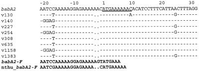
Of the 101 isolates from a central Taiwan population, 100 tested positive for the presence of cagA. The only cagA-negative strain was found in a patient with gastric cancer. Given the high prevalence, no clinical relevance could be drawn between cagA and various diseases, which was consistent with that found in a region in northern Taiwan (29). For babA2 typing, weak amplification was obtained for a number of the strains tested. To examine whether there were mutations in the primer region, the babA2 signal region sequences were determined for eight strains. Sequence analysis of the aligned signal sequence region showed that there were two base deletions near the 3′ end of the forward primer. In addition, there were base substitutions at the babA2-F annealing sites, thus resulting in ambiguous PCR results (Fig. 1). We also determined the region spanning the babA2-R fragment; the babA2-R annealing sites, on the other hand, were much more conservative (data not shown). By use of the modified forward primer (nthu_babA2-F) that contained conserved sequences among Taiwanese isolates, all 101 strains yielded positive PCR amplification. No correlation was found between the babA2 and cagA genotypes. No clinical relevance could be drawn between cagA and various diseases due primarily to the predominant prevalence of cagA-positive and babA2-positive strains. Despite the fact that such a high proportion of babA2 isolates has not been reported elsewhere, the high prevalence of another virulence marker cagA was found in a northern Taiwan population (29). Moreover, the 101 isolates in this study were typed as vacAs1 (data not shown) in accordance with a previous finding (27). No clinical association could be found for babA2 because of its predominance, as opposed to the frequent association of babA2 with severe diseases in other reports (13, 14). Our results thus suggest that babA2, like the cagA and vacAs1-positive phenotype, cannot be a useful virulence predictor in Taiwan (27, 29), similar to the findings from a recent study in Japan (21). These data thus collectively suggest that H. pylori populations vary among different geographic regions and that the relationship of virulence determinants with diseases needs to be assessed for a given region. The triple-positive virulence marker seen in Western countries cannot be feasible for the prediction of high-risk patients in Japan and in Taiwan.
FIG. 1.
Aligned nucleotide sequences of the babA2 signal sequence region. The published sequence of babA2 (GenBank accession number AF033654) and those of eight Taiwanese strains (v130, v140, v227, v254, v308, v635, v1158, and v1383) were aligned and compared. Dashes and dots indicate the nucleotide identity and deletion, respectively, of the babA2 sequences. Underlining indicates the deletion of 10 nucleotides in the babA1 signal sequence. babA2-F, the forward primer used for a mismatch PCR (15); nthu_babA2-F, the modified forward primer containing conserved sequences among Taiwanese isolates.
Acknowledgments
This work was supported by the Veterans General Hospital-National Tsing Hua University-National Yang Ming University Joint Research Program (VTY89-P4-27 and VTY90-P4-23) and the Medical Research Advancement Foundation in memory of Chi-Shuen Tsou and in part by the National Science Council (NSC90-2313-B-007-003), Taipei, Taiwan, and Program for Promoting Academic Excellence of Universities grant 89-B-FA04-1-4, Ministry of Education, Taipei, Taiwan, Republic of China.
REFERENCES
- 1.Alm, R. A., J. Bina, B. M. Andrews, P. Doig, R. E. Hancock, and T. J. Trust. 2000. Comparative genomics of Helicobacter pylori: analysis of the outer membrane protein families. Infect. Immun. 68:4155-4168. [DOI] [PMC free article] [PubMed] [Google Scholar]
- 2.Atherton, J. C., P. Cao, R. M. Peek, Jr., M. K. R. Tummuru, M. J. Blaser, and T. L. Cover. 1995. Mosaicism in vacuolating cytotoxin alleles of Helicobacter pylori. J. Biol. Chem. 270:17771-17777. [DOI] [PubMed] [Google Scholar]
- 3.Atherton, J. C., R. M. Peek, K. T. Tham, T. L. Cover, and M. J. Blaser. 1997. Clinical and pathological importance of heterogeneity in vacA, the vacuolating cytotoxin gene of Helicobacter pylori. Gastroenterology 112:92-99. [DOI] [PubMed] [Google Scholar]
- 4.Blaser, M. J., G. I. Perez-Perez, H. Kleanthous, T. L. Cover, R. M. Peek, P. H. Chyou, G. N. Stemmermann, and A. Nomura. 1995. Infection with Helicobacter pylori strains possessing cagA is associated with an increased risk of developing adenocarcinoma of the stomach. Cancer Res. 55:2111-2115. [PubMed] [Google Scholar]
- 5.Boren, T., P. Falk, K. A. Roth, G. Larson, and S. Normark. 1993. Attachment of Helicobacter pylori to human gastric epithelium mediated by blood group antigens. Science 262:1892-1895. [DOI] [PubMed] [Google Scholar]
- 6.Censini, S., C. Lange, Z. Xiang, J. E. Crabtree, P. Ghiara, M. Borodovsky, R. Rappuoli, and A. Covacci. 1996. cag, a pathogenicity island of Helicobacter pylori, encodes type I-specific and disease-associated virulence factors. Proc. Natl. Acad. Sci. USA 93:14648-14653. [DOI] [PMC free article] [PubMed] [Google Scholar]
- 7.Covacci, A., S. Censini, M. Bugnoli, R. Petracca, D. Burroni, G. Macchia, A. Massone, E. Papini, Z. Xiang, N. Figura, and R. Rappuoli. 1993. Molecular characterization of the 128-kDa immunodominant antigen of Helicobacter pylori associated with cytotoxicity and duodenal ulcer. Proc. Natl. Acad. Sci. USA 90:5791-5795. [DOI] [PMC free article] [PubMed] [Google Scholar]
- 8.Crabtree, J. E., J. D. Taylor, J. I. Wyatt, R. V. Heatley, T. M. Shallcross, and D. S. Tompkins. 1991. Mucosal IgA recognition of Helicobacter pylori 120 kDa protein, peptic ulceration, and gastric pathology. Lancet 338:332-335. [DOI] [PubMed] [Google Scholar]
- 9.Dunn, B. E., H. Cohen, and M. J. Blaser. 1997. Helicobacter pylori. Clin. Microbiol. Rev. 10:720-741. [DOI] [PMC free article] [PubMed] [Google Scholar]
- 10.Engstrand, L., A.-M. H. Nguyen, D. Y. Graham, and F. A. K. el-Zaatari. 1992. Reverse transcription and polymerase chain reaction amplification of rRNA for detection of Helicobacter species. J. Clin. Microbiol. 30:2295-2301. [DOI] [PMC free article] [PubMed] [Google Scholar]
- 11.Falk, P., K. A. Roth, T. Boren, T. U. Westblom, J. I. Gordon, and S. Normark. 1993. An in vitro adherence assay reveals that Helicobacter pylori exhibits cell lineage-specific tropism in the human gastric epithelium. Proc. Natl. Acad. Sci. USA 90:2035-2039. [DOI] [PMC free article] [PubMed] [Google Scholar]
- 12.Falk, P. G., L. Bry, J. Holgersson, and J. I. Gordon. 1995. Expression of a human alpha-1,3/4-fucosyltransferase in the pit cell lineage of FVB/N mouse stomach results in production of Leb-containing glycoconjugates: a potential transgenic mouse model for studying Helicobacter pylori infection. Proc. Natl. Acad. Sci. USA 92:1515-1519. [DOI] [PMC free article] [PubMed] [Google Scholar]
- 13.Gerhard, M., N. Lehn, N. Neumayer, T. Boren, R. Rad, W. Schepp, S. Miehlke, M. Classen, and C. Prinz. 1999. Clinical relevance of the Helicobacter pylori gene for blood-group antigen-binding adhesin. Proc. Natl. Acad. Sci. USA 96:12778-12783. [DOI] [PMC free article] [PubMed] [Google Scholar]
- 14.Ilver, D., A. Arnqvist, J. Ogren, J.-M. Frick, D. Kersulyte, E. T. Incecik, D. E. Berg, A. Covacci, L. Engstrand, and T. Boren. 1998. Helicobacter pylori adhesin binding fucosylated histo-blood group antigens revealed by retagging. Science 279:373-377. [DOI] [PubMed] [Google Scholar]
- 15.Ito, Y., T. Azuma, S. Ito, H. Miyaji, M. Hirai, Y. Yamazaki, F. Sato, T. Kato, Y. Kohli, and M. Kuriyama. 1997. Analysis and typing of the vacA gene from cagA-positive strains of Helicobacter pylori isolated in Japan. J. Clin. Microbiol. 35:1710-1714. [DOI] [PMC free article] [PubMed] [Google Scholar]
- 16.Kuo, C. H., S. K. Poon, Y. C. Su, R. Su, C. S. Chang, and W. C. Wang. 1999. Heterogeneous Helicobacter pylori isolates from H. pylori-infected couples in Taiwan. J. Infect. Dis. 180:2064-2068. [DOI] [PubMed] [Google Scholar]
- 17.Kupiers, E. J. 1999. Review article: exploring the link between Helicobacter pylori and gastric cancer. Aliment. Pharmacol. Ther. 13(Suppl. 1):3-11. [DOI] [PubMed] [Google Scholar]
- 18.Leunk, R. D., P. T. Johnson, B. C. David, W. G. Kraft, and D. R. Morgan. 1988. Cytotoxic activity in broth culture filtrates of Campylobacter pylori. J. Med. Microbiol. 26:93-99. [DOI] [PubMed] [Google Scholar]
- 19.Maeda, S., K. Ogura, H. Yoshida, F. Kanai, T. Ikenoue, N. Kato, Y. Shiratori, and M. Omata. 1998. Major virulence factors, VacA and CagA, are commonly positive in Helicobacter pylori isolates in Japan. Gut 42:338-343. [DOI] [PMC free article] [PubMed] [Google Scholar]
- 20.Mitchell, H. M., S. L. Hazell, Y. Y. Li, and P. J. Hu. 1996. Serological response to specific Helicobacter pylori antigens: antibody against CagA antigen is not predictive of gastric cancer in a developing country. Am. J. Gastroenterol. 91:1785-1788. [PubMed] [Google Scholar]
- 21.Mizushima, T., T. Sugiyama, Y. Komatsu, J. Ishizuka, M. Kato, and M. Asaka. 2001. Clinical relevance of the babA2 genotype of Helicobacter pylori in Japanese clinical isolates. J. Clin. Microbiol. 39:2463-2465. [DOI] [PMC free article] [PubMed] [Google Scholar]
- 22.Pan, Z. J., D. E. Berg, R. W. van der Hulst, W. W. Su, A. Raudonikiene, S. D. Xiao, J. Dankert, G. N. Tytgat, and A. van der Ende. 1998. Prevalence of vacuolating cytotoxin production and distribution of distinct vacA alleles in Helicobacter pylori from China. J. Infect. Dis. 178:220-226. [DOI] [PubMed] [Google Scholar]
- 23.Poon, S. K., C. S. Chang, J. Su, C. H. Lai, C. C. Yang, G. H. Chen, and W. C. Wang. 2002. Primary resistance to antibiotics and its clinical impact on the efficacy of Helicobacter pylori lansoprazole-based triple therapies. Aliment. Pharmacol. Ther. 16:291-296. [DOI] [PubMed] [Google Scholar]
- 24.Segal, E. D., J. Cha, J. Lo, S. Falkow, and L. S. Tompkins. 1999. Altered states: involvement of phosphorylated CagA in the induction of host cellular growth changes by Helicobacter pylori. Proc. Natl. Acad. Sci. USA 96:14559-14564. [DOI] [PMC free article] [PubMed] [Google Scholar]
- 25.Stein, M., R. Rappuoli, and A. Covacci. 2000. Tyrosine phosphorylation of the Helicobacter pylori CagA antigen after cag-driven host cell translocation. Proc. Natl. Acad. Sci. USA 97:1263-1268. [DOI] [PMC free article] [PubMed] [Google Scholar]
- 26.Tummuru, M. K. R., T. L. Cover, and M. J. Blaser. 1993. Cloning and expression of a high-molecular-mass major antigen of Helicobacter pylori: evidence of linkage to cytotoxin production. Infect. Immun. 61:1799-1809. [DOI] [PMC free article] [PubMed] [Google Scholar]
- 27.Wang, H. J., C. H. Kuo, A. A. M. Yeh, P. C. L. Chang, and W. C. Wang. 1998. Vacuolating toxin production in clinical isolates of Helicobacter pylori with different vacA genotypes. J. Infect. Dis. 178:207-212. [DOI] [PubMed] [Google Scholar]
- 28.World Health Organization. 1994. Evaluation of carcinogenic risks to humans, schistosomes, liver flukes and Helicobacter pylori. IARC monographs, vol. 61. International Agency for Research on Cancer, Lyon, France. [PMC free article] [PubMed]
- 29.Yang, J. C., T. H. Wang, H. J. Wang, C. H. Kuo, J. T. Wang, and W. C. Wang. 1997. Genetic analysis of the cytotoxin-associated gene and the vacuolating toxin gene in Helicobacter pylori strains isolated from Taiwanese patients. Am. J. Gastroenterol. 92:1316-1321. [PubMed] [Google Scholar]


