Abstract
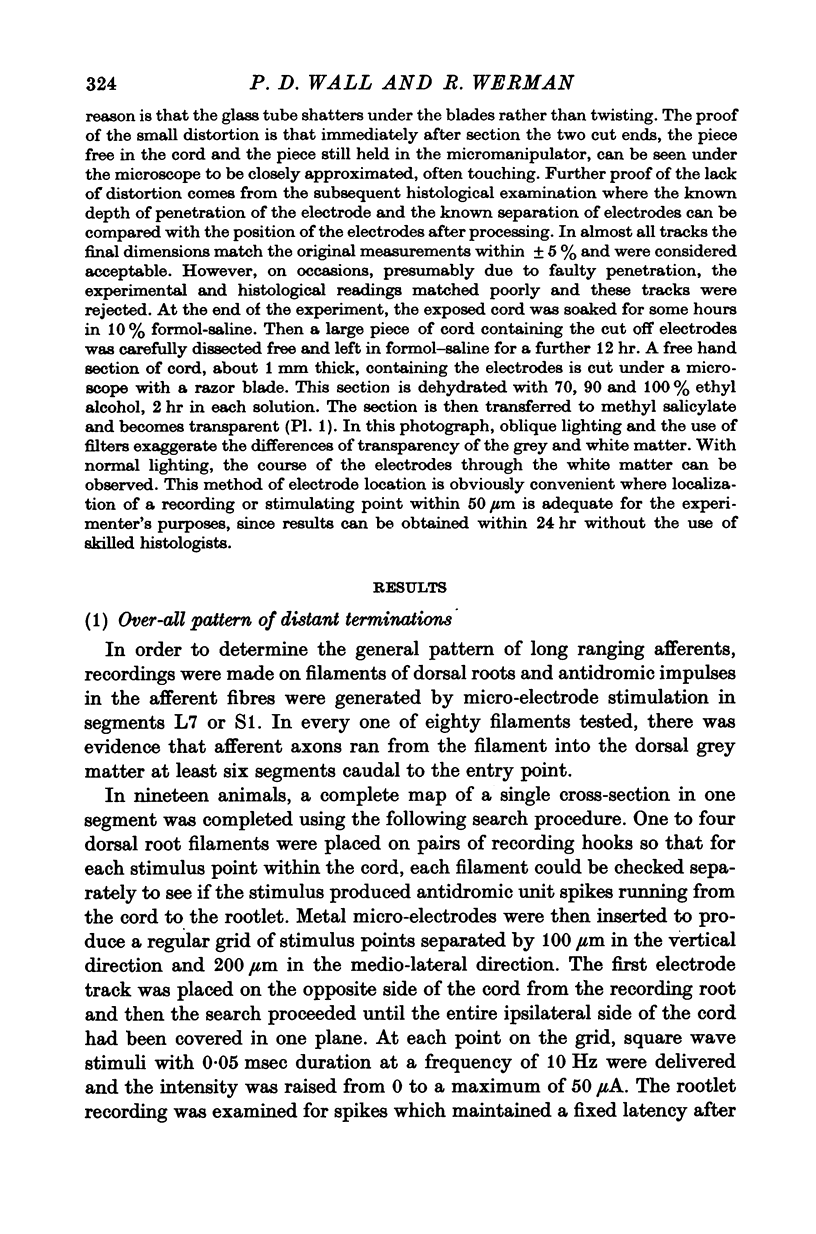
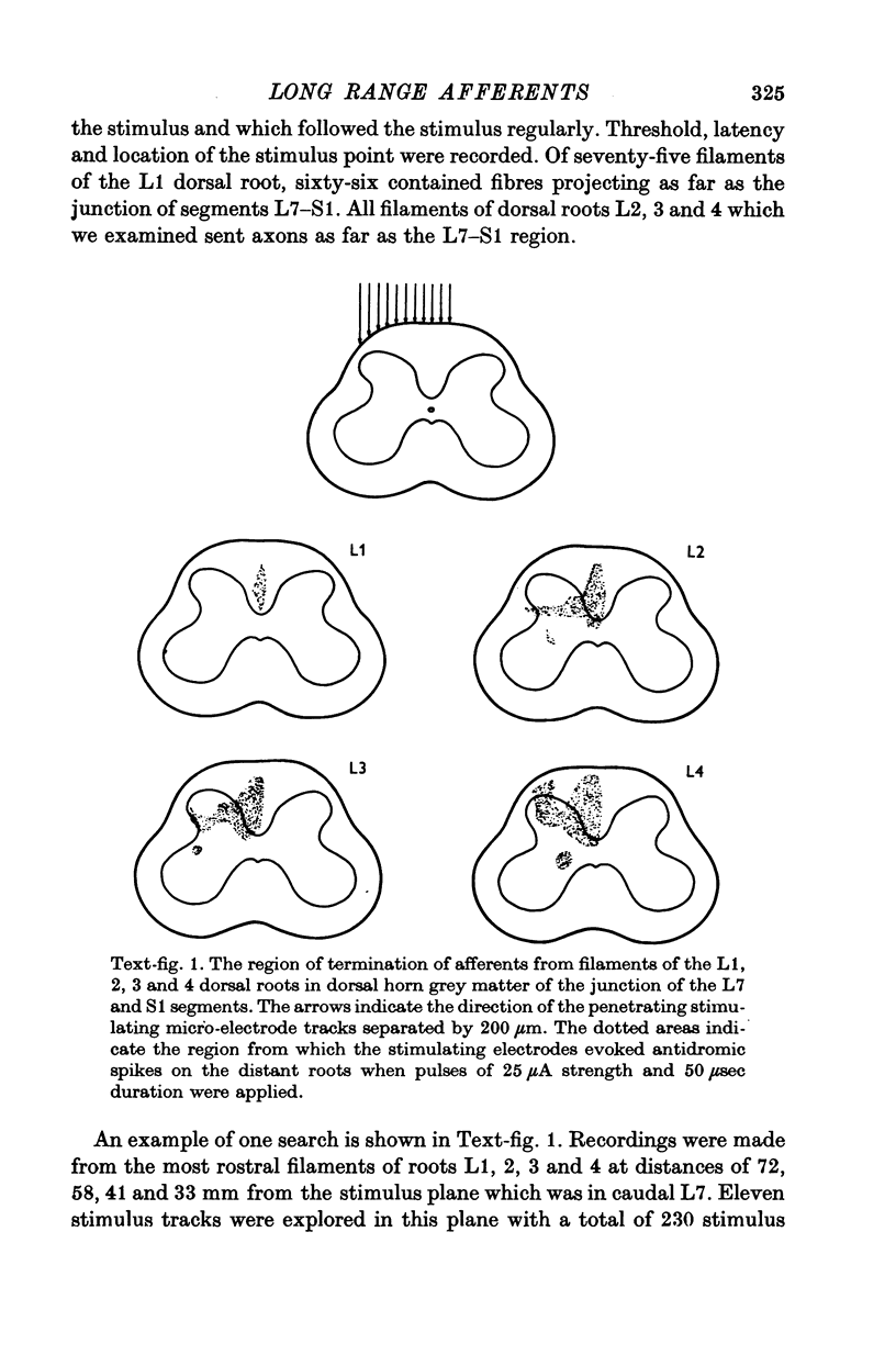
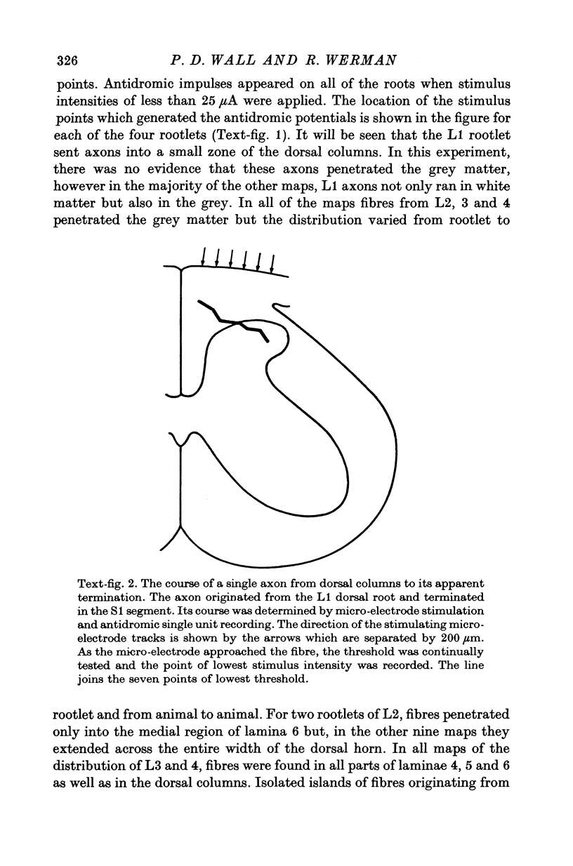
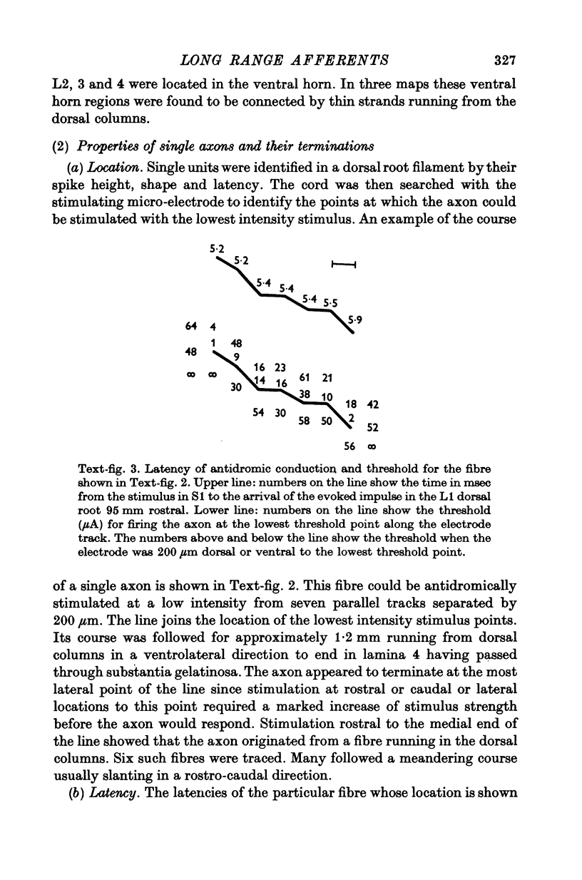
The caudal extent of the terminal arborizations of dorsal root afferents was determined in adult cats. The method used micro-electrode stimulation within the dorsal horn and the recording on a distant dorsal root filament of the antidromic action potentials evoked by the stimulation of axons within the spinal cord. 2. It was found that all filaments examined in the L2, 3 and 4 dorsal roots contained axons which projected at least as far as the S1 segment. The axons descended in or near the dorsal columns and from there penetrated into the grey matter. 3. The course of single fibres was followed to their apparent terminals. Thresholds, latencies and relative and absolute refractory periods were measured for single axons. These measurements confirmed that continuous axons ran from dorsal roots to distant segments and that the action potentials recorded were not dorsal root reflexes. 4. The majority of fibres with long range central arborizations were shown to have normal receptive fields in the dermatome of their parent dorsal root. They were not aberrant fibres leaving the spinal cord. 5. The long range afferents exist in substantial numbers since fifteen of eighty axons isolated by micro-electrode recording in the L2 dorsal root sent their axons as far as the S1 segment. The presence of these afferents from five segments away does not fit the data published on the inhibitory and excitatory receptive fields or dorsal horn cells which appear adequately explained by afferents arriving over nearby dorsal roots up to two segments away.
Full text
PDF
Images in this article
Selected References
These references are in PubMed. This may not be the complete list of references from this article.
- Brown P. B., Fuchs J. L. Somatotopic representation of hindlimb skin in cat dorsal horn. J Neurophysiol. 1975 Jan;38(1):1–9. doi: 10.1152/jn.1975.38.1.1. [DOI] [PubMed] [Google Scholar]
- Chung S. H., Raymond S. A., Lettvin J. Y. Multiple meaning in single visual units. Brain Behav Evol. 1970;3(1):72–101. doi: 10.1159/000125464. [DOI] [PubMed] [Google Scholar]
- Imai Y., Kusama T. Distribution of the dorsal root fibers in the cat. An experimental study with the Nauta method. Brain Res. 1969 Apr;13(2):338–359. doi: 10.1016/0006-8993(69)90292-3. [DOI] [PubMed] [Google Scholar]
- Jankowska E., Roberts W. J. An electrophysiological demonstration of the axonal projections of single spinal interneurones in the cat. J Physiol. 1972 May;222(3):597–622. doi: 10.1113/jphysiol.1972.sp009817. [DOI] [PMC free article] [PubMed] [Google Scholar]
- Merrill E. G., Wall P. D. Factors forming the edge of a receptive field: the presence of relatively ineffective afferent terminals. J Physiol. 1972 Nov;226(3):825–846. doi: 10.1113/jphysiol.1972.sp010012. [DOI] [PMC free article] [PubMed] [Google Scholar]
- SPRAGUE J. M. The distribution of dorsal root fibres on motor cells in the lumbosacral spinal cord of the cat, and the site of excitatory and inhibitory terminals in monosynaptic pathways. Proc R Soc Lond B Biol Sci. 1958 Dec 24;149(937):534–556. doi: 10.1098/rspb.1958.0091. [DOI] [PubMed] [Google Scholar]
- SZENTAGOTHAI J. PROPRIOSPINAL PATHWAYS AND THEIR SYNAPSES. Prog Brain Res. 1964;11:155–177. [PubMed] [Google Scholar]
- Sterling P., Kuypers H. G. Anatomical organization of the brachial spinal cord of the cat. I. The distribution of dorsal root fibers. Brain Res. 1967 Feb;4(1):1–15. doi: 10.1016/0006-8993(67)90144-8. [DOI] [PubMed] [Google Scholar]
- WALL P. D., MCCULLOCH W. S., LETTVIN J. Y., PITTS W. H. The terminal arborisation of the cat's pyramidal tract determined by a new technique. Yale J Biol Med. 1955 Dec;28(3-4):457–464. [PMC free article] [PubMed] [Google Scholar]


