Abstract
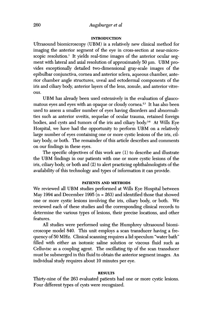
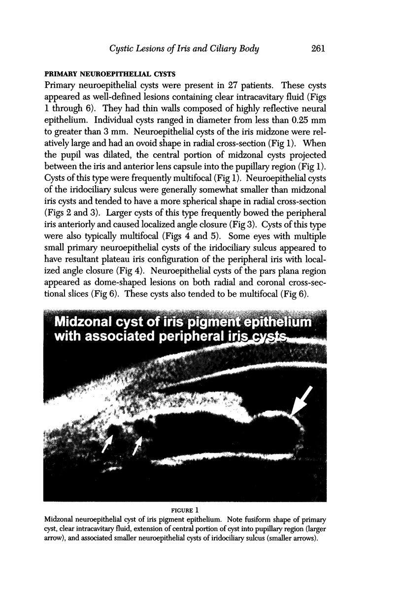
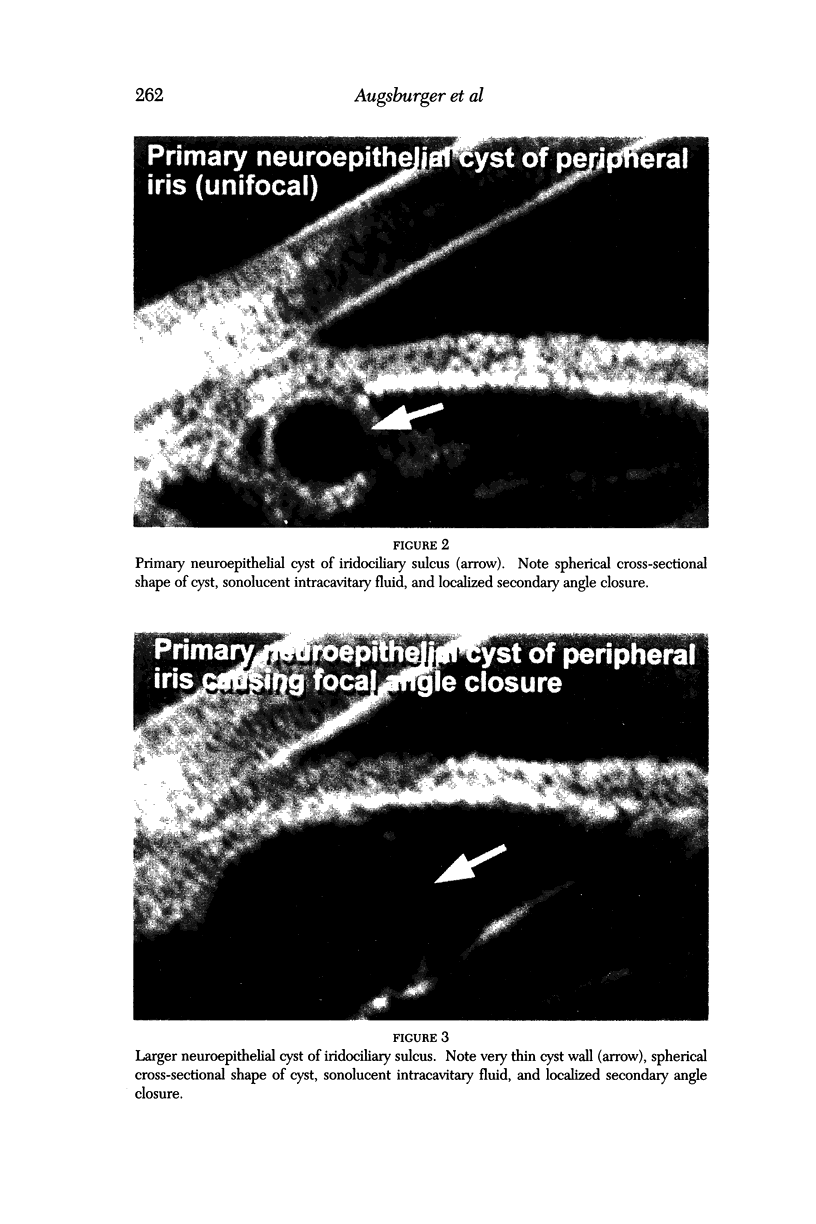
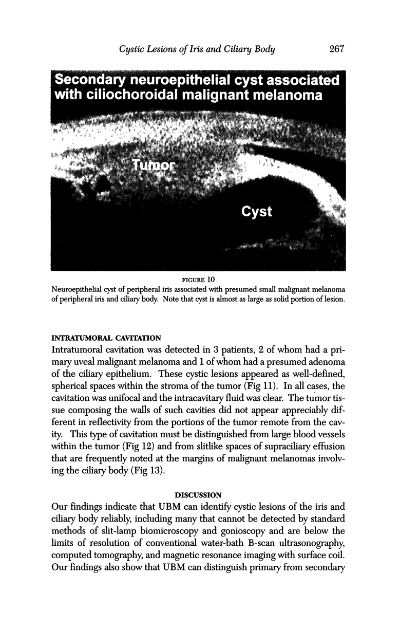
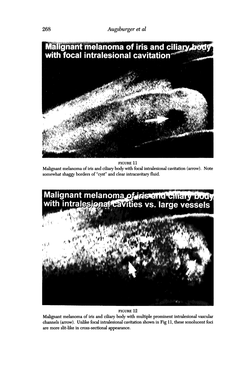
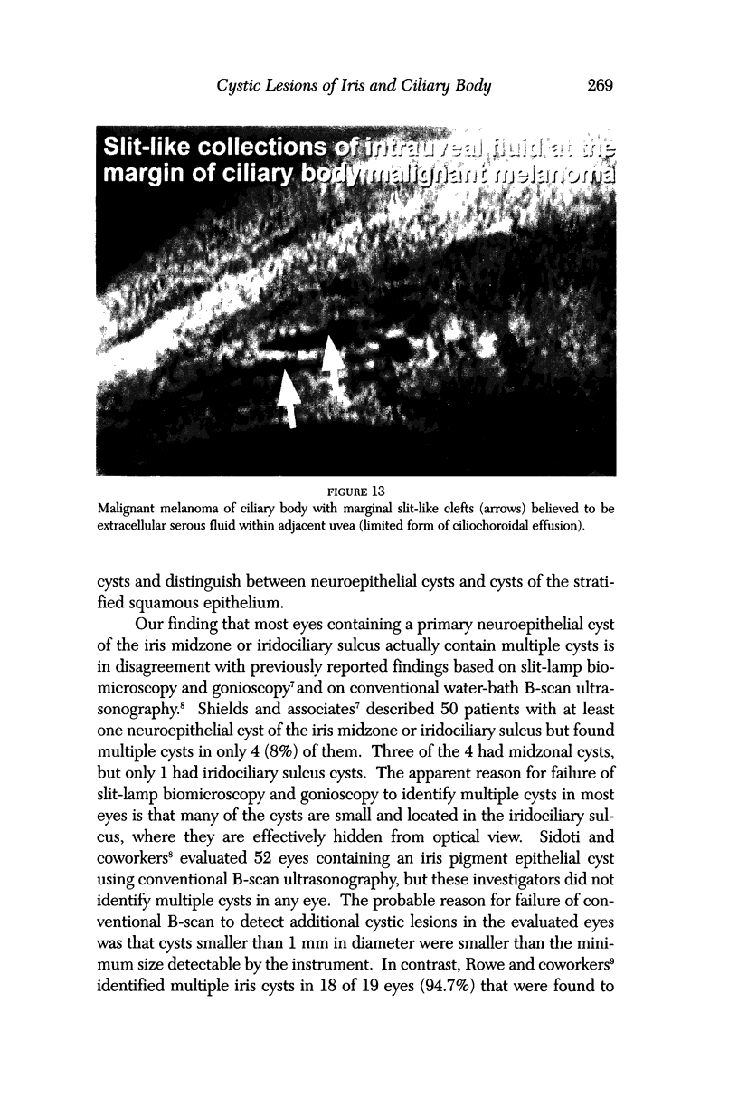
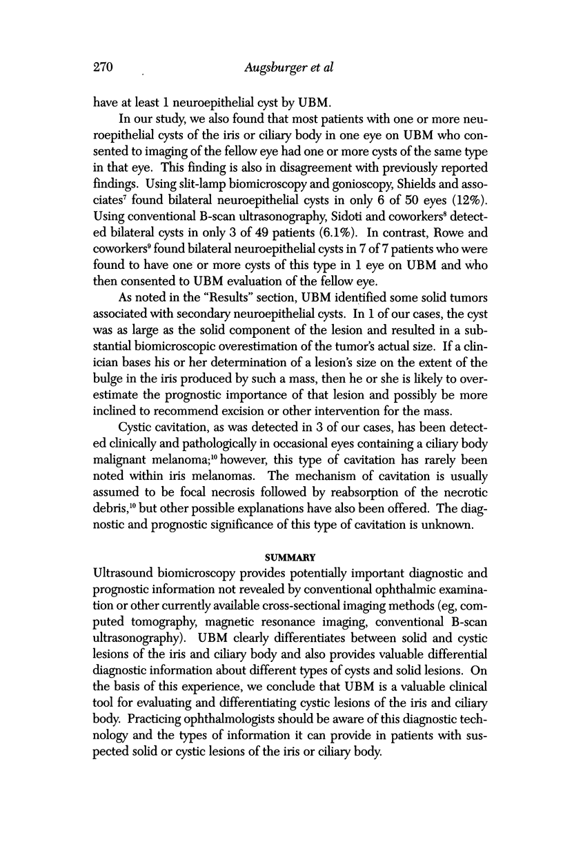
OBJECTIVES: To describe and illustrate the ultrasound biomicroscopic findings in patients with one or more cystic lesions of the iris, ciliary body, or both. METHODS: Retrospective study of 263 ultrasound biomicroscopic studies performed at one institution during a 20-month period, May 1994 through December 1995. All studies were performed using the Humphrey ultrasound biomicroscope model 840. RESULTS: Thirty-nine of the 263 evaluated patients had one or more cystic lesions. Four different types of cysts were detected. Twenty-seven patients had one or more primary neuroepithelial cysts of the iris or ciliary body. These cysts were frequently multifocal and bilateral. All contained clear fluid and had a thin but highly reflective wall. Three patients had a stratified squamous epithelial implantation cyst. These cysts were all unifocal and unilateral. The intracavitary fluid contained multiple suspended particles (presumably desquamated epithelial cells), and the walls were relatively thick. Six patients had a neuroepithelial cyst associated with a solid tumor. Each of these cysts resembled the primary neuroepithelial cysts. Three patients had focal intratumoral cavitation. The cavity in each case contained clear fluid. The surrounding tumor tissue in each case appeared homogeneous. CONCLUSIONS: Ultrasound biomicroscopy appears to be a valuable clinical tool for evaluating and differentiating cystic lesions of the iris and ciliary body.
Full text
PDF
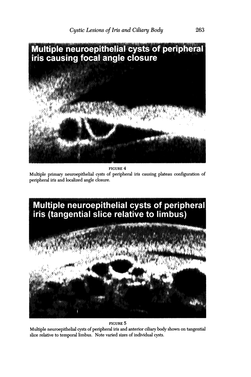
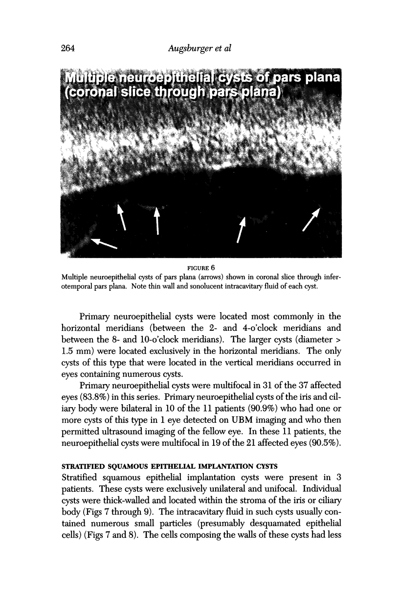
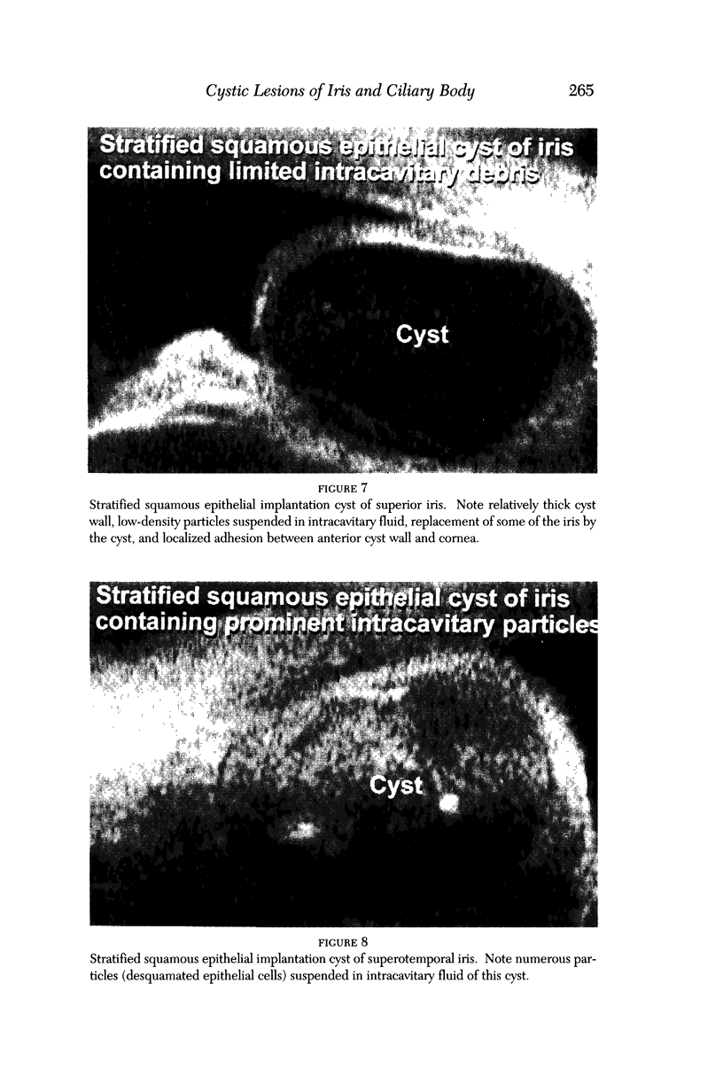
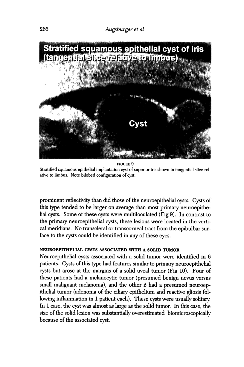
Images in this article
Selected References
These references are in PubMed. This may not be the complete list of references from this article.
- Pavlin C. J., Harasiewicz K., Foster F. S. Ultrasound biomicroscopy of anterior segment structures in normal and glaucomatous eyes. Am J Ophthalmol. 1992 Apr 15;113(4):381–389. doi: 10.1016/s0002-9394(14)76159-8. [DOI] [PubMed] [Google Scholar]
- Pavlin C. J., McWhae J. A., McGowan H. D., Foster F. S. Ultrasound biomicroscopy of anterior segment tumors. Ophthalmology. 1992 Aug;99(8):1220–1228. doi: 10.1016/s0161-6420(92)31820-2. [DOI] [PubMed] [Google Scholar]
- Pavlin C. J., McWhae J. A., McGowan H. D., Foster F. S. Ultrasound biomicroscopy of anterior segment tumors. Ophthalmology. 1992 Aug;99(8):1220–1228. doi: 10.1016/s0161-6420(92)31820-2. [DOI] [PubMed] [Google Scholar]
- Pavlin C. J., Sherar M. D., Foster F. S. Subsurface ultrasound microscopic imaging of the intact eye. Ophthalmology. 1990 Feb;97(2):244–250. doi: 10.1016/s0161-6420(90)32598-8. [DOI] [PubMed] [Google Scholar]
- Shields J. A., Kline M. W., Augsburger J. J. Primary iris cysts: a review of the literature and report of 62 cases. Br J Ophthalmol. 1984 Mar;68(3):152–166. doi: 10.1136/bjo.68.3.152. [DOI] [PMC free article] [PubMed] [Google Scholar]
- Sidoti P. A., Valencia M., Chen N., Baerveldt G., Green R. L. Echographic evaluation of primary cysts of the iris pigment epithelium. Am J Ophthalmol. 1995 Aug;120(2):161–167. doi: 10.1016/s0002-9394(14)72603-0. [DOI] [PubMed] [Google Scholar]
- Zakka K. A., Foos R. Y., Spencer W. H., Kerman B. M., Newman N. M., Pettit T. H. Cavitation in intraocular malignant melanoma. Arch Ophthalmol. 1982 Jan;100(1):112–114. doi: 10.1001/archopht.1982.01030030114012. [DOI] [PubMed] [Google Scholar]


