

Abstract
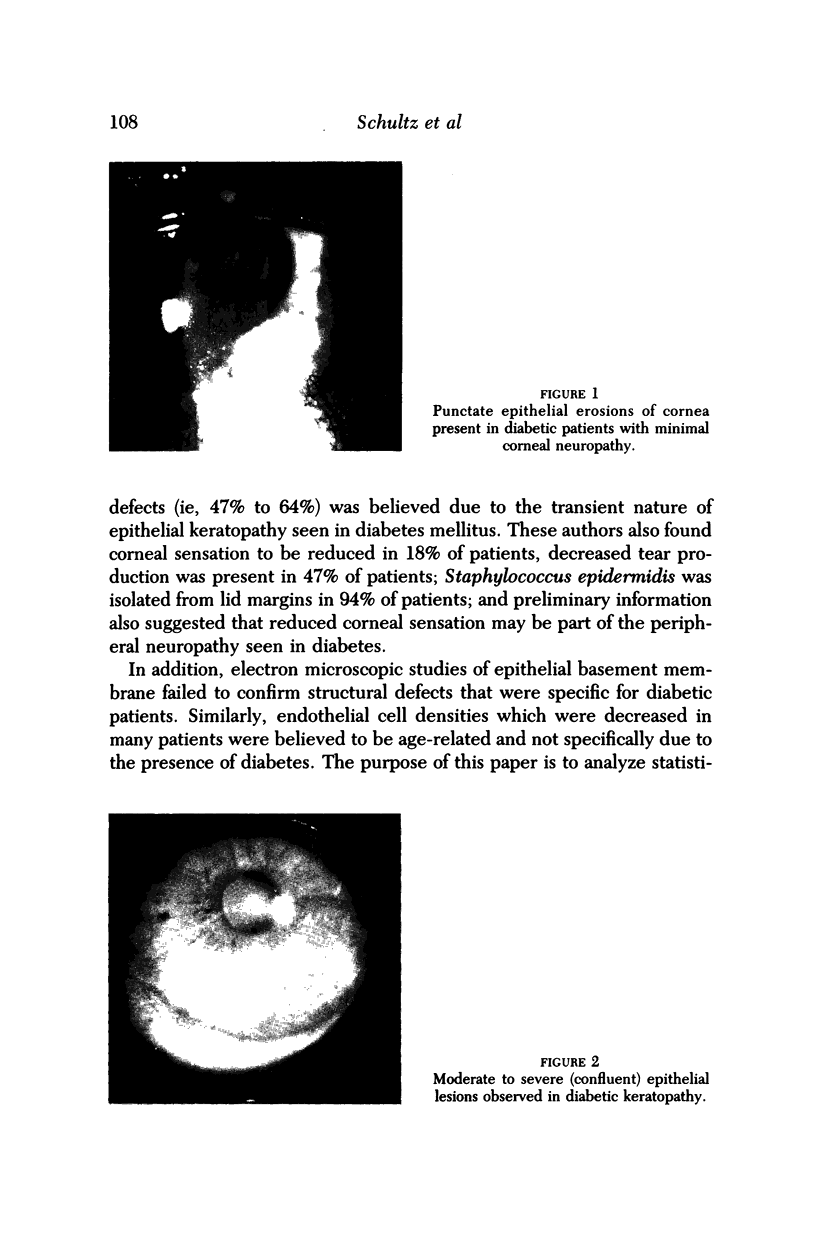
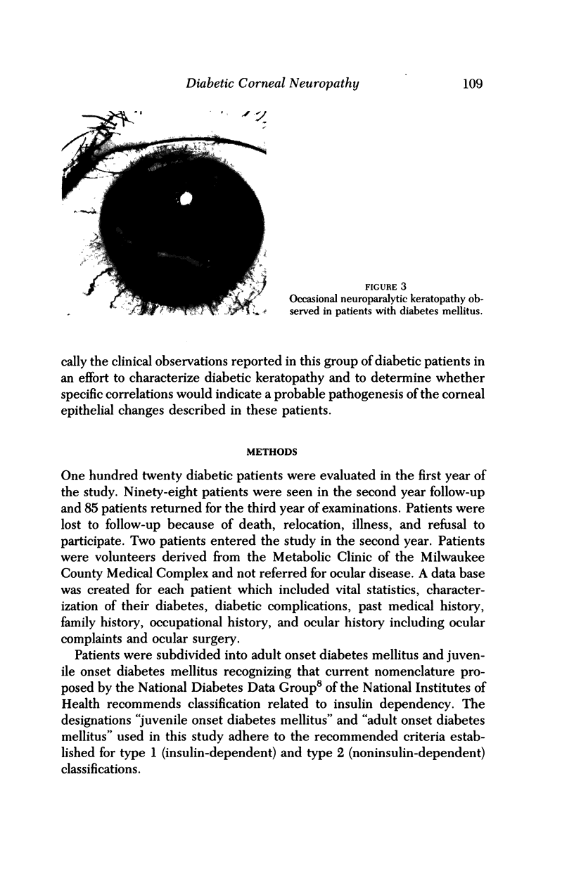
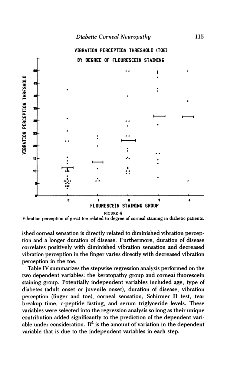
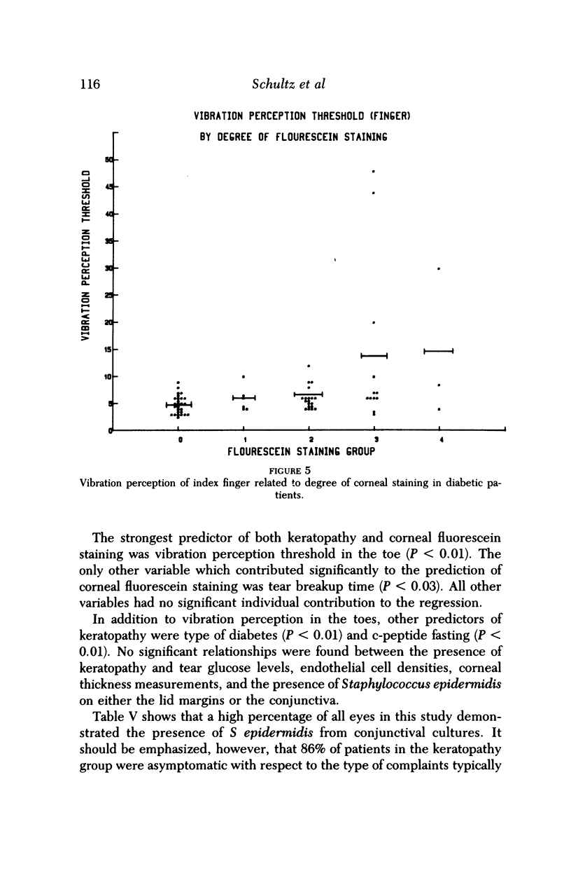
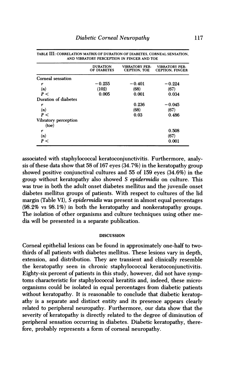
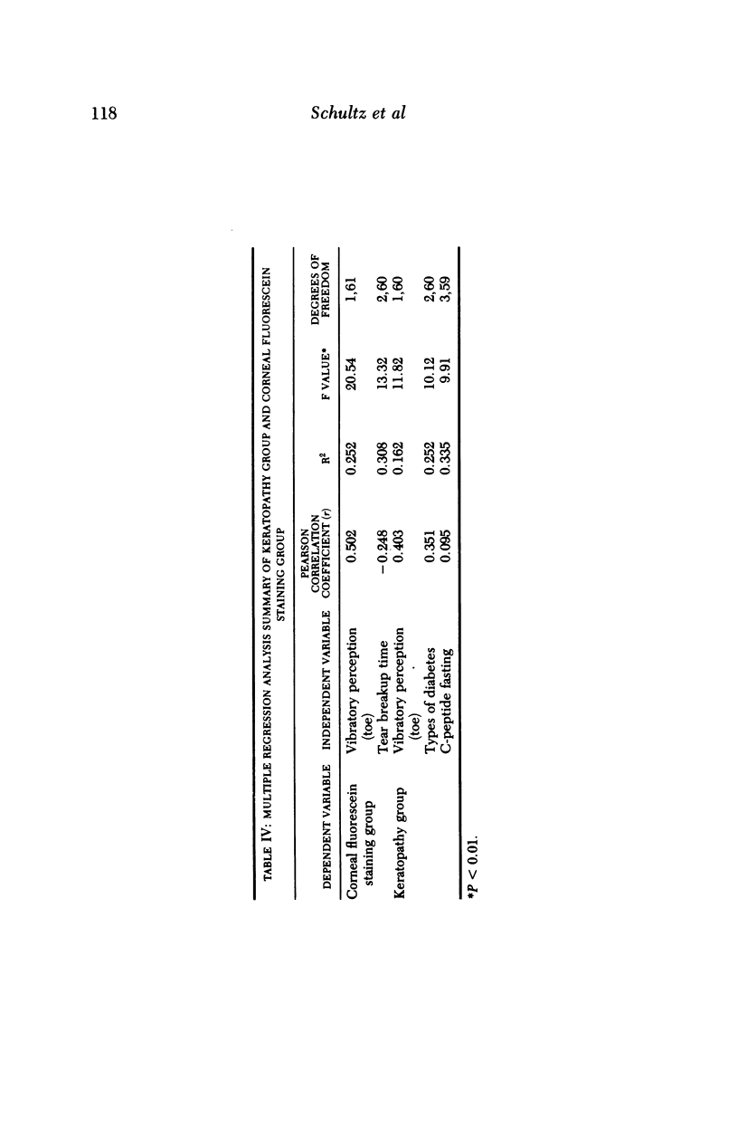
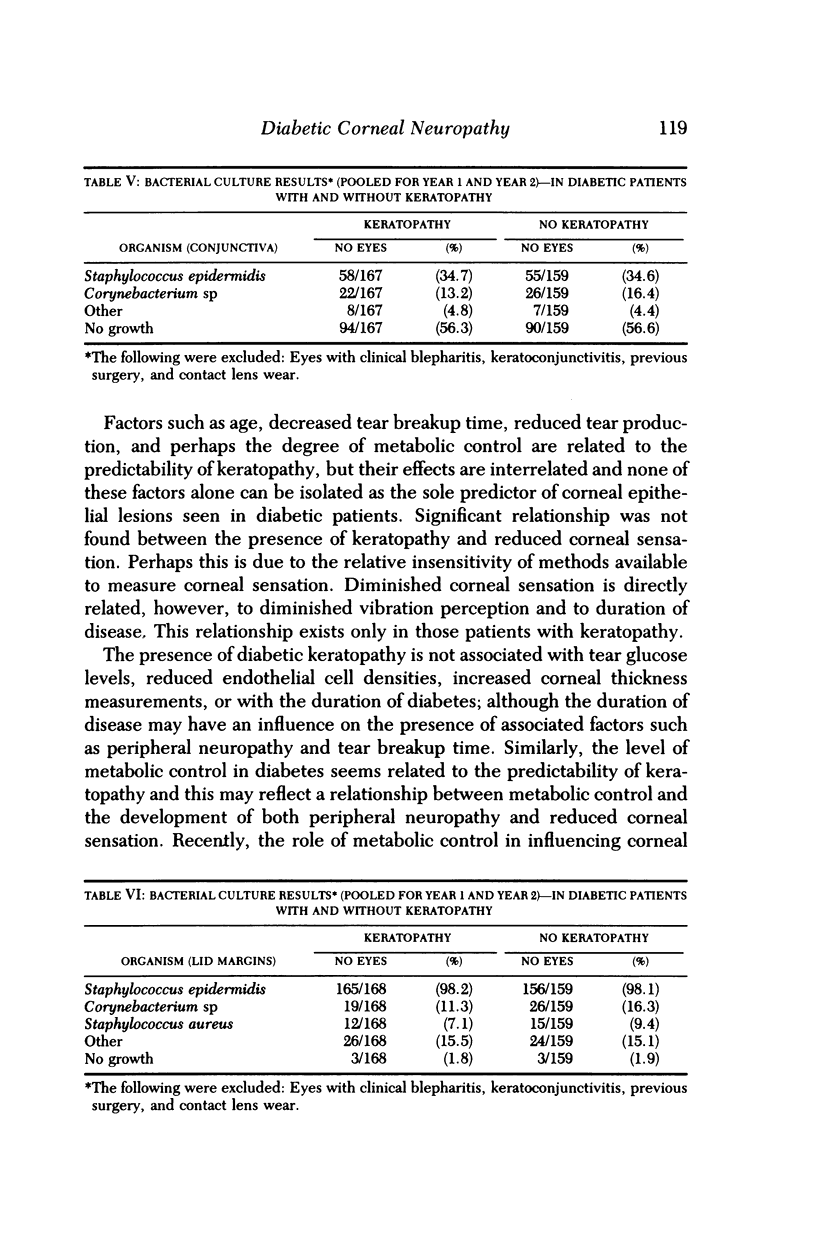
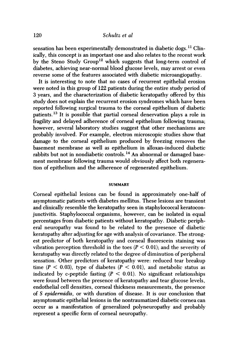
Corneal epithelial lesions can be found in approximately one-half of asymptomatic patients with diabetes mellitus. These lesions are transient and clinically resemble the keratopathy seen in staphylococcal keratoconjunctivitis. Staphylococcal organisms, however, can be isolated in equal percentages from diabetic patients without keratopathy. Diabetic peripheral neuropathy was found to be related to the presence of diabetic keratopathy after adjusting for age with analysis of covariance. The strongest predictor of both keratopathy and corneal fluorescein staining was vibration perception threshold in the toes (P less than 0.01); and the severity of keratopathy was directly related to the degree of diminution of peripheral sensation. Other predictors of keratopathy were: reduced tear breakup time (P less than 0.03), type of diabetes (P less than 0.01), and metabolic status as indicated by c-peptide fasting (P less than 0.01). No significant relationships were found between the presence of keratopathy and tear glucose levels, endothelial cell densities, corneal thickness measurements, the presence of S epidermidis, or with duration of disease. It is our conclusion that asymptomatic epithelial lesions in the nontraumatized diabetic cornea can occur as a manifestation of generalized polyneuropathy and probably represent a specific form of corneal neuropathy.
Full text
PDF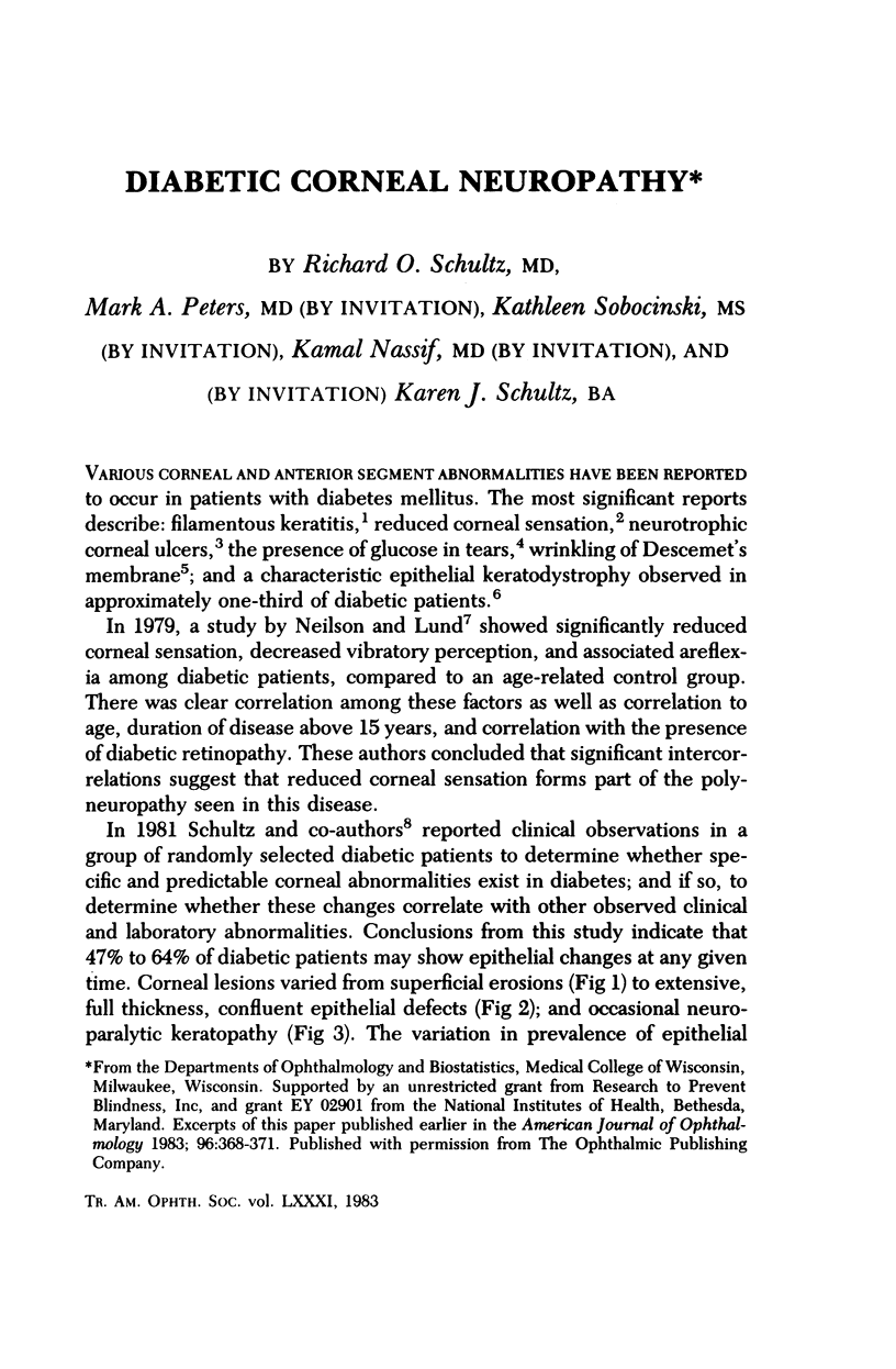
Images in this article
Selected References
These references are in PubMed. This may not be the complete list of references from this article.
- Foulks G. N., Thoft R. A., Perry H. D., Tolentino F. I. Factors related to corneal epithelial complications after closed vitrectomy in diabetics. Arch Ophthalmol. 1979 Jun;97(6):1076–1078. doi: 10.1001/archopht.1979.01020010530002. [DOI] [PubMed] [Google Scholar]
- Gasset A. R., Braverman L. E., Fleming M. C., Arky R. A., Alter B. R. Tear glucose detection of hyperglycemia. Am J Ophthalmol. 1968 Mar;65(3):414–420. doi: 10.1016/0002-9394(68)93093-6. [DOI] [PubMed] [Google Scholar]
- HENKIND P., WISE G. N. Descemet's wrinkles in diabetes. Am J Ophthalmol. 1961 Sep;52:371–374. doi: 10.1016/0002-9394(61)90736-x. [DOI] [PubMed] [Google Scholar]
- Hatchell D. L., Magolan J. J., Jr, Besson M. J., Goldman A. I., Pederson H. J., Schultz K. J. Damage to the epithelial basement membrane in the corneas of diabetic rabbits. Arch Ophthalmol. 1983 Mar;101(3):469–471. doi: 10.1001/archopht.1983.01040010469029. [DOI] [PubMed] [Google Scholar]
- Hyndiuk R. A., Kazarian E. L., Schultz R. O., Seideman S. Neurotrophic corneal ulcers in diabetes mellitus. Arch Ophthalmol. 1977 Dec;95(12):2193–2196. doi: 10.1001/archopht.1977.04450120099012. [DOI] [PubMed] [Google Scholar]
- IOLISPADA G. ULTERIORE CONTRIBUTO ALLO STUDIO DELLA CHERATODISTROFIA EPITELIALE PUNTATA DIABETICA. Boll Ocul. 1964 Nov;43:775–785. [PubMed] [Google Scholar]
- Nielsen N. V., Lund F. S. Diabetic polyneuropathy. Corneal sensitivity, vibratory perception and Achilles tendon reflex in diabetics. Acta Neurol Scand. 1979 Jan;59(1):15–22. doi: 10.1111/j.1600-0404.1979.tb02906.x. [DOI] [PubMed] [Google Scholar]
- Schultz R. O., Van Horn D. L., Peters M. A., Klewin K. M., Schutten W. H. Diabetic keratopathy. Trans Am Ophthalmol Soc. 1981;79:180–199. [PMC free article] [PubMed] [Google Scholar]
- Schwartz D. E. Corneal sensitivity in diabetics. Arch Ophthalmol. 1974 Mar;91(3):174–178. doi: 10.1001/archopht.1974.03900060182003. [DOI] [PubMed] [Google Scholar]
- Trivelli L. A., Ranney H. M., Lai H. T. Hemoglobin components in patients with diabetes mellitus. N Engl J Med. 1971 Feb 18;284(7):353–357. doi: 10.1056/NEJM197102182840703. [DOI] [PubMed] [Google Scholar]