



Abstract
We have examined the effect of covalently crosslinked profilin–actin (PxA), which closely matches the biochemical properties of ordinary profilin–actin and interferes with actin polymerization in vitro and in vivo, on Listeria monocytogenes motility. PxA caused a marked reduction in bacterial motility, which was accompanied by the detachment of bacterial tails. The effect of PxA was dependent on its binding to proline-rich sequences, as shown by the inability of PH133SxA, which cannot interact with such sequences, to impair Listeria motility. PxA did not alter the motility of a Listeria mutant that is unable to recruit Ena (Enabled)/VASP (vasodilator-stimulated phosphoprotein) proteins and profilin to its surface. Finally, PxA did not block the initiation of actin-tail formation, indicating that profilin–actin is only required for the elongation of actin filaments at the bacterial surface. Our findings provide further evidence that profilin–actin is important for actin-based processes, and show that it has a key function in Listeria motility.
Introduction
A rapid and spatially controlled turnover of actin filaments is indispensable for a variety of biological processes, such as lamellipodia extension during cell motility, phagocytosis and the intracellular movement of some bacterial and viral pathogens, such as Listeria monocytogenes (Frischknecht & Way, 2001; Cossart & Bierne, 2001).
Among the proteins that have been implicated in the regulation of actin-filament dynamics is profilin, which was originally discovered as an actin-monomer-binding protein (Carlsson et al., 1977). Profilin is localized to highly dynamic regions of cultured cells, such as the tips of spreading lamellipodia and focal adhesions (Theriot & Mitchison, 1993; Geese et al., 2000), and also accumulates at the rear end of motile Listeria (Theriot et al., 1994; Smith et al., 1996; Geese et al., 2000). The deletion of the genes encoding profilin alters the architecture of the actin cytoskeleton, severely impairs cell migration and leads, in most cases, to lethality (Balasubramanian et al., 1994; Haugwitz et al., 1994; Witke et al., 2001; Schlüter et al., 1997). In addition, profilin binds to cellular components implicated in the control of actin dynamics, such as phosphatidylinositol 4,5-bisphosphate (PtdIns(4,5)P2), Enabled (Ena)/VASP (vasodilatorstimulated phosphoprotein) proteins, N-WASP (Wiskott–Aldrich syndrome protein) and p140mDia (Frazier & Field, 1997; Schlüter et al., 1997; Pollard et al., 2000; Sechi & Wehland, 2000).
In vitro, profilin forms a 1:1 complex with actin (profilin–actin), which can participate in the elongation of actin filaments at their (+) ends (Korenbaum et al., 1998; Kang et al., 1999; Nyman et al., 2002). However, when actin filament (+) ends are capped, profilin acts as an efficient inhibitor of actin nucleation. Moreover, the binding of profilin to actin enhances the exchange rate of ADP to ATP on actin (Goldschmidt-Clermont et al., 1992), and profilin can synergize with other actin-monomer-binding proteins, such as thymosin-β4 and the actin depolymerizing factor (ADF)/cofilin, to increase actin-filament turnover (Pantaloni & Carlier, 1993; Didry et al., 1998). Finally, a crosslinked profilin–actin (PxA) complex, the structure and function of which is similar to that of unmodified profilin–actin, inhibits both the nucleation and the elongation rate of actin filaments in vitro (Nyman et al., 2002). This provides evidence that profilin–actin can bind to the (+) ends of actin filaments as a complex. This is supported by the observation that PxA alters actin-filament organization and blocks lamellipodial extension in cultured cells (Hajkova et al., 2000).
To improve our understanding of the function of profilin–actin in actin-filament dynamics, we have studied the effect of PxA on Listeria motility. Our data provide further evidence that profilin–actin is important for actin-based processes and demonstrate that it has a key function in Listeria motility.
Results
Profilin–actin impairs the intracellular motility of Listeria
To test the effect of PxA on the intracellular actin-based motility of Listeria, we injected PxA at a needle concentration of 5 mg ml−1, which has been found to be effective when used on cultured cells (Hajkova et al., 2000), into PtK2 cells infected with Listeria. Forty minutes after injection, cells were fixed and labelled with fluorescent phalloidin to detect host-cell actin filaments and the bacteria-associated actin tails. In uninjected PtK2 cells or cells that were treated with G-buffer, Listeria were associated with normal actin tails (Fig. 1A; arrowheads in Fig. 1C). Conversely, in PtK2 cells that were injected with 5 mg ml−1 PxA, all bacteria were associated with short actin tails (Fig. 1A; arrowheads in Fig. 1B), indicating that PxA impairs Listeria motility.
Figure 1.
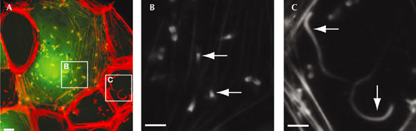
Profilin–actin impairs intracellular Listeria monocytogenes motility. PtK2 cells infected with L. monocytogenes were injected with 5 mg ml−1 profilin–actin (PxA; needle concentration). Oregon 'Green dextran was included in the injection mixture to identify injected cells. Forty minutes after injection, PtK2 cells were fixed and stained with Texas-Red-conjugated phalloidin. The insets in (A) are enlarged in (B) and (C) In uninjected cells, Listeria were associated with normal actin tails (inset (C) in (A), and arrows in (C)), whereas in cells that received PxA, the bacteria were associated with short tails (inset (B) in (A), and arrows in (B)). Scale bars, 10 μm.
PxA reduces Listeria speed and causes tail detachment
To gain further insight into the effect of PxA on Listeria motility, we analysed bacterial movement by video microscopy. Before injection, Listeria were clearly associated with phase-dense actin tails and moved at an average speed of 10.2 ± 2.58 μm min−1 (Fig. 2A–C). Shortly after injection of PxA (needle concentration of 20 mg ml−1), typically within 30 s, bacterial speed was reduced to an average of 2.71 ± 0.73 μm min−1 (Fig. 2A, green and red arrows; Fig. 2B,C; and supplementary information). Moreover, due to the impairment of actin assembly at the bacterial surface, the actin tails depolymerized within 3 min of injection of PxA (Fig. 2A, frames for 130 s and 180 s), the bacteria remained associated with a fuzzy phase-dense material (Fig. 2A, arrows in frame for 180 s), and their movement became irregular. The same result was obtained after injection of PxA at concentrations as low as 3.5 mg ml−1, whereas no perturbation of bacterial motility was observed after injection of G-buffer (Fig. 2B,C; and data not shown).
Figure 2.
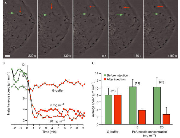
Effect of profilin–actin on Listeria motility. (A) Profilin–actin (PxA) reduces Listeria monocytogenes speed and causes actin-tail detachment. Listeria-infected PtK2 cells were injected with 20 mg ml−1 PxA and were observed by video microscopy. Before injection, Listeria were associated with normal actin tails and moved at an average speed of 10.2 ± 2.58 μm min−1 (arrows in frames for −230 s and −130 s). The injection of PxA caused an impairment of Listeria motility, followed by the rapid disassembly of the actin tails (arrowheads in frames for +130 s and +180 s). Three minutes after injection of PxA, Listeria cells were associated with a fuzzy, phase-dense material, and their movement became slow and irregular. (B,C) Effect of PxA on the instantaneous (B) and average (C) speed of Listeria. The injection of 5 or 20 mg ml−1 PxA caused a decrease in bacterial motility, whereas the injection of G-buffer had no effect. The origin of the axes corresponds to injection time in (B). Error bars in (C) represent one standard deviation above the mean. Numbers in brackets in (C) indicate the number of motile bacteria that were examined. Scale bar, 10 μm.
Next, we analysed the effect of PxA on Listeria motility using MDCK (Madin–Darby canine kidney) cells that stably express green fluorescent protein (GFP)–actin. This actin derivative was incorporated into actin filaments and Listeria actin tails without markedly altering actin dynamics (Choidas et al., 1998), and is therefore a reliable probe for analysing PxA-induced changes in actin tails in detail. Before and shortly after injection of PxA (5 mg ml−1), Listeria cells were associated with normal actin tails (Fig. 3, frame for 10 s; compare with Fig. 1C). Approximately 30 s after injection, a break appeared between the bacterium and its tail (Fig. 3, frame for 32 s) and this, in agreement with the results obtained previously, was followed by the rapid depolymerization of the actin tail (Fig. 3, frames for 76 s and 120 s; and supplementary information). At this stage, the bacterium, which was no longer associated with its tail, was surrounded by a cloud of actin, which presumably corresponded to the fuzzy phase-dense material observed around the bacteria in phase-contrast images (Fig. 2A).
Figure 3.
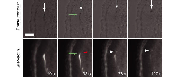
Effects of profilin–actin on Listeria monocytogenes motility analysed in Madin–Darby canine kidney cells expressing green fluorescent protein–actin. MDCK cells that stably expressed green fluorescent protein (GFP)–actin were infected with Listeria and injected with 5 mg ml−1 profilin–actin (PxA). Upper panels show phase-contrast images; lower panels show corresponding GFP–actin fluorescence. Shortly after injection, a break (green arrow; frame for 32 s) appeared between the bacterium (red arrowhead, lower frame for 32 s) and its tail, followed by the complete depolymerization of the actin tail (frames for 76 s and 120 s). The bacterium remained associated with a cloud of actin filaments (white arrows and arrowheads, frames for 76 s and 120 s) and moved slowly. Scale bar, 5 μm.
Overall, the effect of PxA on Listeria motility appears to be characterized by two steps: the impairment of actin-tail assembly at the bacterial surface, and the detachment of the bacteria from their actin tails.
PxA binds proline-rich regions to inhibit Listeria motility
Previous in vitro data have shown that PxA binds to the free (+) ends of actin filaments, thereby inhibiting their elongation (Hajkova et al., 2000; Nyman et al., 2002). Thus, it is possible that the impairment of Listeria motility is due to the binding of PxA to the (+) ends of the actin filaments that abut the bacterial surface. As the targeting of profilin, and possibly profilin–actin complexes, to the bacterial surface is mediated by the interaction with the central proline-rich region of Ena/VASP proteins (Geese et al., 2000, 2002), the localization of which at this site is not affected by PxA (see supplementary information), it is possible that this interaction is required for the inhibitory effect of PxA on bacterial motility.
To test this hypothesis, we generated a PxA complex using the profilin mutant protein, PH133S, which does not bind to poly-L-proline, although it retains the ability to associate with both monomeric actin and PtdIns(4,5)P2 (Björkegren-sjögren et al., 1997). Injection of the complex of PH133S and actin (PH133SxA) at 5 mg ml−1 into Listeria-infected PtK2 cells did not change the motility of the bacteria (Fig. 4A–C; and supplementary information), indicating that poly-L-proline-dependent interactions are essential for targeting PxA to the Listeria surface, where it exerts its inhibitory effect. This was also supported by the observation that PxA does not affect the motility of the Listeria mutant strain ActA5, which expresses a modified version of ActA that lacks the central proline-rich region. As a consequence, these bacteria are unable to recruit Ena/VASP proteins and profilin, and move at a reduced speed (Smith et al., 1996; Niebuhr et al., 1997; Geese et al., 2000; see supplementary information).
Figure 4.
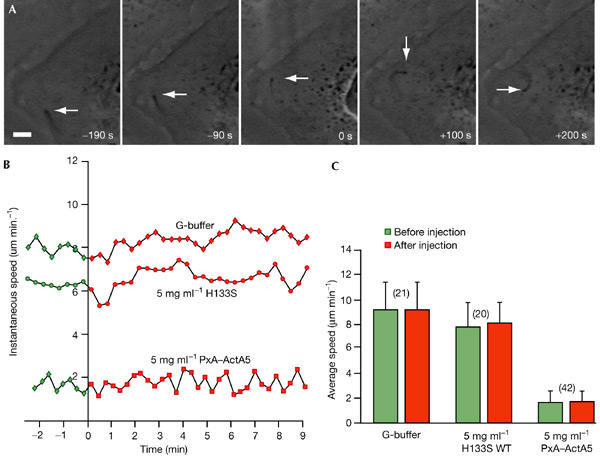
(A) The ability of profilin–actin to interact with proline-rich regions is essential for its inhibitory effect on Listeria monocytogenes motility. PtK2 cells infected with wild-type Listeria were injected with 5 mg ml−1 PH133SxA (a complex of the H133S mutant form of profilin with actin) and observed using video microscopy. Before injection, wild-type Listeria were associated with actin tails and moved at normal speed (arrow in frames for −190 s and −90 s), which was not affected by the injection of PH133SxA (arrow in frames for +100 s and +200 s). (B,C) Quantification of Listeria speed after injection with profilin–actin (PxA) or PH133SxA. The effects of PxA and PH133SxA on the instantaneous (B) and average (C) speed of Listeria ActA5 and wild-type, respectively, are shown. The injection of 5 mg ml−1 PH133SxA or of G-buffer did not alter the motility of wild-type Listeria. Similarly, PxA did not change the motility of Listeria ActA5. The origin of the x axis in (B) corresponds to the time of injection. Error bars in (C) represent one standard deviation above the mean. Numbers in brackets in (C) indicate the number of motile bacteria examined.
PxA does not inhibit formation of Listeria actin tails
Next, we analysed whether PxA affects the initiation of actin-tail formation. Because the rapid and severe effects of PxA on cells (Hajkova et al., 2000) prevented us from carrying out this investigation in living cells, we decided to use a cell-free system based on mouse cytosolic brain extracts that are able to support Listeria motility. This system has been shown to be useful for studying inhibitors of actin cytoskeleton components (May et al., 1999). We incubated mouse cytosolic extracts with various amounts of PxA on ice for 30 min, and then added Listeria to induce the formation of actin tails (incubation with Listeria was carried out at 20 °C for 15–30 min). In control extracts, Listeria induced the formation of actin tails and moved at an average speed of 0.5 ± 0.16 μm min−1 (Fig. 5). Interestingly, in the presence of PxA, Listeria were still able to induce the formation of actin tails, but their motility was impaired by PxA in a concentration-dependent manner (Fig. 5; and supplementary information). This effect was not due to the dilution of the cytosolic extract (its total protein concentration was reduced by a half at the highest PxA concentration tested), as indicated by the observation that an extract diluted with G-buffer supported Listeria motility to the same extent as control extracts (Fig. 5).
Figure 5.
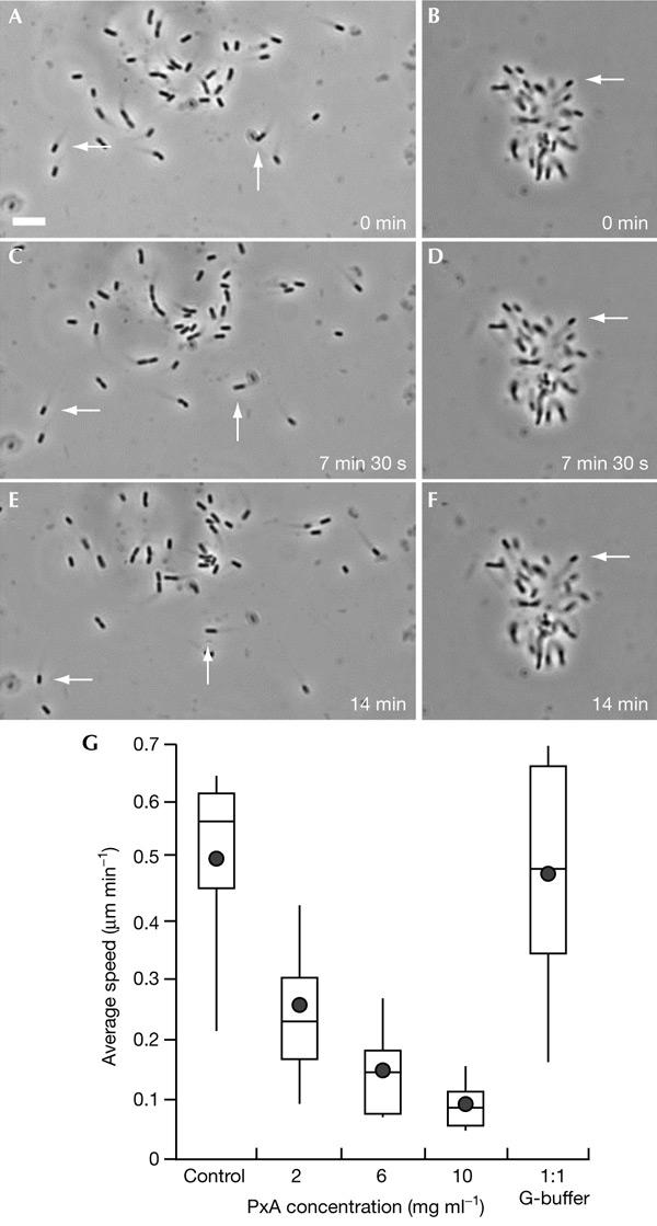
Profilin–actin does not inhibit the initiation of Listeria monocytogenes actin tails in mouse cytosolic brain extracts. Mouse cytosolic brain extracts were incubated with various concentrations of profilin–actin (PxA) on ice for 30 min. Bacteria were then added to the mixture, which was incubated for 15 min at 20 °C. In control extracts, Listeria induced the formation of actin tails and moved at an average speed of 0.5 ± 0.16 μm min−1 (arrows in (A,C,E); (G)). PxA induced a concentration-dependent reduction of bacterial motility (arrows in (B,D,F); (G)), but did not affect the initiation of actin tails. Note that bacteria occasionally aggregated during centrifugation before incubation with the extract. This led to movement diverging from a central point, which is clearly visible in (B,D,F). Scale bar, 10 μm. (G) Box and whiskers plots of bacterial speed. Filled circles indicate the mean; lines in the middle of the boxes indicate the median; the tops of the boxes indicate the 75th quartile and the bottoms of the boxes indicate the 25th quartile; 'whiskers' indicates the 10th and 90th percentiles, respectively.
Discussion
We have shown that profilin–actin has a crucial role in actin-based Listeria motility. The Ena/VASP-dependent recruitment of profilin or profilin–actin at the Listeria surface is thought to mediate the efficient elongation of actin filaments at this site. This notion is supported by two lines of evidence. First, displacement or absence of Ena/VASP proteins from the Listeria surface results in a lack of profilin recruitment to this site and impairs bacterial motility (Smith et al., 1996; Kang et al., 1997; Niebuhr et al., 1997; Geese et al., 2000, 2002). Similarly, Ena/VASP mutants lacking the central proline-rich region cannot target profilin or profilin–actin to the bacterial surface, resulting in decreased and irregular Listeria motility (Geese et al., 2002). Second, the depletion of profilin from cell extracts reduces Listeria motility (Marchand et al., 1995), whereas its addition to a set of purified proteins that can support bacterial motility causes an increase in motility (Loisel et al., 1999). Consistent with these findings, our data indicate that profilin–actin is the source of actin monomers required for efficient filament elongation at the Listeria surface.
Because PxA does not affect the formation of Listeria tails, a process that requires the actin-nucleating activity of the Arp2/3 complex (May et al., 1999; Pistor et al., 2000; Skoble et al., 2000, 2001), we conclude that the initiation of actin tails does not depend on profilin–actin. This is also supported by the observation that PxA has no effect on the motility of Listeria ActA5, the movement of which depends exclusively on the nucleation and inefficient elongation of actin filaments induced by the Arp2/3 complex. Our theory is also consistent with studies showing that Listeria ActA provides the actin monomers necessary for the actin-nucleating activity of the Arp2/3 complex (Skoble et al., 2000, 2001).
Current models for actin-filament elongation predict that profilin–actin interacts with free actin-filament (+) ends. Suggestions as to how profilin–actin might participate in actin polymerization have come from studies of the molecular organization of crystals of profilin–actin (Schutt et al., 1993). Actin monomers are tightly associated in a ribbon-like arrangement, which is suggested to be an intermediate state during actin polymerization (Cedergren-Zeppezauer et al., 1994). In this model, a ribbon-to-helix transformation would take place after the release of profilin, allowing the formation of more actin–actin contacts (Cedergren-Zeppezauer et al., 1994). This is supported by the fact that PxA inhibits the incorporation of actin monomers into filaments in the presence of unmodified profilin–actin, without becoming incorporated into filaments itself (Nyman et al., 2002). Thus, in the initial interaction state, PxA binds weakly to actin filament (+) ends, and the incoming actin must be freed from profilin to allow it to enter into the more stable helix conformation. The more dramatic effect of PxA on actin filament formation in vivo suggests that PxA blocks the actin-polymerizing machinery by binding to one of its components with high affinity. Our observations strongly suggest that the influence of PxA on Listeria motility is due to its high-affinity interaction with Ena/VASP proteins, and that the failure to dislodge profilin prohibits stable incorporation of actin into the growing filaments, resulting in filament breakage. Recently, Dickinson & Purich (2002) proposed a model in which Listeria motility depends on a 'molecular clamp' that controls actin assembly at the interface between bacteria and actin tails. We argue that PxA blocks the 'molecular clamp' and causes actin-tail detachment, as described above. It should be noted that our results do not discriminate between an actin helix formed by a ribbon-to-helix process, as described above, and the Holmes model of F-actin (Holmes et al., 1990); this requires the determination of the atomic structure of F-actin.
In addition to being crucial for Listeria motility, profilin is required for the formin-dependent assembly of actin cables in budding yeasts, and enhances formin-induced actin filament assembly in vitro (Evangelista et al., 2002; Sagot et al., 2002). Moreover, the interaction of profilin with both actin and poly-L-proline is required for the formation of microspikes in fibroblasts (Suetsugu et al., 1998) and actin cables in Saccharomyces cerevisiae (Sagot et al., 2002), and for the intracellular motility of Shigella flexneri (Mimuro et al., 2000; Lommel et al., 2001); it is also essential for the viability of fission yeast (Lu & Pollard, 2001). These findings, together with the present study and the work of Hajkova et al. (2000), indicate that profilin–actin provides a pool of monomeric actin that is ready to support actin filament elongation on interaction with key regulators of actin cytoskeleton dynamics, such as WASP and N-WASP proteins, formins and Ena/VASP proteins.
Methods
Preparation of crosslinked profilin–β-actin.
Bovine profilin I and β- and γ-actin were purified as described in Hajkova et al. (2000) and Nyman et al. (2002). Non-dissociable PxA was prepared as described by Nyman et al. (2002). To generate PH133SxA, human H133S profilin (Korenbaum et al., 1998; and references therein) was crosslinked to β-actin as for wild-type profilin, except that the coupling was done at a molar ratio of 12:1 of H133S profilin to β-actin. The crosslinked profilin–β-actin was concentrated using the Ultrafree-15 Centrifugal Filter Device (Millipore), stored on ice, and used within 2–6 days.
Bacterial and cell culture, transfection and infection.
The L. monocytogenes mutant ActA5 (Niebuhr et al., 1997) and wild-type strains were grown in brain–heart infusion broth (BHI; DIFCO Laboratories) as described previously (Geese et al., 2002). PtK2 and MDCK cells were grown as described in Geese et al. (2000). MDCK cells were transfected with GFP–actin (provided by B. Imhof) using FuGene 6, in accordance with the manufacturer's instructions. One day after transfection, positive cells were selected using 1 mg ml−1 G418. Infection of cells with L. monocytogenes was carried out as described in Geese et al. (2002).
Microinjection of Listeria-infected cells and in vitro motility assays.
Injection of PxA was usually performed 3 h after the start of the infection, using an IM300 microinjector and an oil-driven manipulator (Narishige). We estimated that we injected a volume of PxA that was, on average, one-tenth of the cellular volume. The intracellular concentration of PxA would, therefore, be in the range of 0.5–2.0 mg ml−1, depending on the experiment. Images of injected cells were acquired with an Axiovert 135 TV microscope (Zeiss) equipped with a LD-Achroplan 40×/0.60 NA (numerical aperture) lens using a C3077 CCD camera driven by the Argus 20 image processor (Hamamatsu). Images were recorded on videotape and digitalized using the Scion Frame Grabber VG-5 and Scion Image 1.62 software. The effect of PxA on the motility of Listeria in mouse cytosolic brain extracts was analysed as described by May et al. (1999). Images were acquired as described above.
Immunofluorescence microscopy and analysis of bacterial speed.
Fixation and labelling of Listeria-infected cells and the analysis of bacterial movement were carried out as described in Geese et al. (2002).
Supplementary Material
supplementary information
supplementary information
supplementary information
supplementary information
supplementary information
supplementary information
supplementary information
Acknowledgments
J.W. was supported by Deutsche Forschungsgemeinschaft grant JO 55/153 and by the Fonds der Chemischen Industrie. R.K. was supported by the Swedish Natural Science Research Council (NFR), grant B5101-20005586/2000, and U.L. by NFR grant B5107-20005587/2000, Swedish Cancer Society grant 010351 and Swedish Foundation for International Cooperation in Research and Higher Education (STINT) grant 96/050.
References
- Balasubramanian M.K., Hirani B.R., Burke J.D. & Gould K.L. (1994) The Schizosaccharomyces pombe cdc3+ gene encodes a profilin essential for cytokinesis. J. Cell Biol., 125, 1289–1301. [DOI] [PMC free article] [PubMed] [Google Scholar]
- Björkegren-sjögren C., Korenbaum E., Nordberg P., Lindberg U. & Karlsson R. (1997) Isolation and characterization of two mutants of human profilin I that do not bind poly(L-proline). FEBS Lett., 418, 258–264. [DOI] [PubMed] [Google Scholar]
- Carlsson L., Nystrom L.E., Sundkvist I., Markey F. & Lindberg U. (1977) Actin polymerizability is influenced by profilin, a low molecular weight protein in non-muscle cells. J. Mol. Biol., 115, 465–483. [DOI] [PubMed] [Google Scholar]
- Cedergren-Zeppezauer E.S., Goonesekere N.C., Rozycki M.D., Myslik J.C., Dauter Z., Lindberg U. & Schutt C.E. (1994) Crystallization and structure determination of bovine profilin at 2.0 Å resolution. J. Mol. Biol., 240, 459–475. [DOI] [PubMed] [Google Scholar]
- Choidas A., Jungbluth A., Sechi A., Murphy J., Ullrich A. & Marriott G. (1998) The suitability and application of a GFP–actin fusion protein for long-term imaging of the organization and dynamics of the cytoskeleton in mammalian cells. Eur. J. Cell Biol., 77, 81–90. [DOI] [PubMed] [Google Scholar]
- Cossart P. & Bierne H. (2001) The use of host cell machinery in the pathogenesis of Listeria monocytogenes. Curr. Opin. Immunol., 13, 96–103. [DOI] [PubMed] [Google Scholar]
- Dickinson R.B. & Purich D.L. (2002) Clamped-filament elongation model for actin-based motors. Biophys. J., 82, 605–617. [DOI] [PMC free article] [PubMed] [Google Scholar]
- Didry D., Carlier M.F. & Pantaloni D. (1998) Synergy between actin depolymerizing factor/cofilin and profilin in increasing actin filament turnover. J. Biol. Chem., 273, 25602–25611. [DOI] [PubMed] [Google Scholar]
- Evangelista M., Pruyne D., Amberg D.C., Boone C. & Bretscher A. (2002) Formins direct Arp2/3-independent actin filament assembly to polarize cell growth in yeast. Nature Cell Biol., 4, 32–41. [DOI] [PubMed] [Google Scholar]
- Frazier J.A. & Field C.M. (1997) Actin cytoskeleton: are FH proteins local organizers? Curr. Biol., 7, R414–R417. [DOI] [PubMed] [Google Scholar]
- Frischknecht F. & Way M. (2001) Surfing pathogens and the lessons learned for actin polymerization. Trends Cell Biol., 11, 30–38. [DOI] [PubMed] [Google Scholar]
- Geese M., Schluter K., Rothkegel M., Jockusch B.M., Wehland J. & Sechi A.S. (2000) Accumulation of profilin II at the surface of Listeria is concomitant with the onset of motility and correlates with bacterial speed. J. Cell Sci., 113, 1415–1426. [DOI] [PubMed] [Google Scholar]
- Geese M., Loureiro J.J., Bear J.E., Wehland J., Gertler F.B. & Sechi A.S. (2002) Contribution of Ena/VASP proteins to intracellular motility of Listeria requires phosphorylation and proline-rich core but not F-actin binding or multimerization. Mol. Biol. Cell, 13, 2383–2396. [DOI] [PMC free article] [PubMed] [Google Scholar]
- Goldschmidt-Clermont P.J., Furman M.I., Wachsstock D., Safer D., Nachmias V.T. & Pollard T.D. (1992) The control of actin nucleotide exchange by thymosin-β4 and profilin. A potential regulatory mechanism for actin polymerization in cells. Mol. Biol. Cell, 3, 1015–1024. [DOI] [PMC free article] [PubMed] [Google Scholar]
- Hajkova L., Nyman T., Lindberg U. & Karlsson R. (2000) Effects of cross-linked profilin:β/γ-actin on the dynamics of the microfilament system in cultured cells. Exp. Cell Res., 256, 112–121. [DOI] [PubMed] [Google Scholar]
- Haugwitz M., Noegel A.A., Karakesisoglou J. & Schleicher M. (1994) Dictyostelium amoebae that lack G-actinsequestering profilins show defects in F-actin content, cytokinesis and development. Cell, 79, 303–314. [DOI] [PubMed] [Google Scholar]
- Holmes K.C., Popp D., Gebhard W. & Kabsch W. (1990) Atomic model of the actin filament. Nature, 347, 44–49. [DOI] [PubMed] [Google Scholar]
- Kang F., Laine R.O., Bubb M.R., Southwick F.S. & Purich D.L. (1997) Profilin interacts with the Gly–Pro–Pro–Pro–Pro–Pro sequences of vasodilatorstimulated phosphoprotein (VASP): implications for actin-based Listeria motility. Biochemistry, 36, 8384–8392. [DOI] [PubMed] [Google Scholar]
- Kang F., Purich D.L. & Southwick F.S. (1999) Profilin promotes barbed-end actin filament assembly without lowering the critical concentration. J. Biol. Chem., 274, 36963–36972. [DOI] [PubMed] [Google Scholar]
- Korenbaum E., Nordberg P., Björkegren-sjögren C., Schutt C.E., Lindberg U. & Karlsson R. (1998) The role of profilin in actin polymerization and nucleotide exchange. Biochemistry, 37, 9274–9283. [DOI] [PubMed] [Google Scholar]
- Loisel T.P., Boujemaa R., Pantaloni D. & Carlier M.F. (1999) Reconstitution of actin-based motility of Listeria and Shigella using pure proteins. Nature, 401, 613–616. [DOI] [PubMed] [Google Scholar]
- Lommel S., Benesch S., Rottner K., Franz T., Wehland J. & Kuhn R. (2001) Actin pedestal formation by enteropathogenic Escherichia coli and intracellular motility of Shigella flexneri are abolished in N-WASP- defective cells. EMBO Rep., 2, 850–857. [DOI] [PMC free article] [PubMed] [Google Scholar]
- Lu J. & Pollard T.D. (2001) Profilin binding to poly-L-proline and actin monomers along with ability to catalyze actin nucleotide exchange is required for viability of fission yeast. Mol. Biol. Cell, 12, 1161–1175. [DOI] [PMC free article] [PubMed] [Google Scholar]
- Marchand J.B., Moreau P., Paoletti A., Cossart P., Carlier M.F. & Pantaloni D. (1995) Actin-based movement of Listeria monocytogenes: actin assembly results from the local maintenance of uncapped filament barbed ends at the bacterium surface. J. Cell Biol., 130, 331–343. [DOI] [PMC free article] [PubMed] [Google Scholar]
- May R.C., Hall M.E., Higgs H.N., Pollard T.D., Chakraborty T., Wehland J., Machesky L.M. & Sechi A.S. (1999) The Arp2/3 complex is essential for the actin-based motility of Listeria monocytogenes. Curr. Biol., 9, 759–762. [DOI] [PubMed] [Google Scholar]
- Mimuro H., Suzuki T., Suetsugu S., Miki H., Takenawa T. & Sasakawa C. (2000) Profilin is required for sustaining efficient intra- and intercellular spreading of Shigella flexneri. J. Biol. Chem., 275, 28893–28901. [DOI] [PubMed] [Google Scholar]
- Niebuhr K., Ebel F., Frank R., Reinhard M., Domann E., Carl U.D., Walter U., Gertler F.B., Wehland J. & Chakraborty T. (1997) A novel proline-rich motif present in ActA of Listeria monocytogenes and cytoskeletal proteins is the ligand for the EVH1 domain, a protein module present in the Ena/VASP family. EMBO J., 16, 5433–5444. [DOI] [PMC free article] [PubMed] [Google Scholar]
- Nyman T., Page R., Schutt C.E., Karlsson R. & Lindberg U. (2002) A cross-linked profiling–actin heterodimer interferes with elongation at the fast-growing end of F-actin. J. Biol. Chem., 277, 15828–15833. [DOI] [PubMed] [Google Scholar]
- Pantaloni D. & Carlier M.F. (1993) How profilin promotes actin filament assembly in the presence of thymosin-β4. Cell, 75, 1007–1014. [DOI] [PubMed] [Google Scholar]
- Pistor S., Grobe L., Sechi A.S., Domann E., Gerstel B., Machesky L.M., Chakraborty T. & Wehland J. (2000) Mutations of arginine residues within the 146-KKRRK-150 motif of the ActA protein of Listeria monocytogenes abolish intracellular motility by interfering with the recruitment of the Arp2/3 complex. J. Cell Sci., 113, 3277–3287. [DOI] [PubMed] [Google Scholar]
- Pollard T.D., Blanchoin L. & Mullins R.D. (2000) Molecular mechanisms controlling actin filament dynamics in non-muscle cells. Annu. Rev. Biophys. Biomol. Struct., 29, 545–576. [DOI] [PubMed] [Google Scholar]
- Sagot I., Rodal A.A., Moseley J., Goode B.L. & Pellman D. (2002) An actin nucleation mechanism mediated by Bni1 and profilin. Nature Cell Biol., 4, 626–631. [DOI] [PubMed] [Google Scholar]
- Schlüter K., Jockusch B.M. & Rothkegel M. (1997) Profilins as regulators of actin dynamics. Biochim. Biophys. Acta, 1359, 97–109. [DOI] [PubMed] [Google Scholar]
- Schutt C.E., Myslik J.C., Rozycki M.D., Goonesekere N.C. & Lindberg U. (1993) The structure of crystalline profiling–β-actin. Nature, 365, 810–816. [DOI] [PubMed] [Google Scholar]
- Sechi A.S. & Wehland J. (2000) The actin cytoskeleton and plasma membrane connection: PtdIns(4,5)P(2) influences cytoskeletal protein activity at the plasma membrane. J. Cell Sci., 113, 3685–3695. [DOI] [PubMed] [Google Scholar]
- Skoble J., Portnoy D.A. & Welch M.D. (2000) Three regions within ActA promote Arp2/3 complex-mediated actin nucleation and Listeria monocytogenes motility. J. Cell Biol., 150, 527–538. [DOI] [PMC free article] [PubMed] [Google Scholar]
- Skoble J., Auerbuch V., Goley E.D., Welch M.D. & Portnoy D.A. (2001) Pivotal role of VASP in Arp2/3 complex-mediated actin nucleation, actin branch-formation, and Listeria monocytogenes motility. J. Cell Biol., 155, 89–100. [DOI] [PMC free article] [PubMed] [Google Scholar]
- Smith G.A., Theriot J.A. & Portnoy D.A. (1996) The tandem repeat domain in the Listeria monocytogenes ActA protein controls the rate of actin-based motility, the percentage of moving bacteria, and the localization of vasodilatorstimulated phosphoprotein and profilin. J. Cell Biol., 135, 647–660. [DOI] [PMC free article] [PubMed] [Google Scholar]
- Suetsugu S., Miki H. & Takenawa T. (1998) The essential role of profilin in the assembly of actin for microspike formation. EMBO J., 17, 6516–6526. [DOI] [PMC free article] [PubMed] [Google Scholar]
- Theriot J.A. & Mitchison T.J. (1993) The three faces of profilin. Cell, 75, 835–838. [DOI] [PubMed] [Google Scholar]
- Theriot J.A., Rosenblatt J., Portnoy D.A., Goldschmidt-Clermont P.J. & Mitchison T.J. (1994) Involvement of profilin in the actin-based motility of L. monocytogenes in cells and in cell-free extracts. Cell, 76, 505–517. [DOI] [PubMed] [Google Scholar]
- Witke W., Sutherland J.D., Sharpe A., Arai M. & Kwiatkowski D.J. (2001) Profilin I is essential for cell survival and cell division in early mouse development. Proc. Natl Acad. Sci. USA, 98, 3832–3836. [DOI] [PMC free article] [PubMed] [Google Scholar]
Associated Data
This section collects any data citations, data availability statements, or supplementary materials included in this article.
Supplementary Materials
supplementary information
supplementary information
supplementary information
supplementary information
supplementary information
supplementary information
supplementary information