Abstract
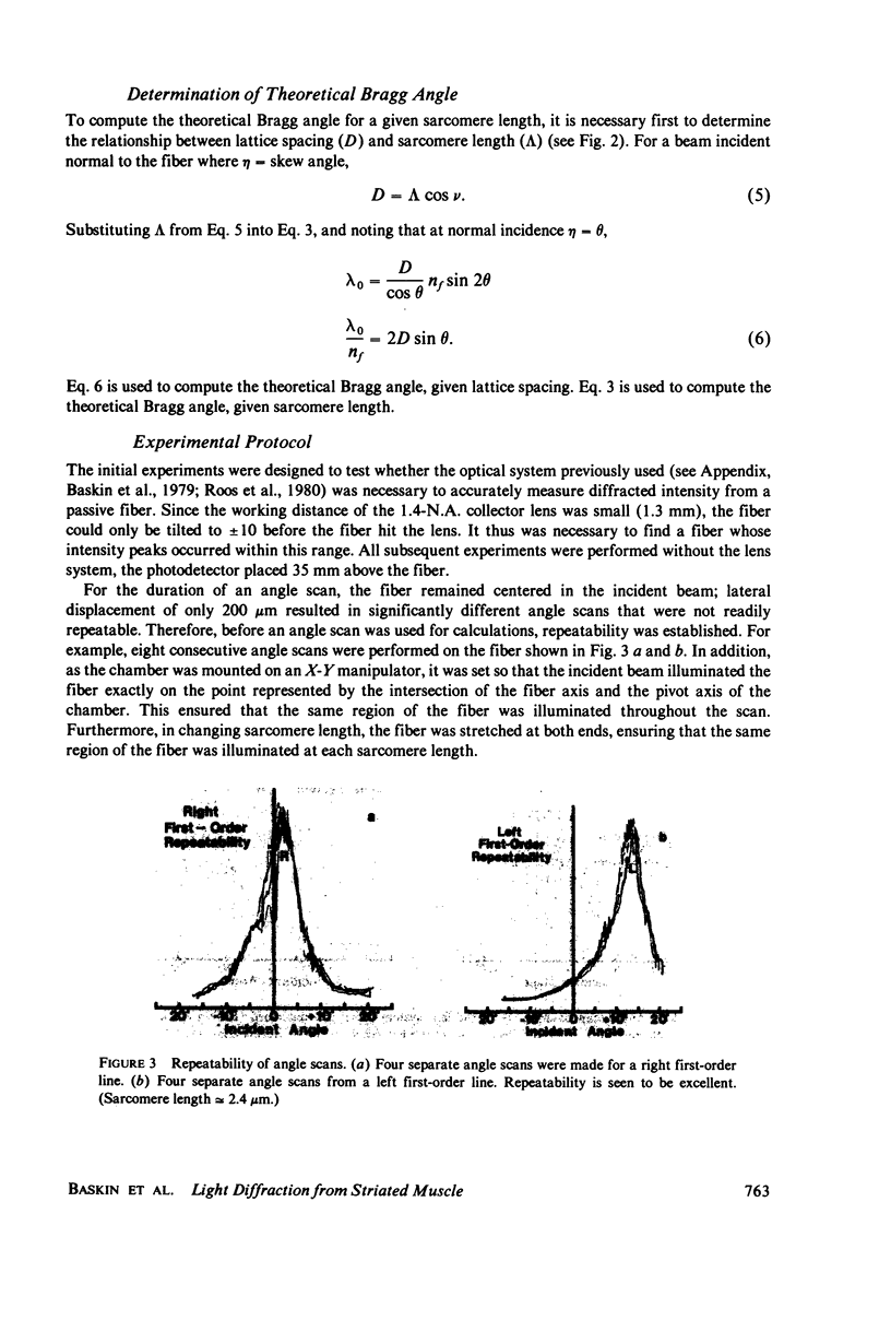
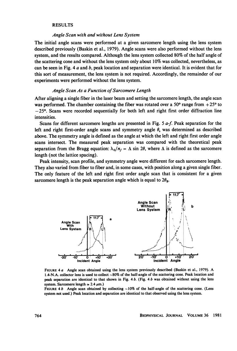
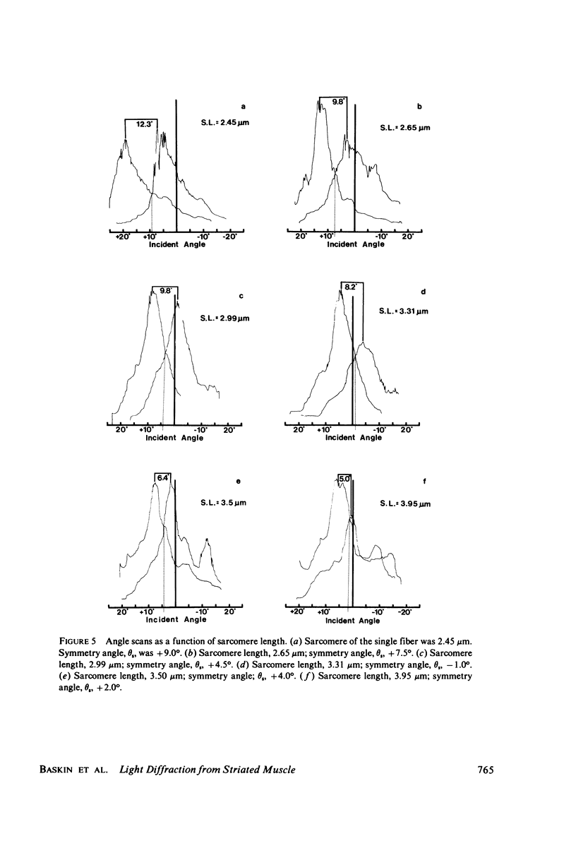
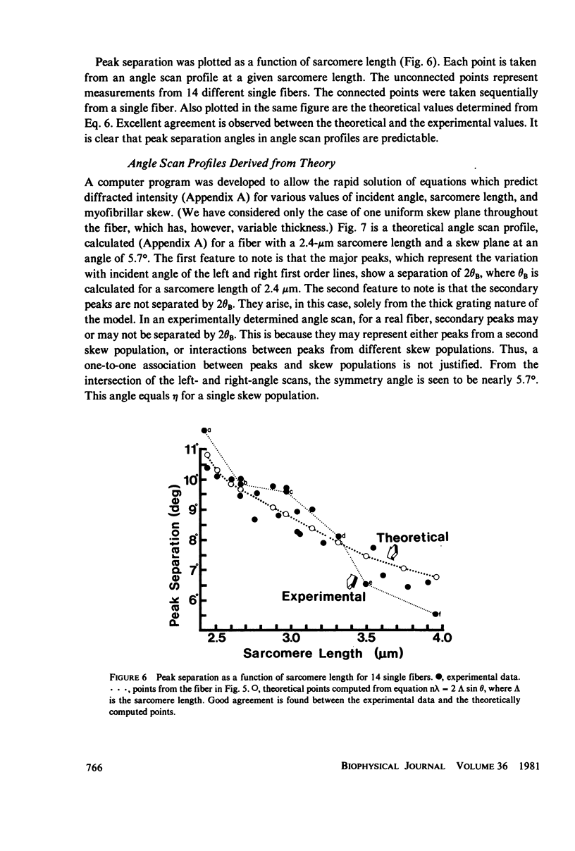
In a recently developed theory of light diffraction by single striated muscle fibers, we considered only the case of normal beam incidence. The present investigation represents both an experimental and theoretical extension of the previous work to arbitrary incident angle. Angle scan profiles over a 50 degrees range of incident angle (+25 degrees to -25 degrees) were obtained at different sarcomere lengths. Left and right first-order scan peak separations were found to be a function of sarcomere length (separation angle = 2 theta B), and good agreement was found between theory and experiment. Our theoretical analysis further showed that a myofibrillar population with a single common skew angle can yield an angle scan profile containing many peaks. Thus, it is not necessary to associate each peak with a different skew population. Finally, we have found that symmetry angle, theta s, also varies with sarcomere length, but not in a regular manner. Its value at a given sarcomere length is a function of a particular region of a given fiber and represents the average skew angle of all the myofibril populations illuminated. The intensity of a diffraction order line is considered to be principally the resultant of two interference phenomena. The first is a volume-grating phenomenon which results from the periodic A-I band structure of the fiber (with some contribution from Z bands and H zones). The second is Bragg reflection from skew planes, if the correct relation between incident angle and skew angle is met. This may result in intensity asymmetry between the left and right first order lines.
Full text
PDF
Selected References
These references are in PubMed. This may not be the complete list of references from this article.
- Baskin R. J., Roos K. P., Yeh Y. Light diffraction study of single skeletal muscle fibres. Biophys J. 1979 Oct;28(1):45–64. doi: 10.1016/S0006-3495(79)85158-9. [DOI] [PMC free article] [PubMed] [Google Scholar]
- Oba T., Baskin R. J., Lieber R. L. Light diffraction studies of active muscles fibres as a function of sarcomere length. J Muscle Res Cell Motil. 1981 Jun;2(2):215–224. doi: 10.1007/BF00711871. [DOI] [PubMed] [Google Scholar]
- Roos K. P., Baskin R. J., Lieber R. L., Cline J. W., Paolini P. J. Digital data acquisition and analysis of striated muscle diffraction patterns with a direct memory access microprocessor system. Rev Sci Instrum. 1980 Jun;51(6):762–767. doi: 10.1063/1.1136308. [DOI] [PubMed] [Google Scholar]
- Rüdel R., Zite-Ferenczy F. Efficiency of light diffraction by cross-striated muscle fibers under stretch and during isometric contraction. Biophys J. 1980 Jun;30(3):507–516. doi: 10.1016/S0006-3495(80)85110-1. [DOI] [PMC free article] [PubMed] [Google Scholar]
- Rüdel R., Zite-Ferenczy F. Interpretation of light diffraction by cross-striated muscle as Bragg reflexion of light by the lattice of contractile proteins. J Physiol. 1979 May;290(2):317–330. doi: 10.1113/jphysiol.1979.sp012773. [DOI] [PMC free article] [PubMed] [Google Scholar]
- Yeh Y., Baskin R. J., Lieber R. L., Roos K. P. Theory of light diffraction by single skeletal muscle fibers. Biophys J. 1980 Mar;29(3):509–522. doi: 10.1016/S0006-3495(80)85149-6. [DOI] [PMC free article] [PubMed] [Google Scholar]


