

Abstract
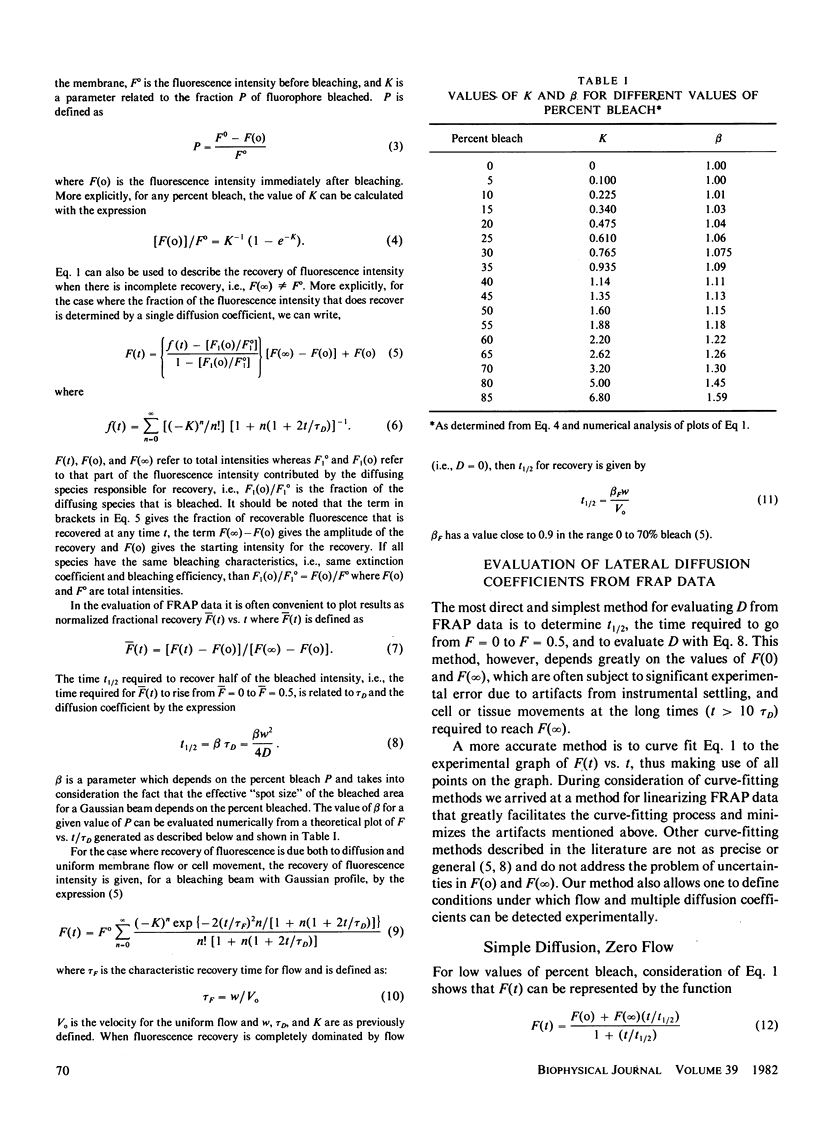
The evaluation of lateral diffusion coefficients of membrane components by the technique of fluorescence recovery after photobleaching (FRAP) is often complicated by uncertainties in the values of the intensities F(O), immediately after bleaching, and F(infinity), after full recovery. These uncertainties arise from instrumental settling time immediately after bleaching and from cell, tissue, microscope, or laser beam movements at the long times required to measure F(infinity). We have developed a method for precise analysis of FRAP data that minimizes these problems. The method is based on the observation that a plot of the reciprocal function R(tau) = F(infinity)/[F(infinity)-F(tau)] is linear over a large time range when (a) the laser beam has a Gaussian profile, (b) recovery involves a single diffusion coefficient, and (c) there is no membrane flow. Moreover, the ratio of intercept to slope of the linear plot is equal to tau 1/2, the time required for the bleached fluorescence to rise to 50% of the full recovery value, F(infinity). The lateral diffusion coefficient D is related to tau 1/2 by tau 1/2 = beta w2/4D where beta is a defined parameter and w is the effective radius of the focused laser beam. These results are shown to indicate that the recovery of fluorescence F(tau) can be represented over a large range of percent bleach, and recovery time tau by the relatively simple expression F(tau) = [ F(o) + F(infinity) (tau/tau 1/2)]/[1 + tau/tau 1/2)]. FRAP data can therefore be easily evaluated by a nonlinear regression analysis with this equation or by a linear fit to the reciprocal function R(tau). It is shown that any error in F(infinity) can be easily detected in a plot of R(tau) vs. tau which deviates significantly from a straight line when F(infinity) is in error by as little as 5%. A scheme for evaluating D by linear analysis is presented. It is also shown that the linear reciprocal plot provides a simple method for detecting flow or multiple diffusion coefficients and for establishing conditions (data precision, differences in multiple diffusion coefficients, magnitude of flow rate compared to lateral diffusion) under which flow or multiple diffusion coefficients can be detected. These aspects are discussed in some detail.
Full text
PDF


Selected References
These references are in PubMed. This may not be the complete list of references from this article.
- Axelrod D., Koppel D. E., Schlessinger J., Elson E., Webb W. W. Mobility measurement by analysis of fluorescence photobleaching recovery kinetics. Biophys J. 1976 Sep;16(9):1055–1069. doi: 10.1016/S0006-3495(76)85755-4. [DOI] [PMC free article] [PubMed] [Google Scholar]
- Barisas B. G. Criticality of beam alignment in fluorescence photobleaching recovery experiments. Biophys J. 1980 Mar;29(3):545–548. doi: 10.1016/S0006-3495(80)85153-8. [DOI] [PMC free article] [PubMed] [Google Scholar]
- Barisas B. G., Leuther M. D. Fluorescence photobleaching recovery measurement of protein absolute diffusion constants. Biophys Chem. 1979 Sep;10(2):221–229. doi: 10.1016/0301-4622(79)85044-9. [DOI] [PubMed] [Google Scholar]
- Edidin M., Zagyansky Y., Lardner T. J. Measurement of membrane protein lateral diffusion in single cells. Science. 1976 Feb 6;191(4226):466–468. doi: 10.1126/science.1246629. [DOI] [PubMed] [Google Scholar]
- Elson H. F., Yguerabide J. Membrane dynamics of differentiating cultured embryonic chick skeletal muscle cells by fluorescence microscopy techniques. J Supramol Struct. 1979;12(1):47–61. doi: 10.1002/jss.400120106. [DOI] [PubMed] [Google Scholar]
- Jacobson K., Derzko Z., Wu E. S., Hou Y., Poste G. Measurement of the lateral mobility of cell surface components in single, living cells by fluorescence recovery after photobleaching. J Supramol Struct. 1976;5(4):565(417)–576(428). doi: 10.1002/jss.400050411. [DOI] [PubMed] [Google Scholar]
- Koppel D. E. Fluorescence redistribution after photobleaching. A new multipoint analysis of membrane translational dynamics. Biophys J. 1979 Nov;28(2):281–291. doi: 10.1016/S0006-3495(79)85176-0. [DOI] [PMC free article] [PubMed] [Google Scholar]
- Peters R., Peters J., Tews K. H., Bähr W. A microfluorimetric study of translational diffusion in erythrocyte membranes. Biochim Biophys Acta. 1974 Nov 15;367(3):282–294. doi: 10.1016/0005-2736(74)90085-6. [DOI] [PubMed] [Google Scholar]
- Smith B. A., McConnell H. M. Determination of molecular motion in membranes using periodic pattern photobleaching. Proc Natl Acad Sci U S A. 1978 Jun;75(6):2759–2763. doi: 10.1073/pnas.75.6.2759. [DOI] [PMC free article] [PubMed] [Google Scholar]
- Woda B. A., Yguerabide J., Feldman J. D. The effect of local anesthetics on the lateral mobility of lymphocyte membrane proteins. Exp Cell Res. 1980 Apr;126(2):327–331. doi: 10.1016/0014-4827(80)90271-2. [DOI] [PubMed] [Google Scholar]