

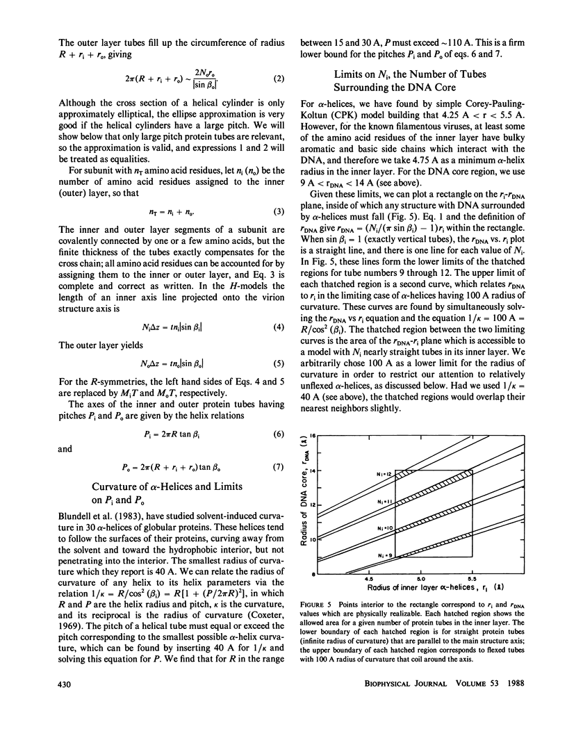
Abstract
A mathematical model is presented which explains the symmetries observed for the protein coats of filamentous bacterial viruses. Three viruses (Ff, IKe, and If1) all have five-start helices with rotation angles of 36 degrees and axial translations of 16 A (Type I symmetry), and three other viruses (Pf1, Xf, and Pf3) all have one-start helices with rotation angles of approximately equal to 67 degrees and translations of approximately 3 A (Type II symmetry). The coat protein subunits in each group diverge from each other in amino acid sequence, and Type II viruses differ dramatically in DNA structure. Regardless of the differences, both Type I and Type II symmetry can be understood as direct, natural consequences of the close-packing of alpha-helical protein subunits. In our treatment, an alpha-helical subunit is modeled as consisting of two interconnected, flexible tubular segments that follow helical paths around the DNA, one in an inner layer and the other in an outer layer. The mathematical model is a set of algebraic equations describing the disposition of the flexible segments. Solutions are described by newly introduced symmetry indices and other parameters. An exhaustive survey over the range of indices has produced a library of all structures that are geometrically feasible within our modeling scheme. Solutions which correspond in their rotation angles to Type I and Type II viruses occur over large ranges of the parameter space. A few solutions with other symmetries are also allowed, and viruses with these symmetries may exist in nature. One solution to the set of equations, obtained without any recourse to the x-ray data, yields a calculated x-ray diffraction pattern for Pf1 which compares reasonably with experimental patterns. The close-packing geometry we have used helps explain the near constant linear mass density of known filamentous phages. Helicoid, rigid cylinder, and maximum entropy structure models proposed by others for Pf1 are reconciled with the flexible tube models and with one another.
Full text
PDF


Selected References
These references are in PubMed. This may not be the complete list of references from this article.
- Anderson W. F., Ohlendorf D. H., Takeda Y., Matthews B. W. Structure of the cro repressor from bacteriophage lambda and its interaction with DNA. Nature. 1981 Apr 30;290(5809):754–758. doi: 10.1038/290754a0. [DOI] [PubMed] [Google Scholar]
- Banner D. W., Nave C., Marvin D. A. Structure of the protein and DNA in fd filamentous bacterial virus. Nature. 1981 Feb 26;289(5800):814–816. doi: 10.1038/289814a0. [DOI] [PubMed] [Google Scholar]
- Beck E., Zink B. Nucleotide sequence and genome organisation of filamentous bacteriophages fl and fd. Gene. 1981 Dec;16(1-3):35–58. doi: 10.1016/0378-1119(81)90059-7. [DOI] [PubMed] [Google Scholar]
- Berkowitz S. A., Day L. A. Turbidity measurements in an analytical ultracentrifuge. Determinations of mass per length for filamentous viruses fd, Xf, and Pf3. Biochemistry. 1980 Jun 10;19(12):2696–2702. doi: 10.1021/bi00553a025. [DOI] [PubMed] [Google Scholar]
- Blundell T., Barlow D., Borkakoti N., Thornton J. Solvent-induced distortions and the curvature of alpha-helices. Nature. 1983 Nov 17;306(5940):281–283. doi: 10.1038/306281a0. [DOI] [PubMed] [Google Scholar]
- Bryan R. K., Bansal M., Folkhard W., Nave C., Marvin D. A. Maximum-entropy calculation of the electron density at 4 A resolution of Pf1 filamentous bacteriophage. Proc Natl Acad Sci U S A. 1983 Aug;80(15):4728–4731. doi: 10.1073/pnas.80.15.4728. [DOI] [PMC free article] [PubMed] [Google Scholar]
- Casadevall A., Day L. A. Silver and mercury probing of deoxyribonucleic acid structures in the filamentous viruses fd, If1, IKe, Xf, Pf1, and Pf3. Biochemistry. 1983 Sep 27;22(20):4831–4842. doi: 10.1021/bi00289a033. [DOI] [PubMed] [Google Scholar]
- Caspar D. L., Makowski L. The symmetries of filamentous phage particles. J Mol Biol. 1981 Jan 25;145(3):611–617. doi: 10.1016/0022-2836(81)90549-0. [DOI] [PubMed] [Google Scholar]
- Chen F. C., Koopmans G., Wiseman R. L., Day L. A., Swinney H. L. Dimensions of Xf virus from its rotational and translational diffusion coefficients. Biochemistry. 1980 Apr 1;19(7):1373–1376. doi: 10.1021/bi00548a016. [DOI] [PubMed] [Google Scholar]
- Chothia C., Levitt M., Richardson D. Helix to helix packing in proteins. J Mol Biol. 1981 Jan 5;145(1):215–250. doi: 10.1016/0022-2836(81)90341-7. [DOI] [PubMed] [Google Scholar]
- Chothia C. Structural invariants in protein folding. Nature. 1975 Mar 27;254(5498):304–308. doi: 10.1038/254304a0. [DOI] [PubMed] [Google Scholar]
- Cross T. A., Tsang P., Opella S. J. Comparison of protein and deoxyribonucleic acid backbone structures in fd and Pf1 bacteriophages. Biochemistry. 1983 Feb 15;22(4):721–726. doi: 10.1021/bi00273a002. [DOI] [PubMed] [Google Scholar]
- Day L. A., Wiseman R. L., Marzec C. J. Structure models for DNA in filamentous viruses with phosphates near the center. Nucleic Acids Res. 1979 Nov 24;7(6):1393–1403. doi: 10.1093/nar/7.6.1393. [DOI] [PMC free article] [PubMed] [Google Scholar]
- Dunker A. K., Klausner R. D., Marvin D. A., Wiseman R. L. Letter: Filamentous bacterial viruses. X. X-ray diffraction studies of the R4-protein mutant. J Mol Biol. 1974 Jan 5;82(1):115–117. doi: 10.1016/0022-2836(74)90579-8. [DOI] [PubMed] [Google Scholar]
- Frangione B., Nakashima Y., Konigsberg W., Wiseman R. L. The amino acid sequence of the major coat protein subunit of the filamentous virus Xf. FEBS Lett. 1978 Dec 15;96(2):381–384. doi: 10.1016/0014-5793(78)80442-6. [DOI] [PubMed] [Google Scholar]
- Hunter G. J., Rowitch D. H., Perham R. N. Interactions between DNA and coat protein in the structure and assembly of filamentous bacteriophage fd. Nature. 1987 May 21;327(6119):252–254. doi: 10.1038/327252a0. [DOI] [PubMed] [Google Scholar]
- Luiten R. G., Putterman D. G., Schoenmakers J. G., Konings R. N., Day L. A. Nucleotide sequence of the genome of Pf3, an IncP-1 plasmid-specific filamentous bacteriophage of Pseudomonas aeruginosa. J Virol. 1985 Oct;56(1):268–276. doi: 10.1128/jvi.56.1.268-276.1985. [DOI] [PMC free article] [PubMed] [Google Scholar]
- Makowski L., Caspar D. L., Marvin D. A. Filamentous bacteriophage Pf1 structure determined at 7A resolution by refinement of models for the alpha-helical subunit. J Mol Biol. 1980 Jun 25;140(2):149–181. doi: 10.1016/0022-2836(80)90101-1. [DOI] [PubMed] [Google Scholar]
- Marvin D. A., Bryan R. K., Nave C. Pf1 Inovirus. Electron density distribution calculated by a maximum entropy algorithm from native fibre diffraction data to 3 A resolution and single isomorphous replacement data to 5 A resolution. J Mol Biol. 1987 Jan 20;193(2):315–343. doi: 10.1016/0022-2836(87)90222-1. [DOI] [PubMed] [Google Scholar]
- Marvin D. A., Pigram W. J., Wiseman R. L., Wachtel E. J., Marvin F. J. Filamentous bacterial viruses. XIL. Molecular architecture of the class I (fd, If1, IKe) virion. J Mol Biol. 1974 Sep 25;88(3):581–598. doi: 10.1016/0022-2836(74)90409-4. [DOI] [PubMed] [Google Scholar]
- Marvin D. A., Wachtel E. J. Structure and assembly of filamentous bacterial viruses. Nature. 1975 Jan 3;253(5486):19–23. doi: 10.1038/253019a0. [DOI] [PubMed] [Google Scholar]
- Marvin D. A., Wiseman R. L., Wachtel E. J. Filamentous bacterial viruses. XI. Molecular architecture of the class II (Pf1, Xf) virion. J Mol Biol. 1974 Jan 15;82(2):121–138. doi: 10.1016/0022-2836(74)90336-2. [DOI] [PubMed] [Google Scholar]
- Marzec C. J., Day L. A. DNA and protein lattice-lattice interactions in the filamentous bacteriophages. Biophys J. 1983 May;42(2):171–180. doi: 10.1016/S0006-3495(83)84383-5. [DOI] [PMC free article] [PubMed] [Google Scholar]
- Nave C., Brown R. S., Fowler A. G., Ladner J. E., Marvin D. A., Provencher S. W., Tsugita A., Armstrong J., Perham R. N. Pf1 filamentous bacterial virus. X-ray fibre diffraction analysis of two heavy-atom derivatives. J Mol Biol. 1981 Jul 15;149(4):675–707. doi: 10.1016/0022-2836(81)90353-3. [DOI] [PubMed] [Google Scholar]
- Nave C., Fowler A. G., Malsey S., Marvin D. A., Siegrist H., Wachtel E. J. Macromolecular structural transitions in Pf1 filamentous bacterial virus. Nature. 1979 Sep 20;281(5728):232–234. doi: 10.1038/281232a0. [DOI] [PubMed] [Google Scholar]
- Newman J., Day L. A., Dalack G. W., Eden D. Hydrodynamic determination of molecular weight, dimensions, and structural parameters of Pf3 virus. Biochemistry. 1982 Jul 6;21(14):3352–3358. doi: 10.1021/bi00257a016. [DOI] [PubMed] [Google Scholar]
- Newman J., Swinney H. L., Day L. A. Hydrodynamic properties and structure of fd virus. J Mol Biol. 1977 Nov 5;116(3):593–603. doi: 10.1016/0022-2836(77)90086-9. [DOI] [PubMed] [Google Scholar]
- Opella S. J., Stewart P. L., Valentine K. G. Protein structure by solid-state NMR spectroscopy. Q Rev Biophys. 1987 Feb;19(1-2):7–49. doi: 10.1017/s0033583500004017. [DOI] [PubMed] [Google Scholar]
- Peterson C., Winter W. T., Dalack G. W., Day L. A. Structure of the filamentous bacteriophage, Pf3, by X-ray fiber diffraction. J Mol Biol. 1982 Dec 25;162(4):877–881. doi: 10.1016/0022-2836(82)90552-6. [DOI] [PubMed] [Google Scholar]
- Putterman D. G., Casadevall A., Boyle P. D., Yang H. L., Frangione B., Day L. A. Major coat protein and single-stranded DNA-binding protein of filamentous virus Pf3. Proc Natl Acad Sci U S A. 1984 Feb;81(3):699–703. doi: 10.1073/pnas.81.3.699. [DOI] [PMC free article] [PubMed] [Google Scholar]
- Specthrie L., Greenberg J., Glucksman M. J., Diaz J., Makowski L. Structural responsiveness of filamentous bacteriophage Pf1: comparison of virion structure in fibers and solution. The effect of temperature and ionic strength. Biophys J. 1987 Aug;52(2):199–214. doi: 10.1016/S0006-3495(87)83207-1. [DOI] [PMC free article] [PubMed] [Google Scholar]
- Thomas G. J., Jr, Agard D. A. Quantitative analysis of nucleic acids, proteins, and viruses by Raman band deconvolution. Biophys J. 1984 Dec;46(6):763–768. doi: 10.1016/S0006-3495(84)84074-6. [DOI] [PMC free article] [PubMed] [Google Scholar]
- Thomas G. J., Jr, Day L. A. Conformational transitions in Pf3 and their implications for the structure and assembly of filamentous bacterial viruses. Proc Natl Acad Sci U S A. 1981 May;78(5):2962–2966. doi: 10.1073/pnas.78.5.2962. [DOI] [PMC free article] [PubMed] [Google Scholar]
- Thomas G. J., Jr, Prescott B., Day L. A. Structure similarity, difference and variability in the filamentous viruses fd, If1, IKe, Pf1 and Xf. Investigation by laser Raman spectroscopy. J Mol Biol. 1983 Apr 5;165(2):321–356. doi: 10.1016/s0022-2836(83)80260-5. [DOI] [PubMed] [Google Scholar]
- Torbet J., Maret G. High-field magnetic birefringence study of the structure of rodlike phages Pf1 and fd in solution. Biopolymers. 1981 Dec;20(12):2657–2669. doi: 10.1002/bip.1981.360201212. [DOI] [PubMed] [Google Scholar]
- Torbet J. Neutron scattering study of the solution structure of bacteriophages Pf1 and fd. FEBS Lett. 1979 Dec 1;108(1):61–65. doi: 10.1016/0014-5793(79)81179-5. [DOI] [PubMed] [Google Scholar]
- Wachtel E. J., Marvin F. J., Marvin D. A. Structural transition in a filamentous protein. J Mol Biol. 1976 Nov 5;107(3):379–383. doi: 10.1016/s0022-2836(76)80011-3. [DOI] [PubMed] [Google Scholar]
- Wiseman R. L., Day L. A. Different packaging of DNA in the filamentous viruses Pf1 and Xf. J Mol Biol. 1977 Nov 5;116(3):607–611. doi: 10.1016/0022-2836(77)90088-2. [DOI] [PubMed] [Google Scholar]
- Zamyatnin A. A. Protein volume in solution. Prog Biophys Mol Biol. 1972;24:107–123. doi: 10.1016/0079-6107(72)90005-3. [DOI] [PubMed] [Google Scholar]