Abstract
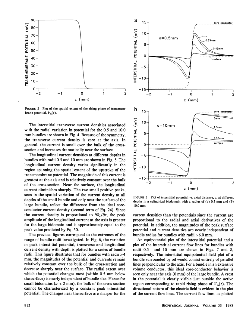
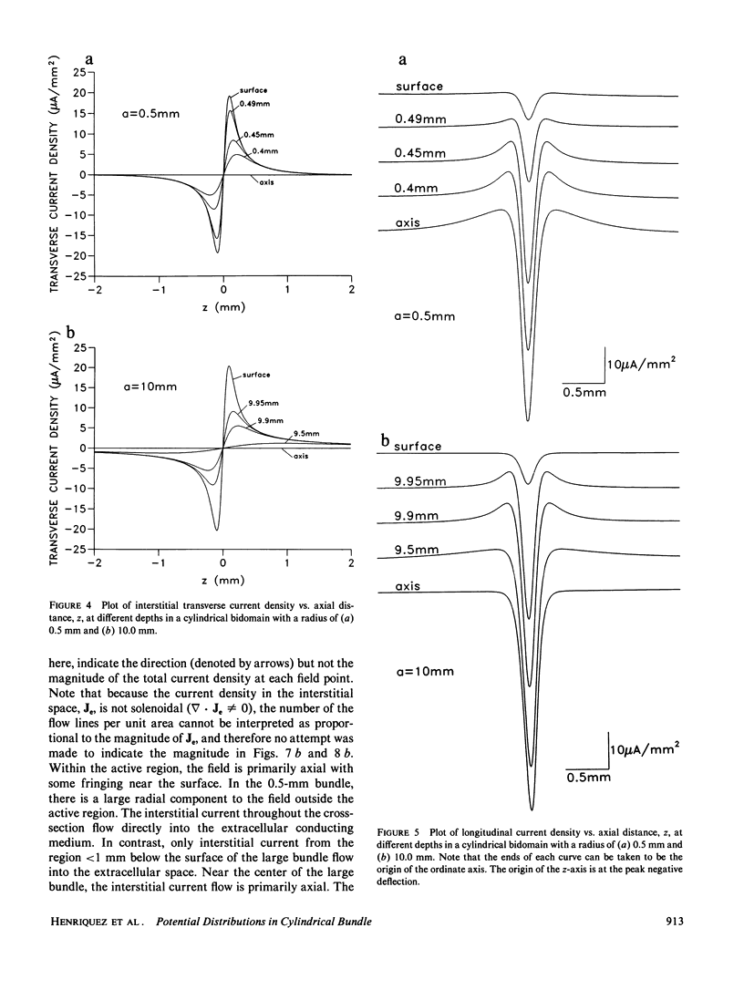
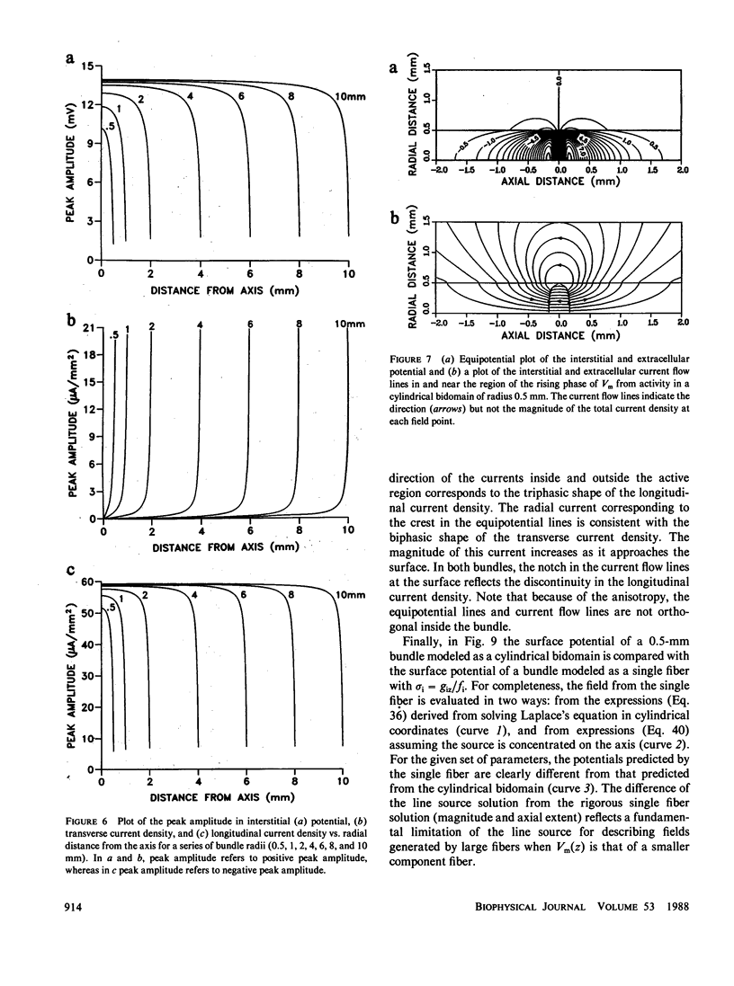
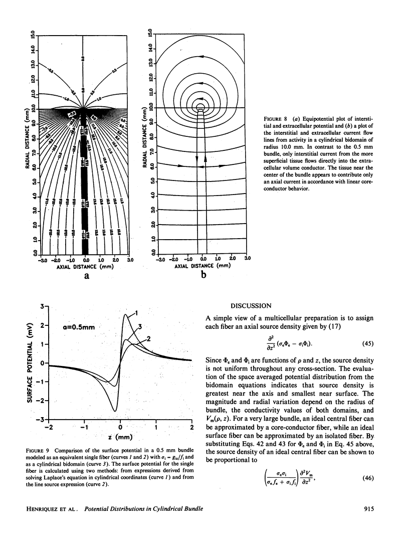
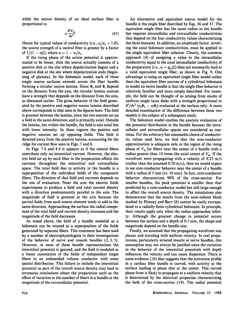
The intracellular and interstitial potentials associated with each cell or fiber in multicellular preparations carrying a uniformly propagating wave are important for characterizing the electrophysiological behavior of the preparation and in particular, for evaluating the source contributed by each fiber. The aforementioned potentials depend on a number of factors including the conductivities characterizing the intracellular, interstitial, and extracellular domains, the thickness of the tissue, and the distance (depth) of the field point from the surface of the tissue. A model study is presented describing the extracellular and interstitial potential distribution and current flow in a cylindrical bundle of cardiac muscle arising from a planar wavefront. For simplicity, the bundle is considered as a bidomain. Using typical values of conductivity, the results show that the intracellular and interstitial potential of fibers near the center of a very large bundle (greater than 10 mm) may be approximated by the potentials of a single fiber surrounded by a limited extracellular space (a fiber in oil), hence justifying a core-conductor model. For smaller bundles, the peak interstitial potential is less than that predicted by the core-conductor model but still large enough to affect the overall source strength. The magnitude of the source strength is greatest for fibers lying near the center of the bundle and diminishes sharply for fibers within 50 microns of the surface.
Full text
PDF
Selected References
These references are in PubMed. This may not be the complete list of references from this article.
- Clerc L. Directional differences of impulse spread in trabecular muscle from mammalian heart. J Physiol. 1976 Feb;255(2):335–346. doi: 10.1113/jphysiol.1976.sp011283. [DOI] [PMC free article] [PubMed] [Google Scholar]
- Ganapathy N., Clark J. W., Jr, Wilson O. B., Giles W. Forward and inverse potential field solutions for cardiac strands of cylindrical geometry. IEEE Trans Biomed Eng. 1985 Aug;32(8):566–577. doi: 10.1109/TBME.1985.325481. [DOI] [PubMed] [Google Scholar]
- Henriquez C. S., Plonsey R. Effect of resistive discontinuities on waveshape and velocity in a single cardiac fibre. Med Biol Eng Comput. 1987 Jul;25(4):428–438. doi: 10.1007/BF02443364. [DOI] [PubMed] [Google Scholar]
- Nandedkar S., Stålberg E. Simulation of macro EMG motor unit potentials. Electroencephalogr Clin Neurophysiol. 1983 Jul;56(1):52–62. doi: 10.1016/0013-4694(83)90006-8. [DOI] [PubMed] [Google Scholar]
- Plonsey R., Barr R. C. Interstitial potentials and their change with depth into cardiac tissue. Biophys J. 1987 Apr;51(4):547–555. doi: 10.1016/S0006-3495(87)83380-5. [DOI] [PMC free article] [PubMed] [Google Scholar]
- Plonsey R. Quantitative formulations of electrophysiological sources of potential fields in volume conductors. IEEE Trans Biomed Eng. 1984 Dec;31(12):868–872. doi: 10.1109/TBME.1984.325250. [DOI] [PubMed] [Google Scholar]
- Plonsey R. The active fiber in a volume conductor. IEEE Trans Biomed Eng. 1974 Sep;21(5):371–381. doi: 10.1109/TBME.1974.324406. [DOI] [PubMed] [Google Scholar]
- Roth B. J., Wikswo J. P., Jr A bidomain model for the extracellular potential and magnetic field of cardiac tissue. IEEE Trans Biomed Eng. 1986 Apr;33(4):467–469. doi: 10.1109/TBME.1986.325804. [DOI] [PubMed] [Google Scholar]
- Rudy Y., Quan W. L. A model study of the effects of the discrete cellular structure on electrical propagation in cardiac tissue. Circ Res. 1987 Dec;61(6):815–823. doi: 10.1161/01.res.61.6.815. [DOI] [PubMed] [Google Scholar]
- Schoonhoven R., Stegeman D. F., van Oosterom A. The forward problem in electroneurography. II: Comparison of models. IEEE Trans Biomed Eng. 1986 Mar;33(3):335–341. doi: 10.1109/TBME.1986.325719. [DOI] [PubMed] [Google Scholar]
- Sommer J. R., Scherer B. Geometry of cell and bundle appositions in cardiac muscle: light microscopy. Am J Physiol. 1985 Jun;248(6 Pt 2):H792–H803. doi: 10.1152/ajpheart.1985.248.6.H792. [DOI] [PubMed] [Google Scholar]
- Spach M. S., Dolber P. C. Relating extracellular potentials and their derivatives to anisotropic propagation at a microscopic level in human cardiac muscle. Evidence for electrical uncoupling of side-to-side fiber connections with increasing age. Circ Res. 1986 Mar;58(3):356–371. doi: 10.1161/01.res.58.3.356. [DOI] [PubMed] [Google Scholar]
- Stegeman D. F., de Weerd J. P., Eijkman E. G. A volume conductor study of compound action potentials of nerves in situ: the forward problem. Biol Cybern. 1979 Jun;33(2):97–111. doi: 10.1007/BF00355258. [DOI] [PubMed] [Google Scholar]
- Suenson M. Interaction between ventricular cells during the early part of excitation in the ferret heart. Acta Physiol Scand. 1985 Sep;125(1):81–90. doi: 10.1111/j.1748-1716.1985.tb07694.x. [DOI] [PubMed] [Google Scholar]


