Abstract
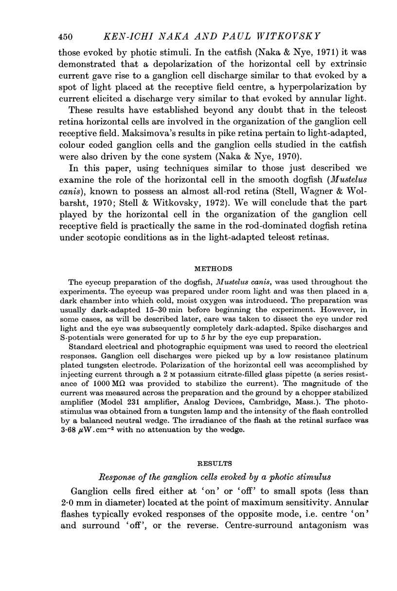
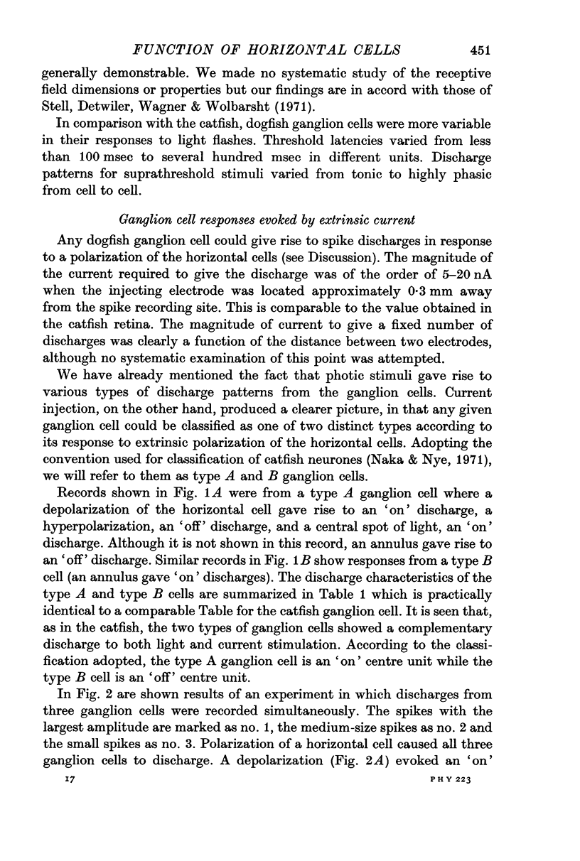
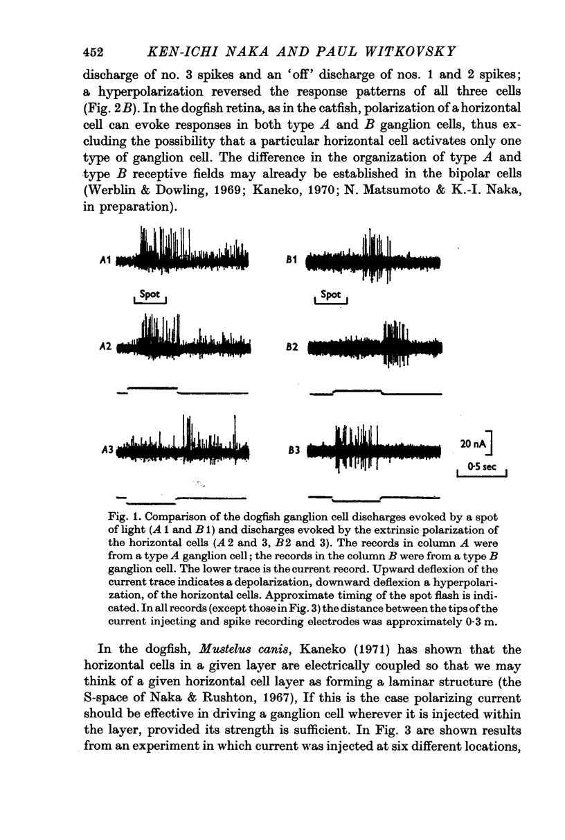
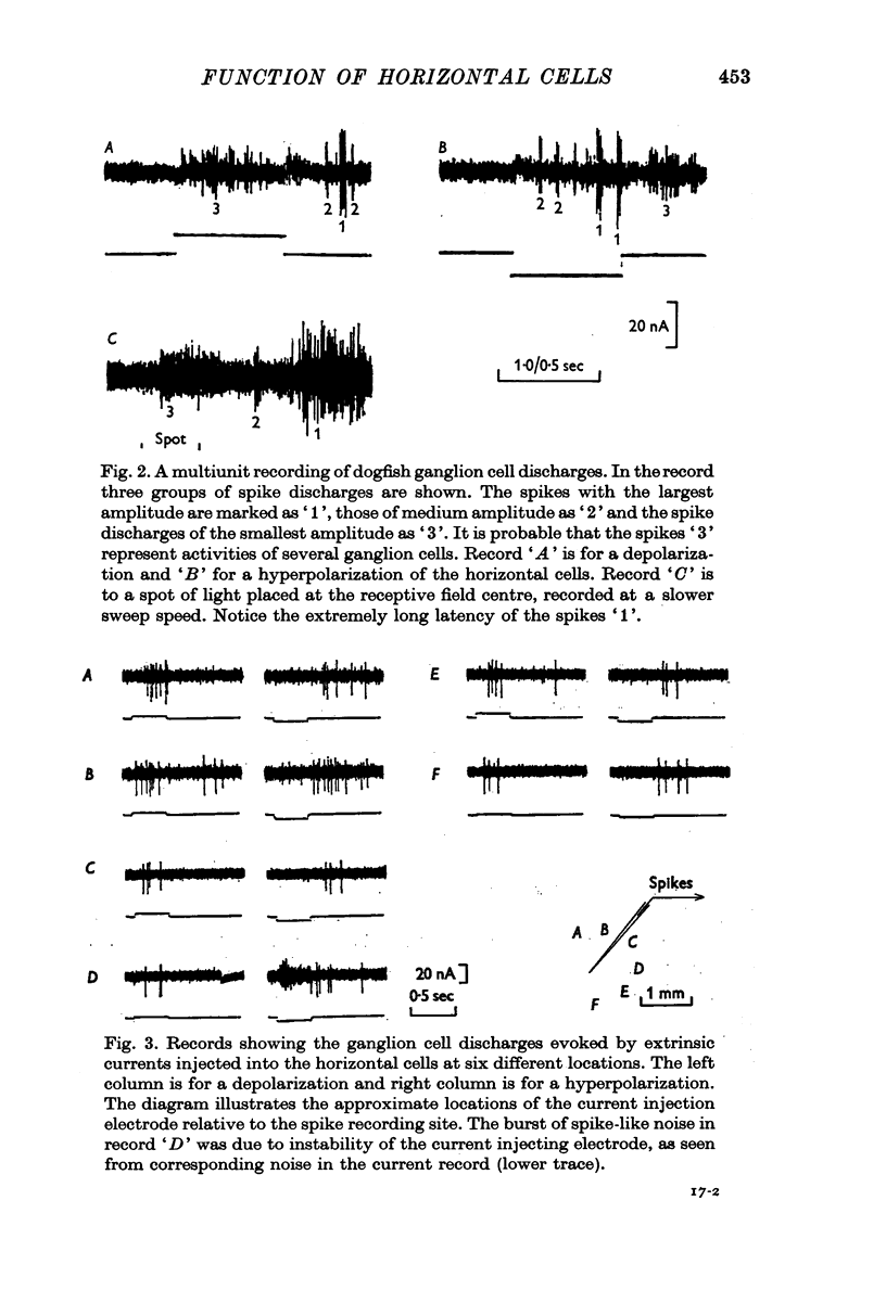
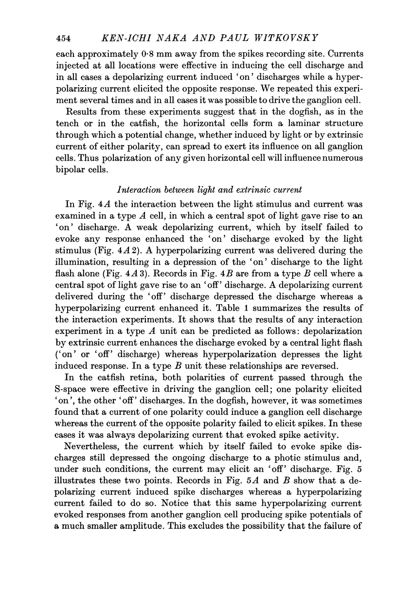
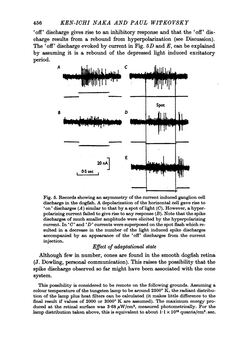
1. Ganglion cell discharges were evoked by extrinsic polarization of the horizontal cells in the retina of the smooth dogfish (Mustelus canis). Depolarization of the horizontal cell gave rise to a discharge similar to that evoked by a spot of light (centre type response) and hyperpolarization of the horizontal cell, a discharge similar to that by an annulus (surround type response).
2. Procion dye injection established that the current-passing electrode was sometimes located in the external horizontal cell. Other possibilities, such as middle and internal horizontal cells, were neither confirmed nor excluded.
3. Activation of ganglion cells by current was possible under completely dark-adapted conditions and for several log units above this level.
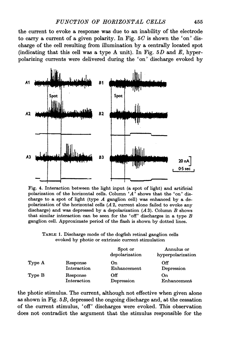
4. Depolarizing current enhanced the ganglion cell response evoked by a light spot in the centre of its receptive field; hyperpolarizing current antagonized the response to the same flash.
5. The results are consistent with the supposition that a potential change in the horizontal cell, irrespective of its polarity, or whether produced by light or current, spreads within a laminar layer (the S-space). The effect of the potential change is to modulate the response of bipolar cells and their input into the ganglion cell.
Full text
PDF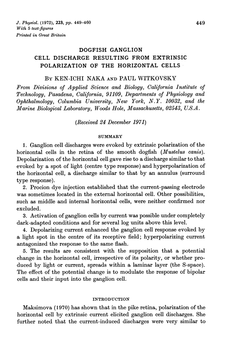
Selected References
These references are in PubMed. This may not be the complete list of references from this article.
- Kaneko A. Electrical connexions between horizontal cells in the dogfish retina. J Physiol. 1971 Feb;213(1):95–105. doi: 10.1113/jphysiol.1971.sp009370. [DOI] [PMC free article] [PubMed] [Google Scholar]
- Kaneko A. Physiological and morphological identification of horizontal, bipolar and amacrine cells in goldfish retina. J Physiol. 1970 May;207(3):623–633. doi: 10.1113/jphysiol.1970.sp009084. [DOI] [PMC free article] [PubMed] [Google Scholar]
- Naka K. I., Nye P. W. Receptive-field organization of the catfish retina: are at least two lateral mechanisms involved? J Neurophysiol. 1970 Sep;33(5):625–642. doi: 10.1152/jn.1970.33.5.625. [DOI] [PubMed] [Google Scholar]
- Naka K. I., Nye P. W. Role of horizontal cells in organization of the catfish retinal receptive field. J Neurophysiol. 1971 Sep;34(5):785–801. doi: 10.1152/jn.1971.34.5.785. [DOI] [PubMed] [Google Scholar]
- Naka K. I. Receptive field mechanism in the vertebrate retina. Science. 1971 Feb 19;171(3972):691–693. doi: 10.1126/science.171.3972.691. [DOI] [PubMed] [Google Scholar]
- Naka K. I., Rushton W. A. The generation and spread of S-potentials in fish (Cyprinidae). J Physiol. 1967 Sep;192(2):437–461. doi: 10.1113/jphysiol.1967.sp008308. [DOI] [PMC free article] [PubMed] [Google Scholar]
- Werblin F. S., Dowling J. E. Organization of the retina of the mudpuppy, Necturus maculosus. II. Intracellular recording. J Neurophysiol. 1969 May;32(3):339–355. doi: 10.1152/jn.1969.32.3.339. [DOI] [PubMed] [Google Scholar]


