

Abstract
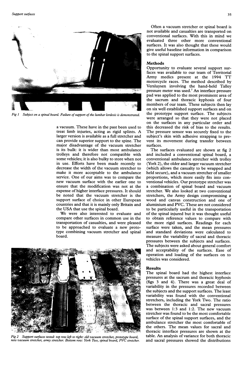
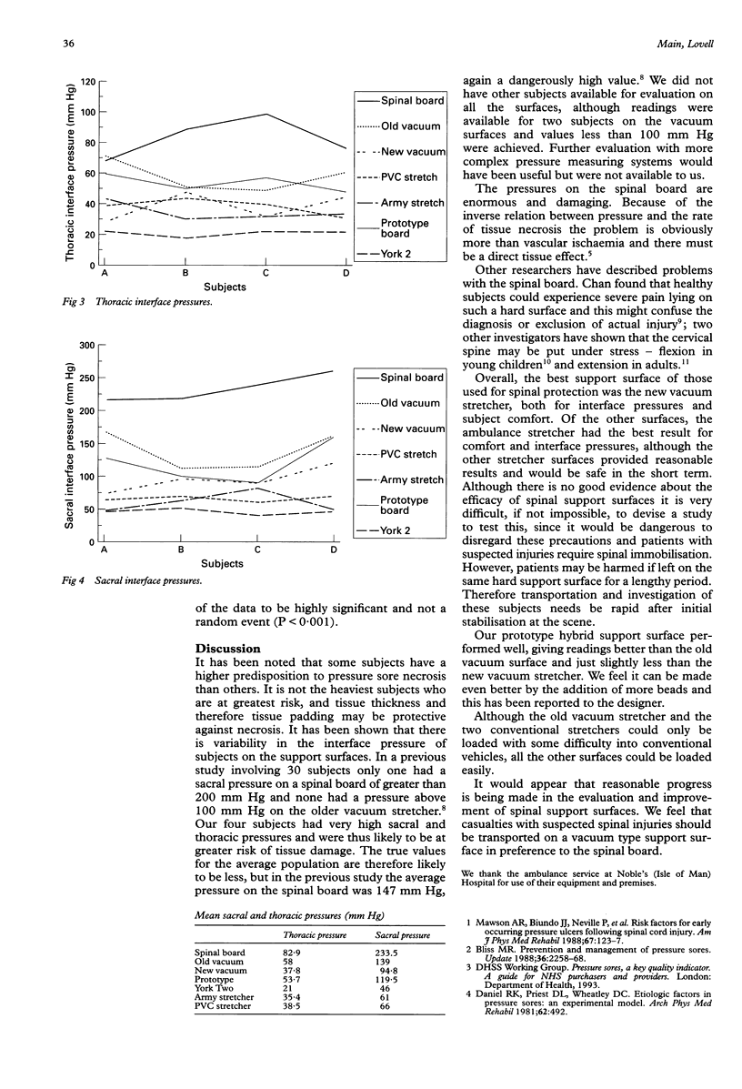
The aim was to evaluate seven evacuation support surfaces. These included the conventional spinal board, two designs of vacuum stretcher, a prototype support surface which was a combination of both principles, and three conventional stretchers. Interface pressures were evaluated in four healthy volunteers. The sacral and thoracic interface pressures were measured. Mean sacral readings were: spinal board 233.5 mm Hg, old vacuum stretcher 139 mm Hg, new design 94.8 mm Hg, prototype board 119.5 mm Hg, York Two stretcher 46 mm Hg, Army stretcher 61 mm Hg, and the PVC and aluminium stretcher 66 mm Hg. Thoracic pressure readings were: spinal board 82.9 mm Hg, old design vacuum stretcher 58 mm Hg, new design 37.8 mm Hg, prototype board 53.7 mm Hg, York two 21 mm Hg, army stretcher 35.4 mm Hg, and PVC stretcher 38.5 mm Hg. Analysis of variance showed both distributions to be highly significant (P < 0.001). The spinal board has several deficiencies, including lack of support for the lumbar lordosis. It should not be the preferred surface for the transfer of patients with spinal injuries.
Full text
PDF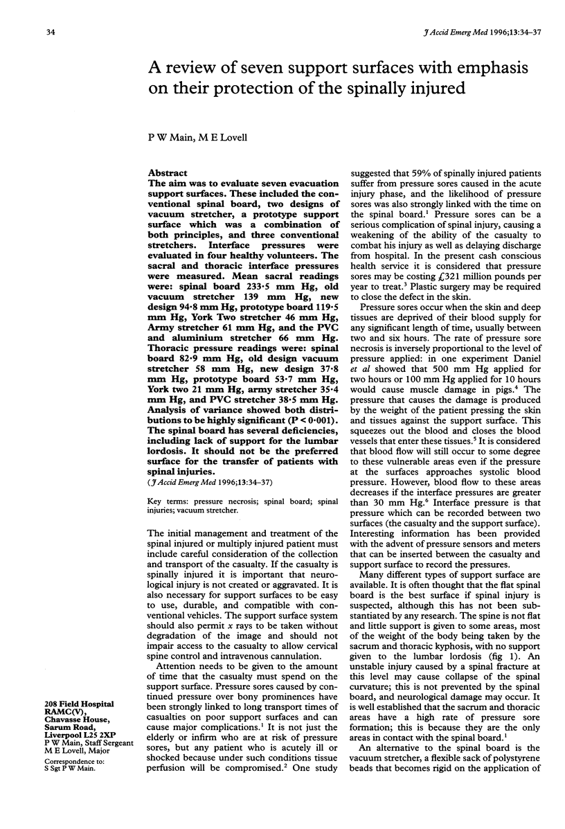

Images in this article
Selected References
These references are in PubMed. This may not be the complete list of references from this article.
- Bader D. L., Gant C. A. Changes in transcutaneous oxygen tension as a result of prolonged pressures at the sacrum. Clin Phys Physiol Meas. 1988 Feb;9(1):33–40. doi: 10.1088/0143-0815/9/1/002. [DOI] [PubMed] [Google Scholar]
- Chan D., Goldberg R., Tascone A., Harmon S., Chan L. The effect of spinal immobilization on healthy volunteers. Ann Emerg Med. 1994 Jan;23(1):48–51. doi: 10.1016/s0196-0644(94)70007-9. [DOI] [PubMed] [Google Scholar]
- Daniel R. K., Priest D. L., Wheatley D. C. Etiologic factors in pressure sores: an experimental model. Arch Phys Med Rehabil. 1981 Oct;62(10):492–498. [PubMed] [Google Scholar]
- Herzenberg J. E., Hensinger R. N., Dedrick D. K., Phillips W. A. Emergency transport and positioning of young children who have an injury of the cervical spine. The standard backboard may be hazardous. J Bone Joint Surg Am. 1989 Jan;71(1):15–22. [PubMed] [Google Scholar]
- Lovell M. E., Evans J. H. A comparison of the spinal board and the vacuum stretcher, spinal stability and interface pressure. Injury. 1994 Apr;25(3):179–180. doi: 10.1016/0020-1383(94)90158-9. [DOI] [PubMed] [Google Scholar]
- Mawson A. R., Biundo J. J., Jr, Neville P., Linares H. A., Winchester Y., Lopez A. Risk factors for early occurring pressure ulcers following spinal cord injury. Am J Phys Med Rehabil. 1988 Jun;67(3):123–127. doi: 10.1097/00002060-198806000-00007. [DOI] [PubMed] [Google Scholar]
- Reuler J. B., Cooney T. G. The pressure sore: pathophysiology and principles of management. Ann Intern Med. 1981 May;94(5):661–666. doi: 10.7326/0003-4819-94-5-661. [DOI] [PubMed] [Google Scholar]
- Versluysen M. How elderly patients with femoral fracture develop pressure sores in hospital. Br Med J (Clin Res Ed) 1986 May 17;292(6531):1311–1313. doi: 10.1136/bmj.292.6531.1311. [DOI] [PMC free article] [PubMed] [Google Scholar]