Abstract
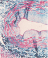
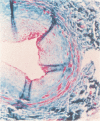
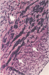
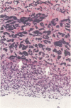
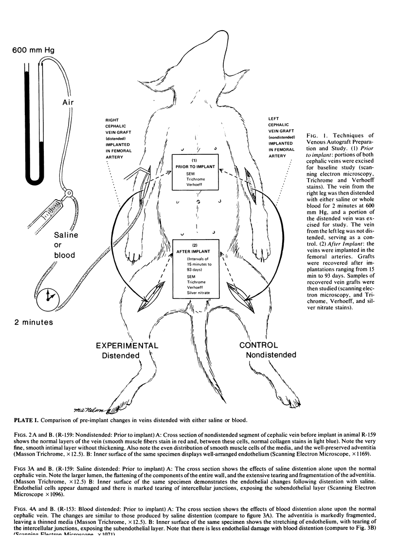
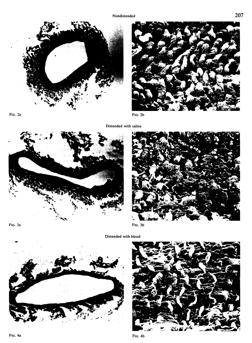
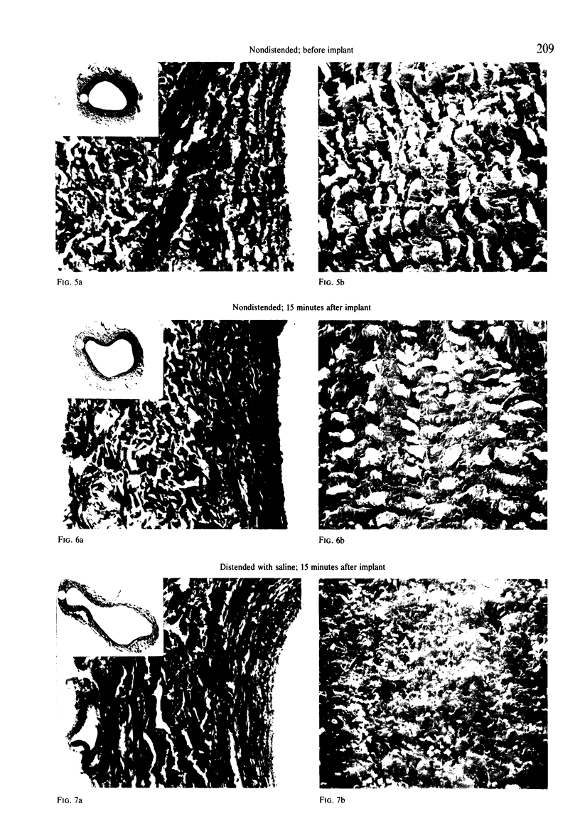
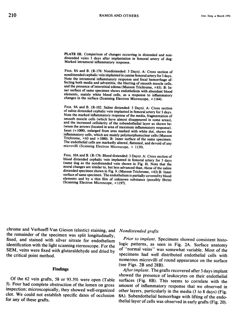
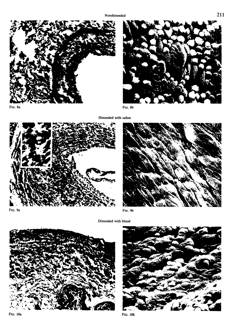
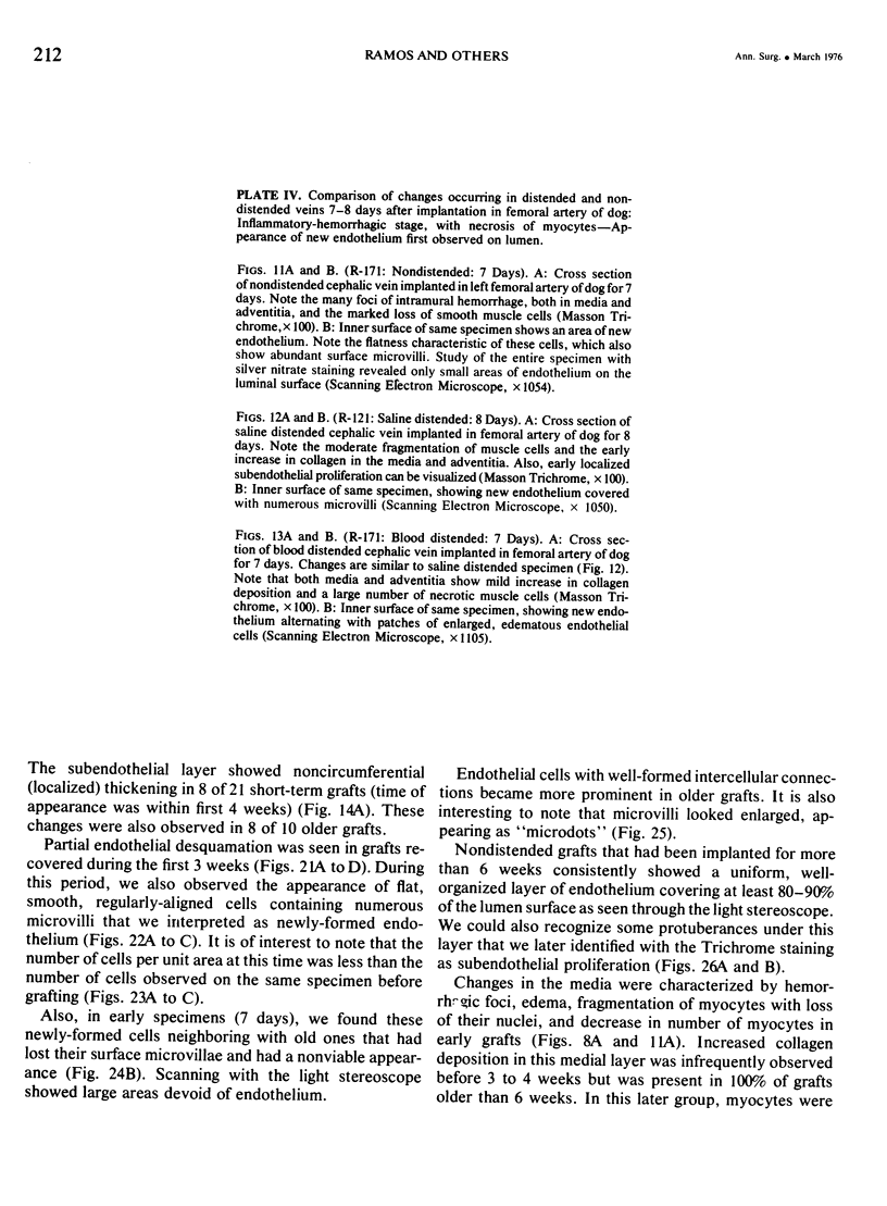
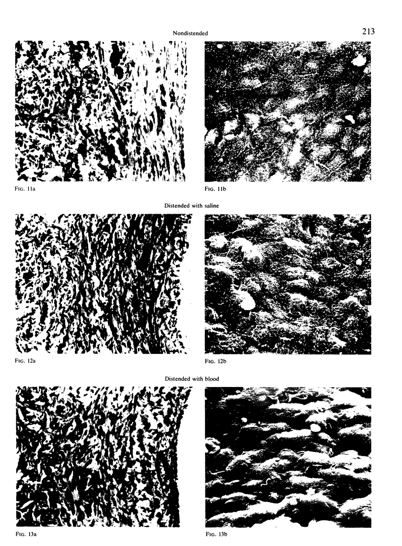
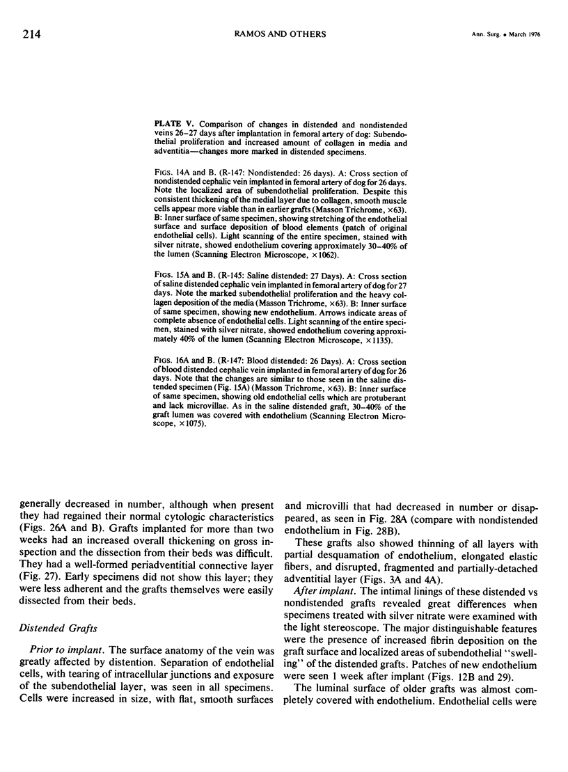
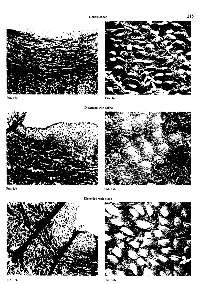
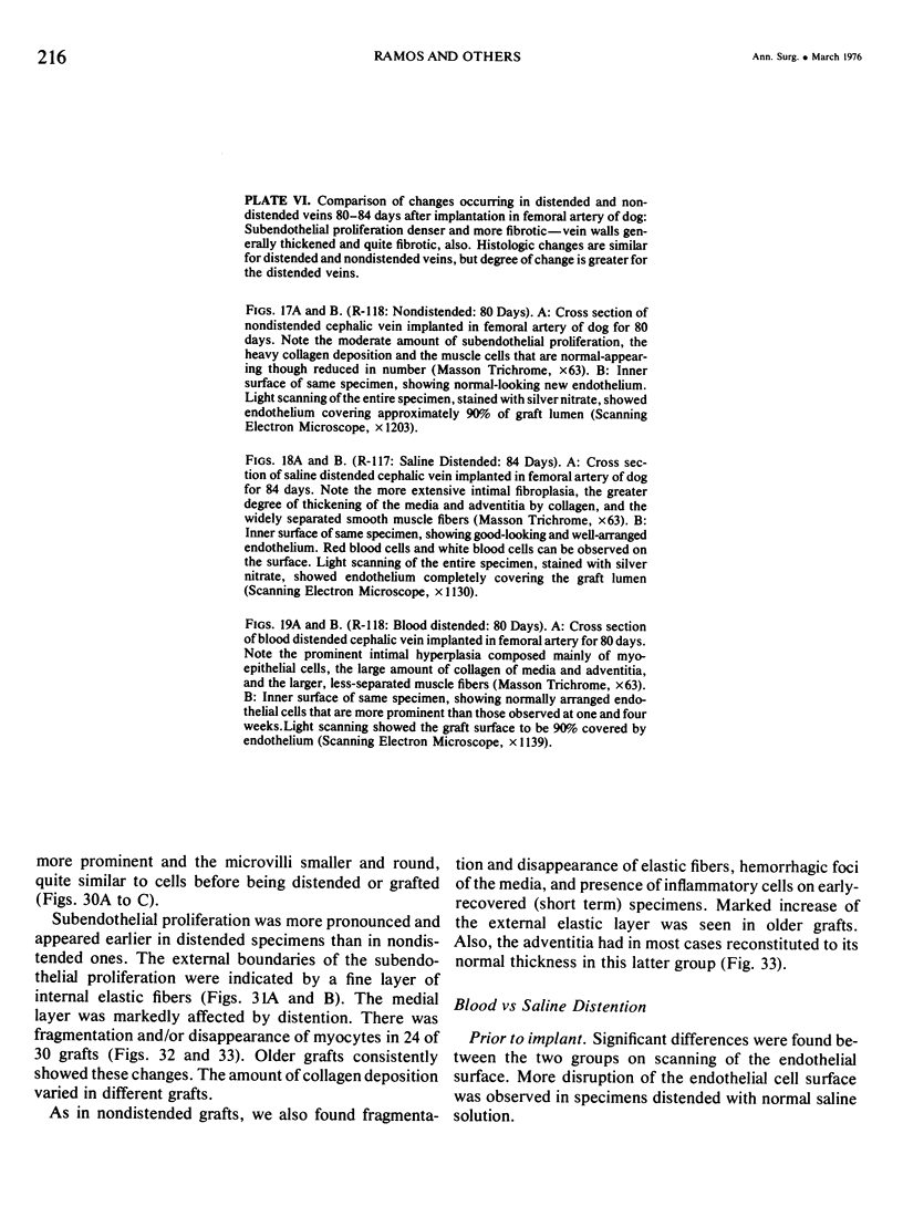
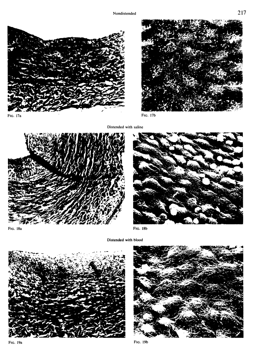
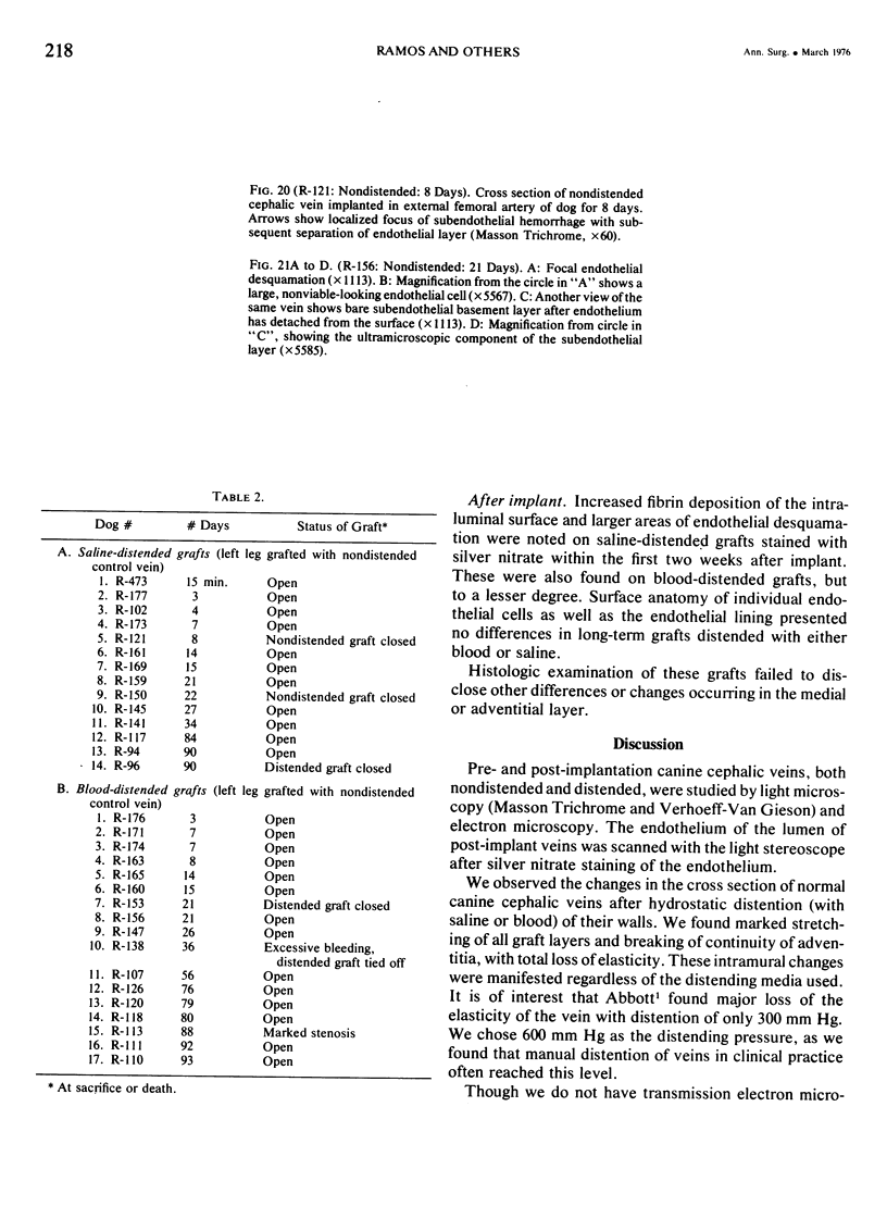
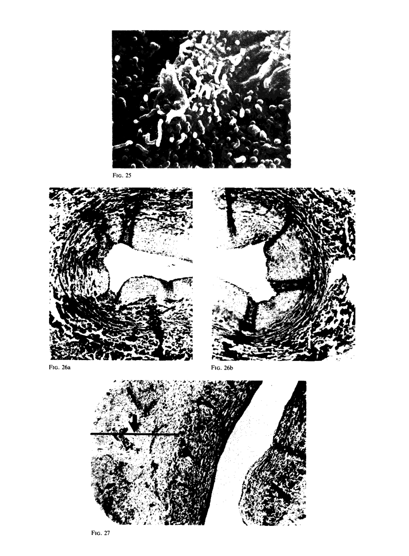
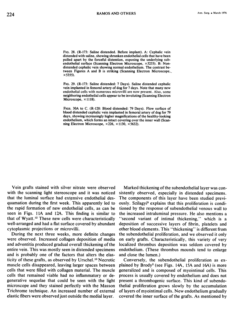
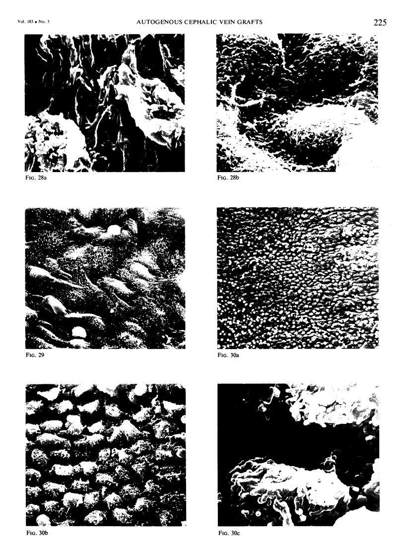
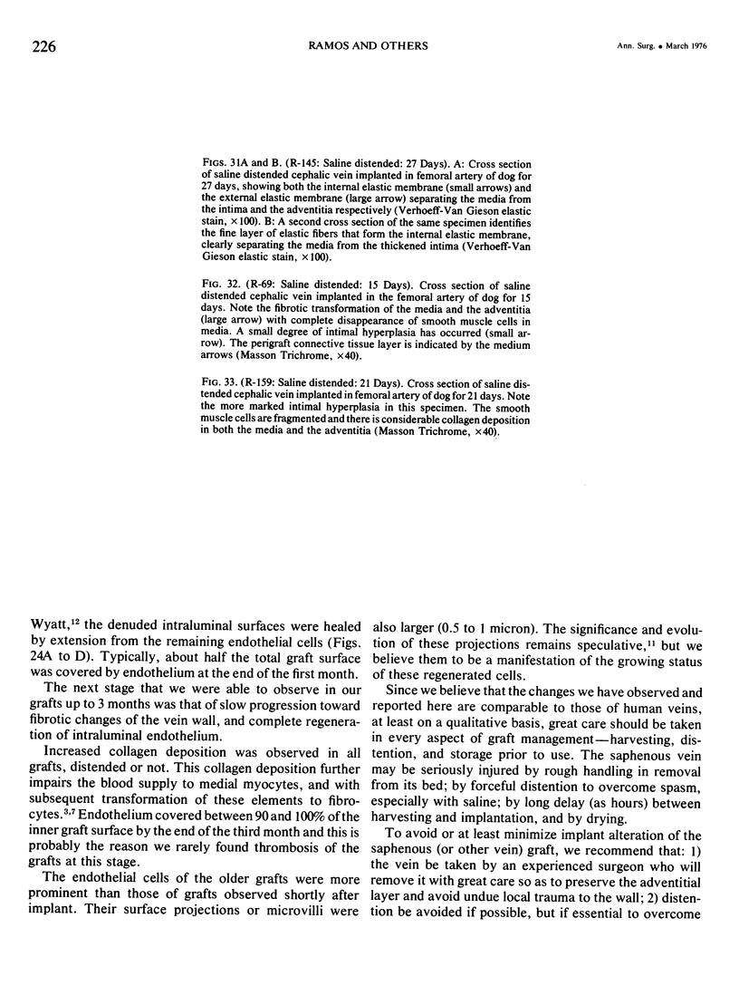
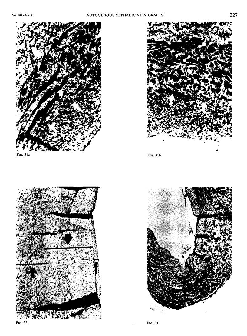
Sixty-two autogenous cephalic vein segments were grafted into the femoral arteries of 31 mongrel dogs, the left side receiving non-distended (control) grafts and the right side distended (experimental) grafts. Distending media were heparinized blood and saline. Veins were distended at 600 mm Hg for 2 minutes. Specimens were taken at intervals from 15 minutes to 3 months, and were studied by gross inspection, surface observations (light scanning stereoscope to X70 scanning electron microscope to X6,000) and routine histologic techniques (light microscope to X 1000). In general, grafting of veins in the arterial system was followed by progressive degenerative changes in all layers of the vein, including endothelial cell involution, desquamation and re-endothelialization. Often a variable degree of subendothelial fibrous and/or myoepithelial proliferation occurred which might compromise even a lumen lined by healthy endothelium. Distention caused these changes to occur earlier (2-4 weeks) and to be more pronounced. Distention with saline caused more damage to the endothelium than did distention with blood. We conclude that preimplant distention of vein grafts (to overcome spasm) should be employed sparingly, as it adversely affects the endothelial covering of the flow surface, accelerates the development of degenerative changes, and may predispose the graft to early thrombotic complications.
Full text
PDF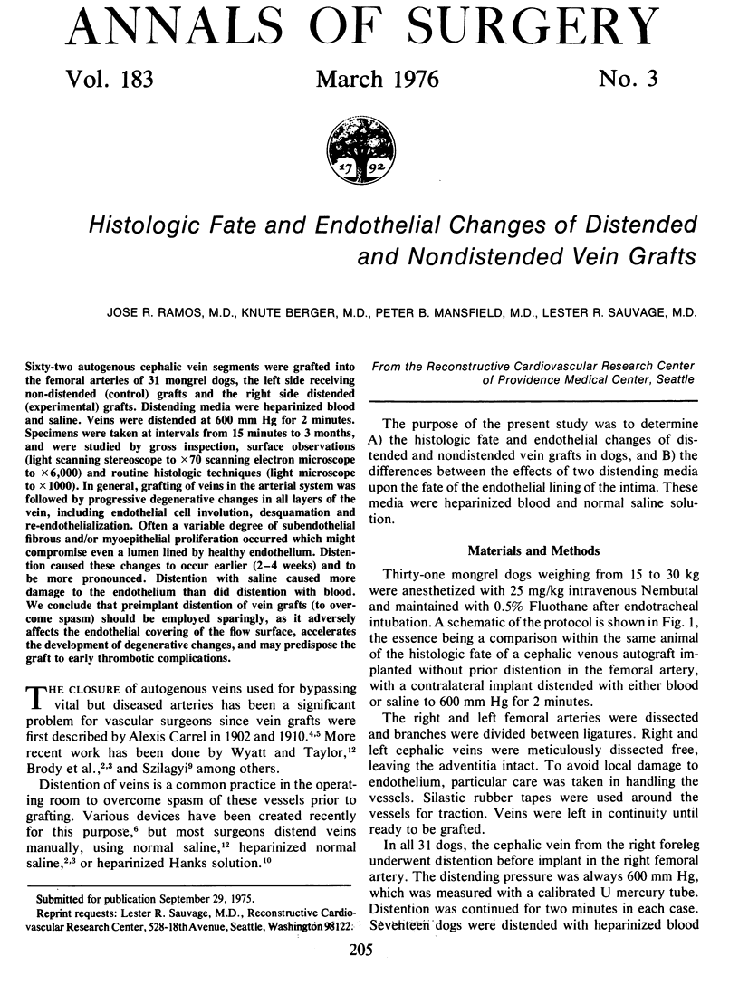
Images in this article
Selected References
These references are in PubMed. This may not be the complete list of references from this article.
- Abbott W. M., Wieland S., Austen W. G. Structural changes during preparation of autogenous venous grafts. Surgery. 1974 Dec;76(6):1031–1040. [PubMed] [Google Scholar]
- Brody W. R., Angeli W. W., Kosek J. C. Histologic fate of the venous coronary artery bypass in dogs. Am J Pathol. 1972 Jan;66(1):111–130. [PMC free article] [PubMed] [Google Scholar]
- Carrel A. III. Graft of the Vena Cava on the Abdominal Aorta. Ann Surg. 1910 Oct;52(4):462–470. doi: 10.1097/00000658-191010000-00003. [DOI] [PMC free article] [PubMed] [Google Scholar]
- Hairston P. Vein-support frame for aorto-coronary bypass grafting with an autogenous vein. J Thorac Cardiovasc Surg. 1972 Nov;64(5):820–821. [PubMed] [Google Scholar]
- McCabe M., Cunningham G. J., Wyatt A. P., Rothnie N. G., Taylor G. W. A histological and histochemical examination of autogenous vein grafts. Br J Surg. 1967 Feb;54(2):147–155. doi: 10.1002/bjs.1800540216. [DOI] [PubMed] [Google Scholar]
- Szilagyi D. E., Elliott J. P., Hageman J. H., Smith R. F., Dall'olmo C. A. Biologic fate of autogenous vein implants as arterial substitutes: clinical, angiographic and histopathologic observations in femoro-popliteal operations for atherosclerosis. Ann Surg. 1973 Sep;178(3):232–246. doi: 10.1097/00000658-197309000-00002. [DOI] [PMC free article] [PubMed] [Google Scholar]
- Urschel H. C., Razzuk M. A., Wood R. E., Paulson D. L. Factors influencing patency of aortocoronary artery saphenous vein grafts. Surgery. 1972 Dec;72(6):1048–1063. [PubMed] [Google Scholar]
- Wechezak A. R., Mansfield P. B. Isolation and growth characteristics of cell lines from bovine venous endothelium. In Vitro. 1973 Jul-Aug;9(1):39–45. doi: 10.1007/BF02615988. [DOI] [PubMed] [Google Scholar]
- Wyatt A. P., Taylor G. W. Vein grafts: changes in the endothelium of autogenous free vein grafts used as arterial replacements. Br J Surg. 1966 Nov;53(11):943–947. doi: 10.1002/bjs.1800531107. [DOI] [PubMed] [Google Scholar]


