Abstract
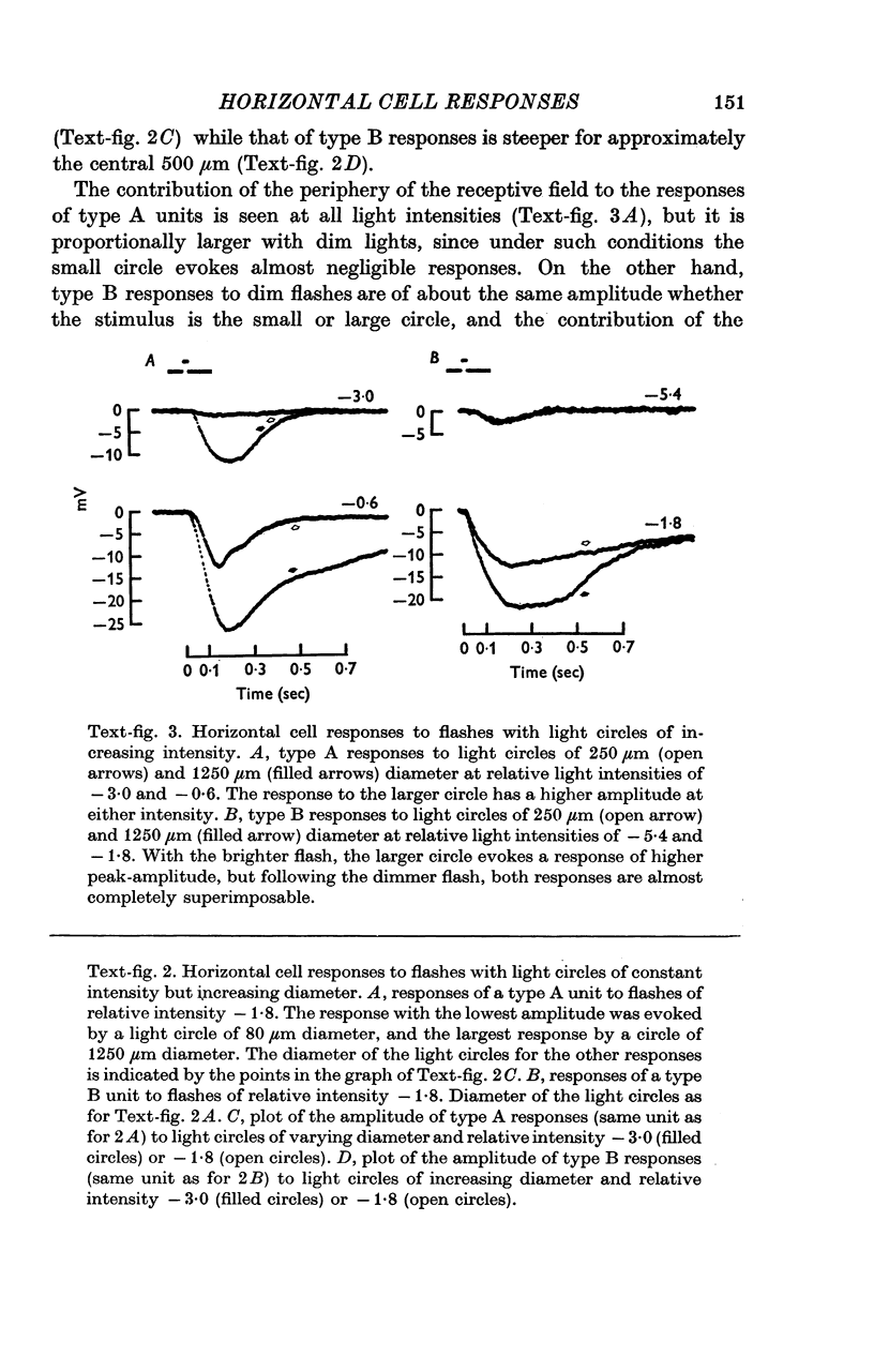
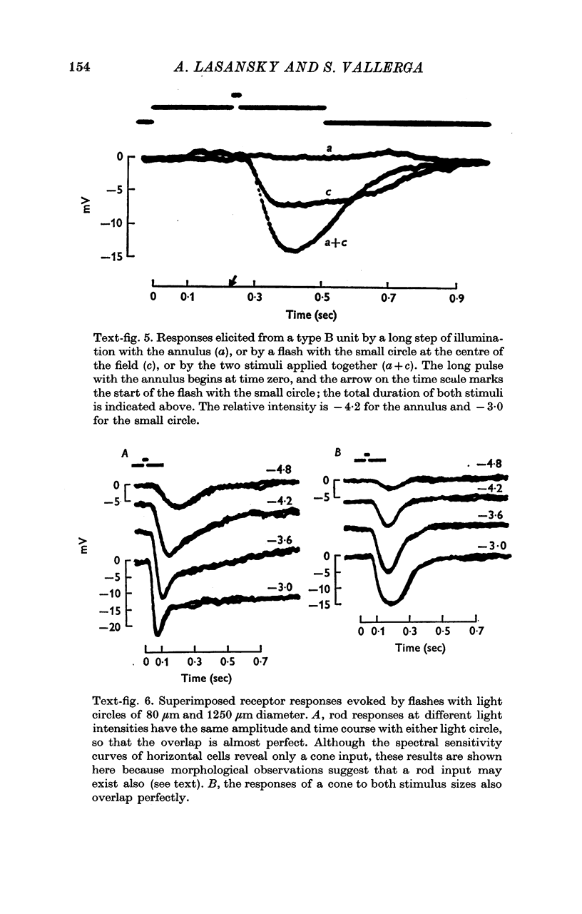
The responses to light of horizontal cells were recorded intracellularly in the retina of the larval tiger salamander. 2. All the units studied had a large summation area and were hyperpolarized by circles of light of any wave-length centred on the recording electrode, but two types could be distinguished according to the properties of their receptive fields. Type A units were hyperpolarized following illumination of any portion of their receptive field, while type B units were not hyperpolarized by illumination of their surround unless the centre was simultaneously illuminated, stimulation of the surround alone resulting in either a small depolarization or virtually no response. 3. Procion yellow injections showed that type A responses are recorded from thick and long processes not directly continuous with an identifiable cell body, while type B responses originate from the cell body of cells that send very fine and tortuous processes towards the receptors. The histological observations also suggested that the type A units represent expansions or swellings of one or more of the fine processes originating from the type B units. Therefore, it seems possible that both types of units are just different parts of a single kind of horizontal cell, and that a majority of the dye injections failed to stain them simultaneously because of the small diameter of the connecting process. 4. The large summation area of type A units can be explained, just as for horizontal cells in other retinae, by supposing that they are electrically coupled to other units of the same type. The receptive field properties of type B units, however, can only be partly explained by electrical coupling, and then only if the existence of voltage-dependent junctions is postulated. Instead, the reversal of the polarity of responses to an annulus of light during steady illumination of the centre, plus the available electron microscopic evidence, suggest that the effect of the surround on the type B units is due to a chemical synaptic impingement from the type A units.
Full text
PDF


Images in this article
Selected References
These references are in PubMed. This may not be the complete list of references from this article.
- Baylor D. A., Fuortes M. G., O'Bryan P. M. Receptive fields of cones in the retina of the turtle. J Physiol. 1971 Apr;214(2):265–294. doi: 10.1113/jphysiol.1971.sp009432. [DOI] [PMC free article] [PubMed] [Google Scholar]
- Kaneko A. Electrical connexions between horizontal cells in the dogfish retina. J Physiol. 1971 Feb;213(1):95–105. doi: 10.1113/jphysiol.1971.sp009370. [DOI] [PMC free article] [PubMed] [Google Scholar]
- Kolb H. The connections between horizontal cells and photoreceptors in the retina of the cat: electron microscopy of Golgi preparations. J Comp Neurol. 1974 May 1;155(1):1–14. doi: 10.1002/cne.901550102. [DOI] [PubMed] [Google Scholar]
- Lasansky A. Cell junctions at the outer synaptic layer of the retina. Invest Ophthalmol. 1972 May;11(5):265–275. [PubMed] [Google Scholar]
- Lasansky A., Marchiafava P. L. Light-induced resistance changes in retinal rods and cones of the tiger salamander. J Physiol. 1974 Jan;236(1):171–191. doi: 10.1113/jphysiol.1974.sp010429. [DOI] [PMC free article] [PubMed] [Google Scholar]
- Lasansky A. Organization of the outer synaptic layer in the retina of the larval tiger salamander. Philos Trans R Soc Lond B Biol Sci. 1973;265(872):471–489. doi: 10.1098/rstb.1973.0033. [DOI] [PubMed] [Google Scholar]
- Marchiafava P. L., Pasino E. The spatial dependent characteristics of the fish S-potentials evoked by brief flashes. Vision Res. 1973 Jul;13(7):1355–1365. doi: 10.1016/0042-6989(73)90211-3. [DOI] [PubMed] [Google Scholar]
- Naka K. I., Rushton W. A. The generation and spread of S-potentials in fish (Cyprinidae). J Physiol. 1967 Sep;192(2):437–461. doi: 10.1113/jphysiol.1967.sp008308. [DOI] [PMC free article] [PubMed] [Google Scholar]
- Norton A. L., Spekreijse H., Wolbarsht M. L., Wagner H. G. Receptive field organization of the S-potential. Science. 1968 May 31;160(3831):1021–1022. doi: 10.1126/science.160.3831.1021. [DOI] [PubMed] [Google Scholar]
- SVAETICHIN G., MACNICHOL E. F., Jr Retinal mechanisms for chromatic and achromatic vision. Ann N Y Acad Sci. 1959 Nov 12;74(2):385–404. doi: 10.1111/j.1749-6632.1958.tb39560.x. [DOI] [PubMed] [Google Scholar]
- Simon E. J. Two types of luminosity horizontal cells in the retina of the turtle. J Physiol. 1973 Apr;230(1):199–211. doi: 10.1113/jphysiol.1973.sp010183. [DOI] [PMC free article] [PubMed] [Google Scholar]
- Stretton A. O., Kravitz E. A. Neuronal geometry: determination with a technique of intracellular dye injection. Science. 1968 Oct 4;162(3849):132–134. doi: 10.1126/science.162.3849.132. [DOI] [PubMed] [Google Scholar]
- Werblin F. S., Dowling J. E. Organization of the retina of the mudpuppy, Necturus maculosus. II. Intracellular recording. J Neurophysiol. 1969 May;32(3):339–355. doi: 10.1152/jn.1969.32.3.339. [DOI] [PubMed] [Google Scholar]
- Witkovsky P., Dowling J. E. Synaptic relationships in the plexiform layers of carp retina. Z Zellforsch Mikrosk Anat. 1969;100(1):60–82. doi: 10.1007/BF00343821. [DOI] [PubMed] [Google Scholar]
- Yamada E., Ishikawa T. The fine structure of the horizontal cells in some vertebrate retinae. Cold Spring Harb Symp Quant Biol. 1965;30:383–392. doi: 10.1101/sqb.1965.030.01.038. [DOI] [PubMed] [Google Scholar]


