Abstract
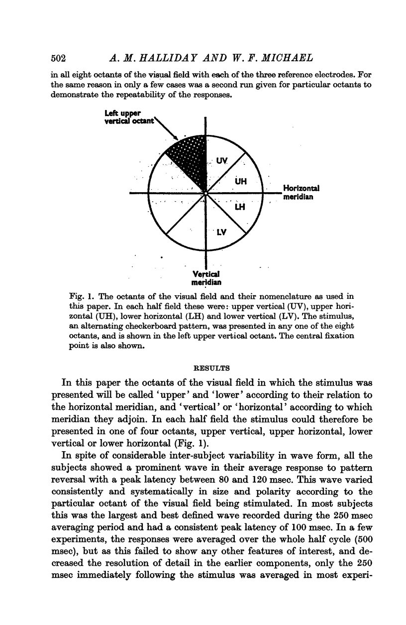
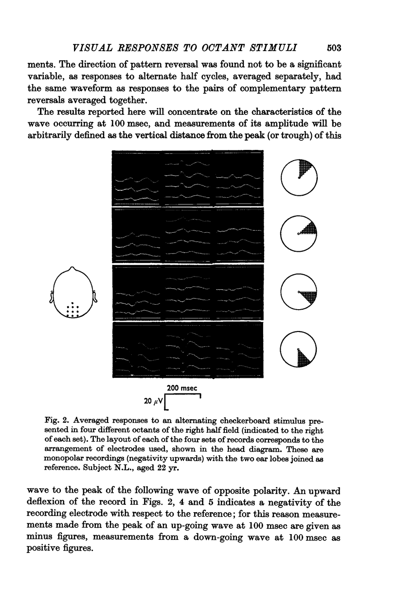
1. Averaged responses have been recorded from an array of ten scalp electrodes over the occipital cortex in man to the reversal of a black-and-white checkerboard pattern, presented in different octants of the visual field.
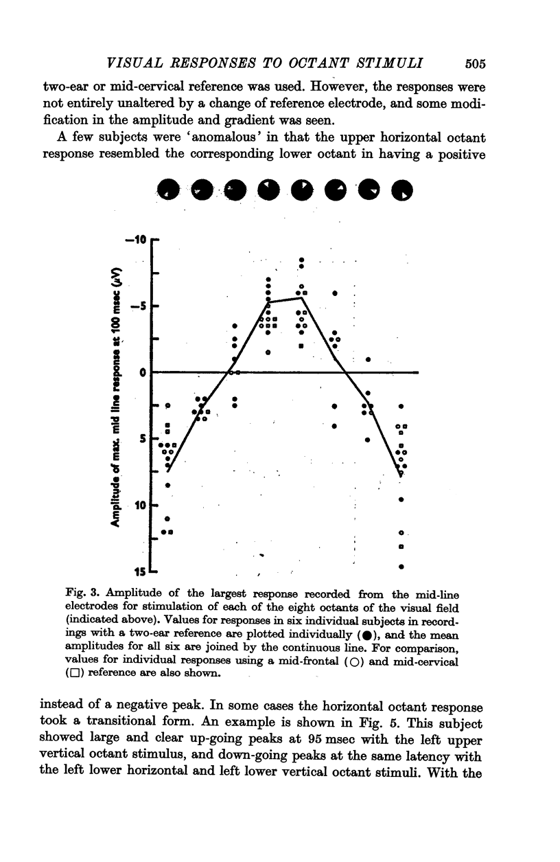
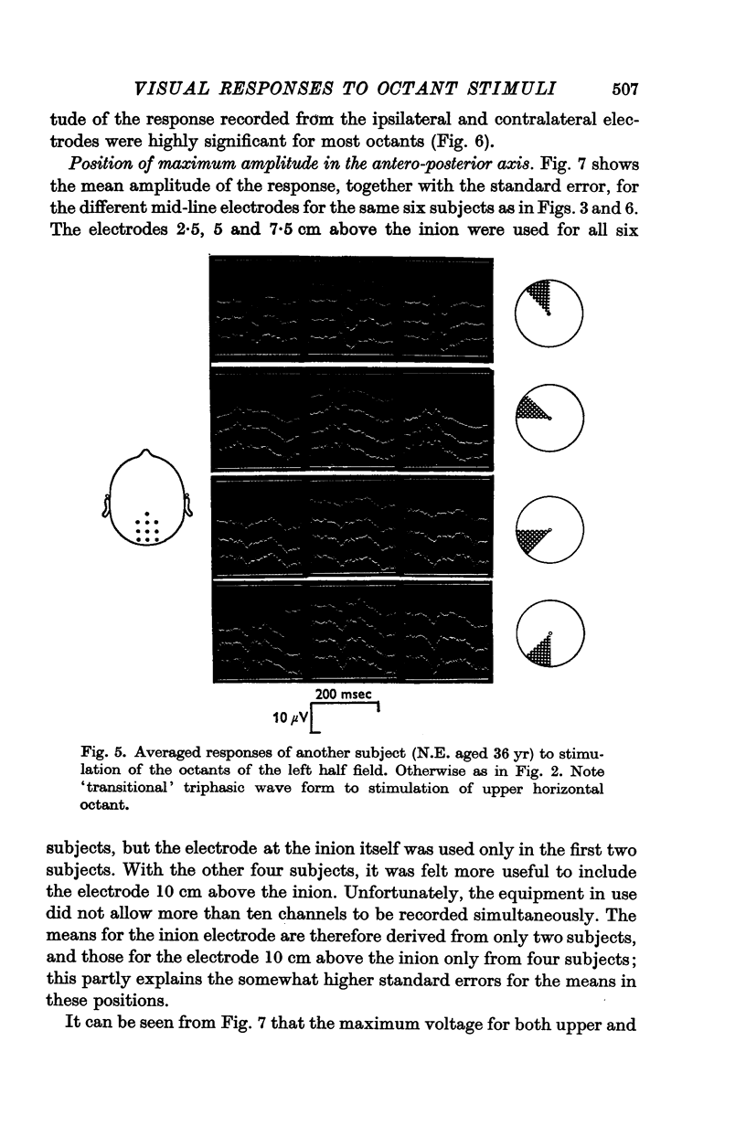
2. In all subjects a prominent wave was seen, with a peak latency of about 100 msec, which showed consistent and systematic changes with variation in the position of the stimulus in the visual field.
3. With stimulation of the octants next to the vertical meridian, this component was of large amplitude, while with stimulation of the octants next to the horizontal meridian, it was small and inconspicuous.
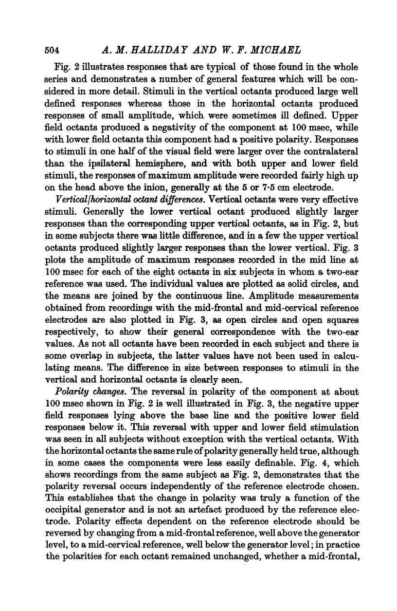
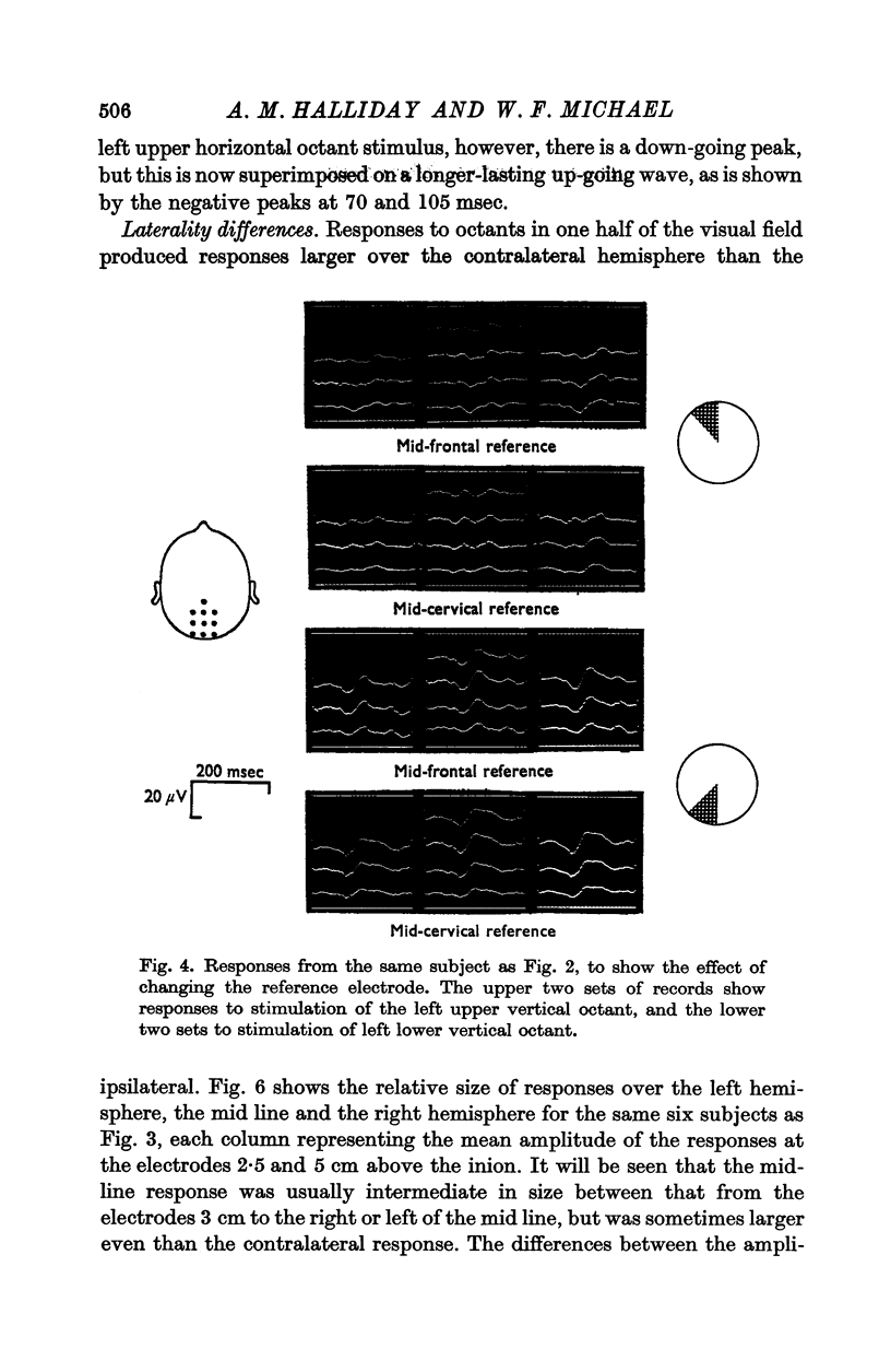
4. With upper field octants, the peak at 100 msec was surface-negative, while with lower field octants it was reversed in polarity.
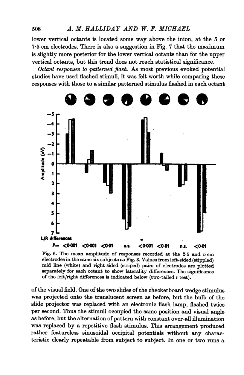
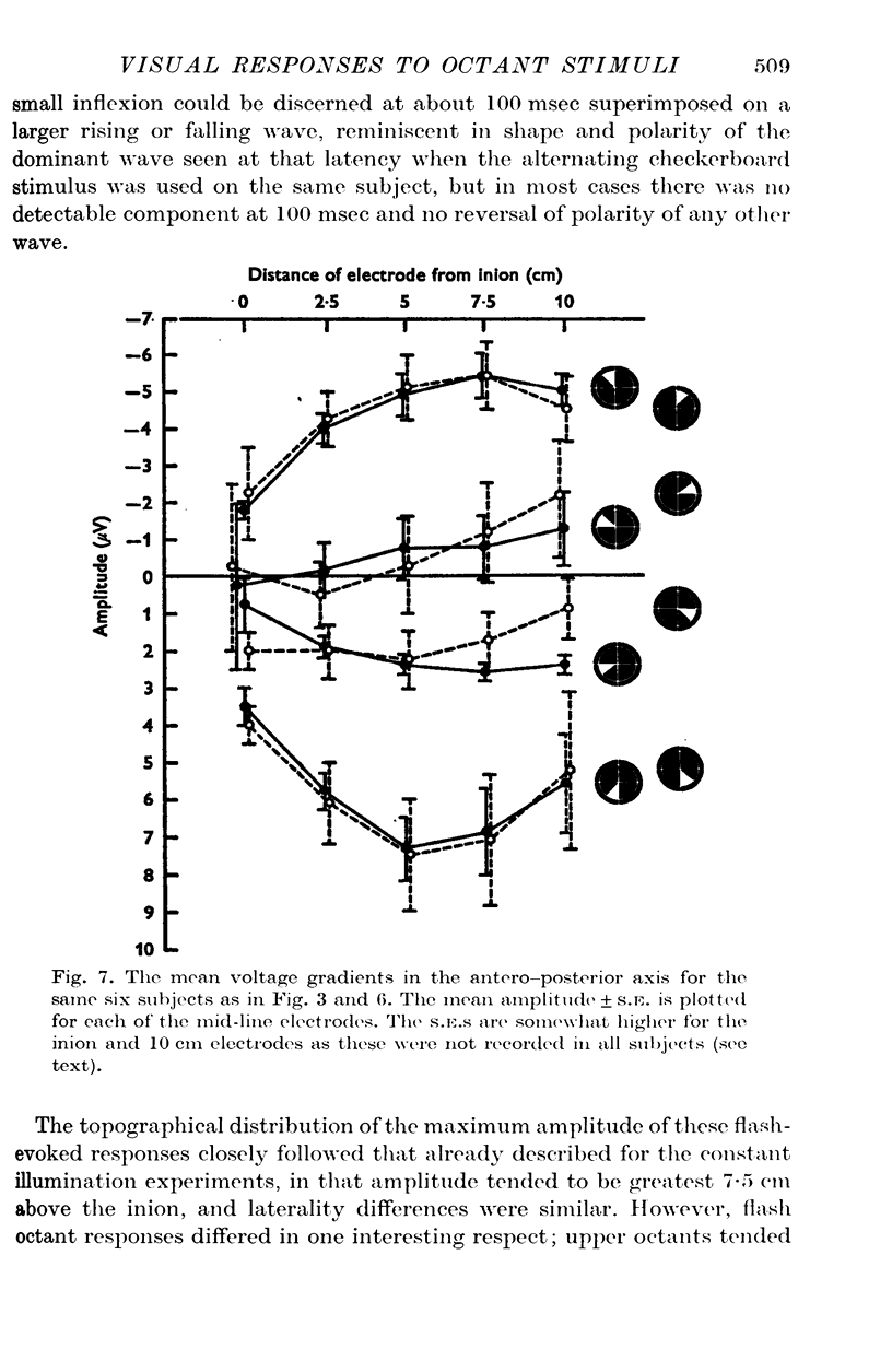
5. The occipital response was largest 5 or 7·5 cm above the inion, and the amplitude recorded 3 cm lateral to the mid line was larger over the hemisphere contralateral to the half field being stimulated than ipsilaterally.
6. These findings are discussed in relation to the underlying anatomy of the visual cortex, and it is concluded that these responses are likely to arise mainly from extra-striate areas.
Full text
PDF
Selected References
These references are in PubMed. This may not be the complete list of references from this article.
- BISHOP G. H., CLARE M. H. [Responses of cortex to direct electrical stimuli applied at different depths]. J Neurophysiol. 1953 Jan;16(1):1–19. doi: 10.1152/jn.1953.16.1.1. [DOI] [PubMed] [Google Scholar]
- Brindley G. S., Lewin W. S. The sensations produced by electrical stimulation of the visual cortex. J Physiol. 1968 May;196(2):479–493. doi: 10.1113/jphysiol.1968.sp008519. [DOI] [PMC free article] [PubMed] [Google Scholar]
- COPENHAVER R. M., BEINHOCKER G. D. EVOKED OCCIPITAL POTENTIALS RECORDED FROM SCALP ELECTRODES IN RESPONSE TO FOCAL VISUAL ILLUMINATION. Invest Ophthalmol. 1963 Aug;2:393–406. [PubMed] [Google Scholar]
- COPENHAVER R. M., PERRY N. W., Jr FACTORS AFFECTING VISUALLY EVOKED CORTICAL POTENTIALS SUCH AS IMPAIRED VISION OF VARYING ETIOLOGY. Invest Ophthalmol. 1964 Dec;3:665–675. [PubMed] [Google Scholar]
- Cragg B. G. The topography of the afferent projections in the circumstriate visual cortex of the monkey studied by the Nauta method. Vision Res. 1969 Jul;9(7):733–747. doi: 10.1016/0042-6989(69)90011-x. [DOI] [PubMed] [Google Scholar]
- DANIEL P. M., WHITTERIDGE D. The representation of the visual field on the cerebral cortex in monkeys. J Physiol. 1961 Dec;159:203–221. doi: 10.1113/jphysiol.1961.sp006803. [DOI] [PMC free article] [PubMed] [Google Scholar]
- DOTY R. W. Potentials evoked in cat cerebral cortex by diffuse and by punctiform photic stimuli. J Neurophysiol. 1958 Sep;21(5):437–464. doi: 10.1152/jn.1958.21.5.437. [DOI] [PubMed] [Google Scholar]
- Eason R. G., Oden D., White C. T. Visullay evoked cortical potentials and reaction time in relation to site of retinal stimulation. Electroencephalogr Clin Neurophysiol. 1967 Apr;22(4):313–324. doi: 10.1016/0013-4694(67)90201-5. [DOI] [PubMed] [Google Scholar]
- Holmes G. DISTURBANCES OF VISION BY CEREBRAL LESIONS. Br J Ophthalmol. 1918 Jul;2(7):353–384. doi: 10.1136/bjo.2.7.353. [DOI] [PMC free article] [PubMed] [Google Scholar]
- Hubel D. H., Wiesel T. N. Cortical and callosal connections concerned with the vertical meridian of visual fields in the cat. J Neurophysiol. 1967 Nov;30(6):1561–1573. doi: 10.1152/jn.1967.30.6.1561. [DOI] [PubMed] [Google Scholar]
- MYERS R. E. Commissural connections between occipital lobes of the monkey. J Comp Neurol. 1962 Feb;118:1–16. doi: 10.1002/cne.901180102. [DOI] [PubMed] [Google Scholar]
- VAUGHAN H. G., Jr, KATZMAN R., TAYLOR J. ALTERATIONS OF VISUAL EVOKED RESPONSE IN THE PRESENCE OF HOMONYMOUS VISUAL DEFECTS. Electroencephalogr Clin Neurophysiol. 1963 Oct;15:737–746. doi: 10.1016/0013-4694(63)90164-0. [DOI] [PubMed] [Google Scholar]
- Zeki S. M. Representation of central visual fields in prestriate cortex of monkey. Brain Res. 1969 Jul;14(2):271–291. doi: 10.1016/0006-8993(69)90110-3. [DOI] [PubMed] [Google Scholar]


