

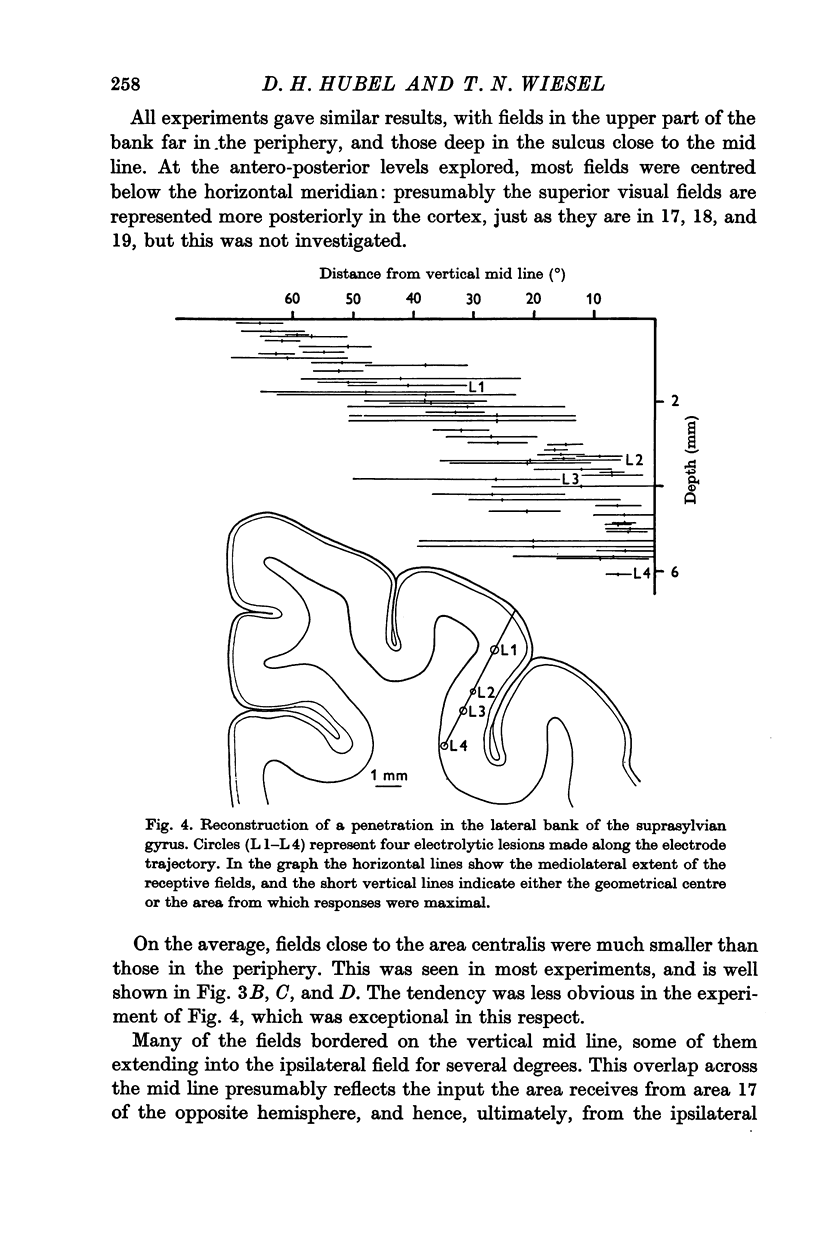
Abstract
On anatomical and physiological grounds a zone of cat cortex deep in the medial bank of the suprasylvian sulcus (the Clare—Bishop area) is known to receive strong visual projections both from the lateral geniculate body and area 17. We have mapped receptive fields of single cells in this area in eight cats.
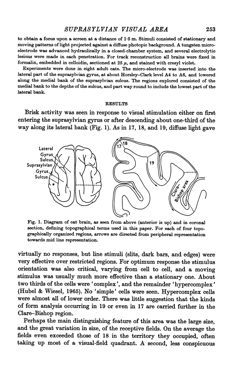
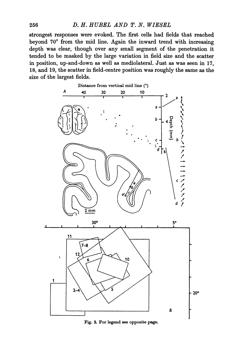
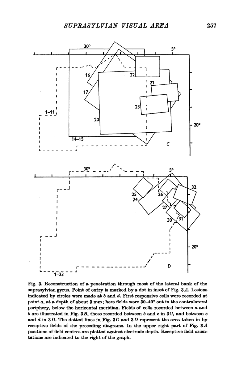
Active responses to visual stimuli were found over most of the medial bank of the suprasylvian sulcus extending to the depths and over to the lowest part of the lateral bank. The area is clearly topographically arranged. The first responsive cells, recorded over the lateral convexity and 2-3 mm down the medial bank, had receptive fields in the far periphery of the contralateral visual fields. The receptive fields tended to be large, but showed considerable variation in size and scatter in their positions. As the electrode advanced down the bank, fields of successively recorded cells gradually tended to move inwards, so that in the depths of the sulcus the inner borders of many of the fields reached the vertical mid line. Here the fields were smaller, though they still varied very much in size.
Receptive fields were larger than in 17, 18, or 19, but otherwise were not obviously different from the complex and lower-order hypercomplex fields in those areas. No simple fields, or concentric fields of the retino-geniculate type, were seen. Cells with common receptive-field orientation were grouped together, but whether or not the grouping occurs in columns was not established.
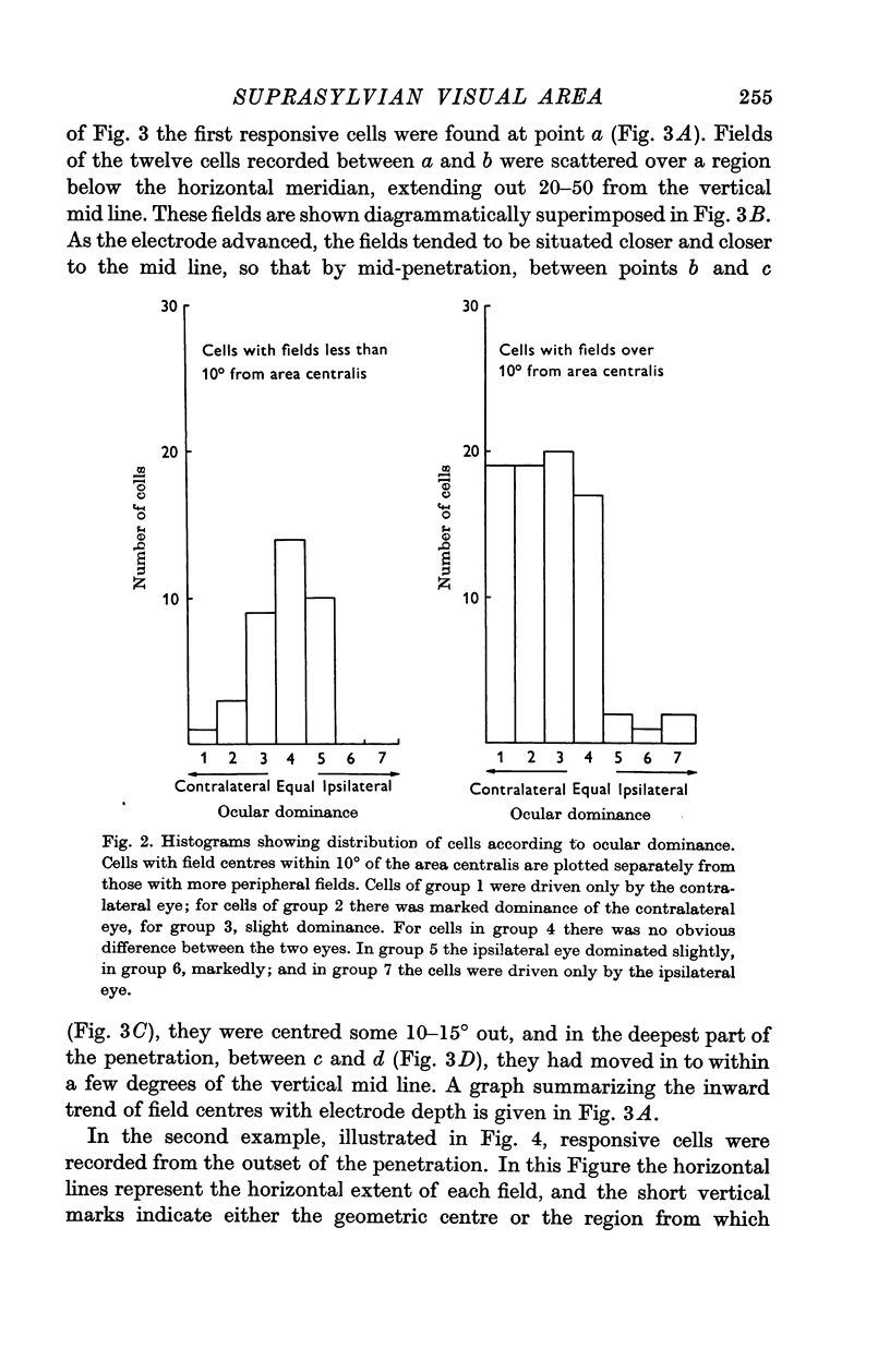
Most cells were driven independently by the two eyes. Fields in the two eyes seemed to be identical in organization. Cells dominated by the contralateral eye were much more common than ipsilaterally dominated ones, but when cells with parafoveal and peripheral fields were considered separately, the asymmetry was seen to apply mainly to cells with peripheral fields.
Full text
PDF
Selected References
These references are in PubMed. This may not be the complete list of references from this article.
- CLARE M. H., BISHOP G. H. Responses from an association area secondarily activated from optic cortex. J Neurophysiol. 1954 May;17(3):271–277. doi: 10.1152/jn.1954.17.3.271. [DOI] [PubMed] [Google Scholar]
- HUBEL D. H., WIESEL T. N. RECEPTIVE FIELDS AND FUNCTIONAL ARCHITECTURE IN TWO NONSTRIATE VISUAL AREAS (18 AND 19) OF THE CAT. J Neurophysiol. 1965 Mar;28:229–289. doi: 10.1152/jn.1965.28.2.229. [DOI] [PubMed] [Google Scholar]
- HUBEL D. H., WIESEL T. N. Receptive fields, binocular interaction and functional architecture in the cat's visual cortex. J Physiol. 1962 Jan;160:106–154. doi: 10.1113/jphysiol.1962.sp006837. [DOI] [PMC free article] [PubMed] [Google Scholar]
- Hubel D. H., Wiesel T. N. Cortical and callosal connections concerned with the vertical meridian of visual fields in the cat. J Neurophysiol. 1967 Nov;30(6):1561–1573. doi: 10.1152/jn.1967.30.6.1561. [DOI] [PubMed] [Google Scholar]
- OTSUKA R., HASSLER R. [On the structure and segmentation of the cortical center of vision in the cat]. Arch Psychiatr Nervenkr Z Gesamte Neurol Psychiatr. 1962;203:212–234. doi: 10.1007/BF00352744. [DOI] [PubMed] [Google Scholar]
- Sterling P., Wickelgren B. G. Visual receptive fields in the superior colliculus of the cat. J Neurophysiol. 1969 Jan;32(1):1–15. doi: 10.1152/jn.1969.32.1.1. [DOI] [PubMed] [Google Scholar]
- Wickelgren B. G., Sterling P. Influence of visual cortex on receptive fields in the superior colliculus of the cat. J Neurophysiol. 1969 Jan;32(1):16–23. doi: 10.1152/jn.1969.32.1.16. [DOI] [PubMed] [Google Scholar]
- Wilson M. E. Cortico-cortical connexions of the cat visual areas. J Anat. 1968 Mar;102(Pt 3):375–386. [PMC free article] [PubMed] [Google Scholar]
- Wilson M. E., Cragg B. G. Projections from the lateral geniculate nucleus in the cat and monkey. J Anat. 1967 Sep;101(Pt 4):677–692. [PMC free article] [PubMed] [Google Scholar]