

Abstract
1. Spectral, spatial and temporal properties of receptive fields of dark-adapted, on—off retinal ganglion cells in the intact eye of the plaice, were analysed by recording from their axon terminals in the superficial layers of the optic tectum with indium micro-electrodes.
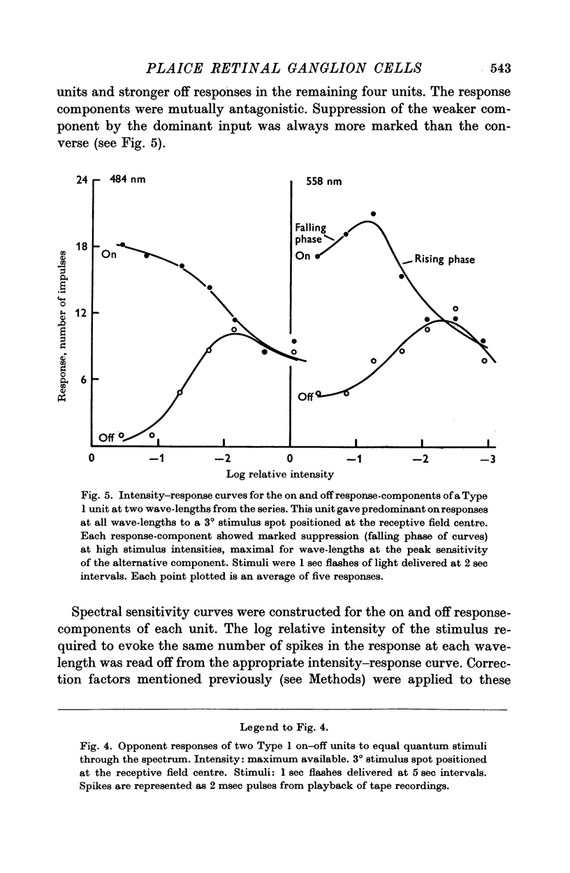
2. Two cell-types were identified. The first gave fast-adapting, spectrally opponent on—off responses without centre-surround subdivisions of the receptive field. On and off response-components were mutually antagonistic. The second type gave slow-adapting on—off or off responses for different stimulus positions within the receptive field, with centre-surround or adjacent field configurations. Only on—off centre cells, showing mutual antagonism between field centre and surround, or off centre cells with inhibitory centres, were found. These cells had weak opponent or non-opponent properties.
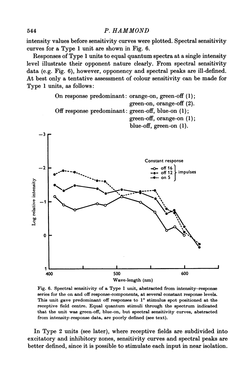
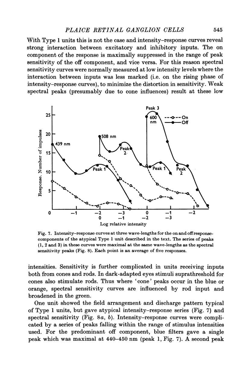
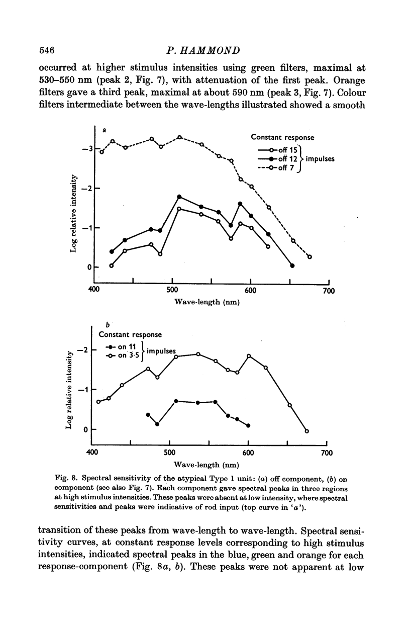
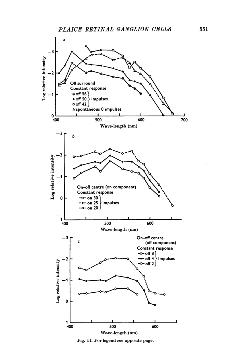
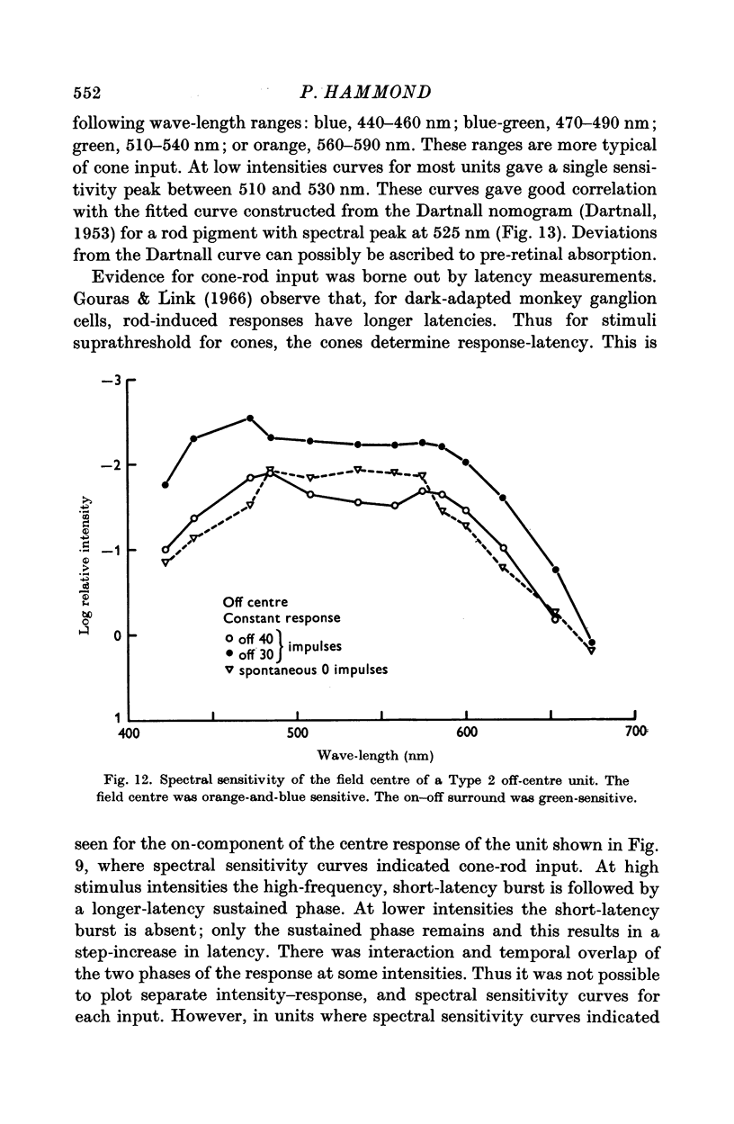
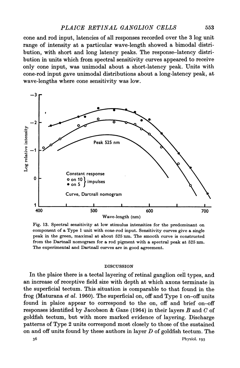
3. Most cells of each type received inputs both from cones and rods. At stimulus intensities suprathreshold for cones, response-components gave spectral peaks which have been classified into one of four wave-length ranges; blue, 440-460 nm; blue-green, 470-490 nm; green, 510-540 nm; and orange, 560-590 nm. No cells analysed gave sensitivity maxima in the red. At low stimulus intensities all cells with rod input gave a single spectral peak between 510 and 530 nm.
Full text
PDF
Images in this article


Selected References
These references are in PubMed. This may not be the complete list of references from this article.
- CRONLY-DILLON J. R. UNITS SENSITIVE TO DIRECTION OF MOVEMENT IN GOLDFISH OPTIC TECTUM. Nature. 1964 Jul 11;203:214–215. doi: 10.1038/203214a0. [DOI] [PubMed] [Google Scholar]
- DARTNALL H. J. A. The interpretation of spectral sensitivity curves. Br Med Bull. 1953;9(1):24–30. doi: 10.1093/oxfordjournals.bmb.a074302. [DOI] [PubMed] [Google Scholar]
- Dowling J. E., Boycott B. B. Organization of the primate retina: electron microscopy. Proc R Soc Lond B Biol Sci. 1966 Nov 15;166(1002):80–111. doi: 10.1098/rspb.1966.0086. [DOI] [PubMed] [Google Scholar]
- Gouras P., Link K. Rod and cone interaction in dark-adapted monkey ganglion cells. J Physiol. 1966 May;184(2):499–510. doi: 10.1113/jphysiol.1966.sp007928. [DOI] [PMC free article] [PubMed] [Google Scholar]
- JACOBSON M., GAZE R. M. TYPES OF VISUAL RESPONSE FROM SINGLE UNITS IN THE OPTIC TECTUM AND OPTIC NERVE OF THE GOLDFISH. Q J Exp Physiol Cogn Med Sci. 1964 Apr;49:199–209. doi: 10.1113/expphysiol.1964.sp001720. [DOI] [PubMed] [Google Scholar]
- KUFFLER S. W. Discharge patterns and functional organization of mammalian retina. J Neurophysiol. 1953 Jan;16(1):37–68. doi: 10.1152/jn.1953.16.1.37. [DOI] [PubMed] [Google Scholar]
- MARKS W. B. VISUAL PIGMENTS OF SINGLE GOLDFISH CONES. J Physiol. 1965 May;178:14–32. doi: 10.1113/jphysiol.1965.sp007611. [DOI] [PMC free article] [PubMed] [Google Scholar]
- MATURANA H. R., LETTVIN J. Y., MCCULLOCH W. S., PITTS W. H. Anatomy and physiology of vision in the frog (Rana pipiens). J Gen Physiol. 1960 Jul;43(6):129–175. doi: 10.1085/jgp.43.6.129. [DOI] [PMC free article] [PubMed] [Google Scholar]
- SCHWASSMAN H. O., KRUGER L. ORGANIZATION OF THE VISUAL PROJECTION UPON THE OPTIC TECTUM OF SOME FRESHWATER FISH. J Comp Neurol. 1965 Feb;124:113–126. doi: 10.1002/cne.901240109. [DOI] [PubMed] [Google Scholar]
- Wiesel T. N., Hubel D. H. Spatial and chromatic interactions in the lateral geniculate body of the rhesus monkey. J Neurophysiol. 1966 Nov;29(6):1115–1156. doi: 10.1152/jn.1966.29.6.1115. [DOI] [PubMed] [Google Scholar]
- Witkovsky P. The spectral sensitivity of retinal ganglion cells in the carp. Vision Res. 1965 Dec;5(11):603–614. doi: 10.1016/0042-6989(65)90034-9. [DOI] [PubMed] [Google Scholar]