Abstract
The perception of microbial signal molecules is part of the strategy evolved by plants to survive attacks by potential pathogens. To gain a more complete understanding of the early signaling events involved in these responses, we used radioactive orthophosphate to pulse-label suspension-cultured cells of Arabidopsis in conjunction with two-dimensional gel electrophoresis and mass spectrometry to identify proteins that are phosphorylated rapidly in response to bacterial and fungal elicitors. One of these proteins, AtPhos43, and related proteins in tomato and rice, are phosphorylated within minutes after treatment with flagellin or chitin fragments. By measuring 32P incorporation into AtPhos43 immunoprecipitated from extracts of elicitor-treated hormone and defense-response mutants, we found that phosphorylation of AtPhos43 after flagellin treatment but not chitin treatment is dependent on FLS2, a receptor-like kinase involved in flagellin perception. Induction by both elicitors is not dependent on salicylic acid or EDS1, a putative lipase involved in defense signaling.
INTRODUCTION
The ability of plants to defend themselves against the majority of potential pathogens depends on sensitive perception mechanisms that recognize microbial invaders and subsequently activate defense responses. Although genetic approaches have shown that the various resistance genes activate multiple signal transduction pathways and that common defense responses can be activated via independent pathways (Innes, 1998), they have been far less successful in identifying the signaling components involved (Glazebrook et al., 1997; Innes, 1998). The inability to identify these components may be the result of redundancy in path-ways, redundancy in components of pathways, or lethality conferred by mutations in genes encoding these proteins. Thus, alternative methods may be necessary to complement existing genetic studies to elucidate the complex patterns of signaling after the recognition of microbial elicitors.
Genetic evidence, however, does indicate that protein phosphorylation plays an important role in defense signaling. Mutations in the Pto serine/threonine kinase in tomato (Martin et al., 1993) and Xa21 leucine-rich repeat (LRR) kinase in rice (Song et al., 1995) compromise the plant's resistance to race-specific pathogens. Similarly, mutations in the FLS2 LRR-kinase in Arabidopsis render the plant insensitive to the bacterial elicitor, flagellin (Gómez-Gómez and Boller, 2000). Thus, phosphorylation is required to initiate responses to diverse microbial signals. Recently, negative regulation of defense pathways also was demonstrated to be under the control of phosphorylation. Mutations in EDR1, a MAP kinase kinase kinase (Frye et al., 2001), and in MAP kinase 4 (Petersen et al., 2000) result in Arabidopsis plants that are more resistant to virulent pathogens. In both cases, resistance in the mutants involves derepression of the salicyclic acid (SA)-induced defense pathway, indicating that inactivation of phosphorylation cascades also is required for the induction of defense responses.
In addition to the direct genetic evidence listed above, there is an abundance of correlative data supporting the importance of phosphorylation in defense signaling. A family of mitogen-activated protein kinases (MAPKs) are activated within minutes after race-specific and nonrace-specific elicitation in a variety of plant species (Ligterink et al., 1997; Zhang et al., 1998; Romeis et al., 1999; Nühse et al., 2000). Although no target for these MAPKs is known, the speed and specificity of activation indicate that they play a role in signaling. Pharmacological inhibitors of protein kinases block a broad spectrum of early defense responses, and phosphatase inhibitors induce many of the same responses induced by microbial elicitors (Boller, 1995; Scheel, 1998; McDowell and Dangl, 2000). Together, these results indicate that dynamic changes in protein phosphorylation are involved at an early step in the pathway. Thus, proteins differentially phosphorylated in response to microbial elicitors most likely are components of signal transduction pathways connecting perception (e.g., by the LRR-containing proteins) with defense responses.
Although it has been shown that the phosphorylation pattern of proteins changes rapidly after treatment of cells with elicitors (Dietrich et al., 1990; Felix et al., 1991; Viard et al., 1994; Lecourieux-Ouaked et al., 2000), relatively few of these phosphoproteins have been identified. The phosphorylation of the Pti1 serine/threonine kinase (Zhou et al., 1995) and Pti4 putative transcription factor (Gu et al., 2000) by Pto kinase in tomato represents the only case in which a kinase involved in defense responses has been connected to its substrates. In addition to these proteins, only a calcium-dependent protein kinase in tobacco (Romeis et al., 2000), a bZIP DNA binding protein in soybean (Droge-Laser et al., 1997), and the p67-phox and p47-phox components of NADPH oxidase in tomato (Xing et al., 1997) have been shown to be phosphorylated during elicitor responses.
To gain a better understanding of the events that occur rapidly after elicitor perception, we used a “directed proteomics” approach to identify proteins that are phosphorylated rapidly in the response of Arabidopsis cells to microbial elicitors. Directed proteomics focuses on a particular subset of proteins using two-dimensional (2D) gel electrophoresis to separate radoioactively labeled phosphoproteins and mass spectrometry to identify these target proteins. One of the proteins identified, AtPhos43, is a plant-specific protein phosphorylated in several plant species within minutes after treatment with bacterial or fungal elicitors. Using an in planta phosphorylation assay, we determined that flagellin-induced phosphorylation of AtPhos43 requires the FLS2 kinase. Neither chitin- nor flagellin-induced phosphorylation is dependent on salicylic acid or EDS1.
RESULTS
Phosphoprotein Proteome of flg22-Treated Arabidopsis Cells
To take full advantage of the sensitivity of nanoelectrospray ionization tandem mass spectrometry (nanoESI-MS-MS) for protein identification, we established a phospholabeling method using Arabidopsis because of its substantial sequence database. Suspension cultured cells of Arabidopsis responded to treatment with the flagellin 22 peptide (flg22) as described for tomato cells (Felix et al., 1999) with only small differences in response kinetics and in concentrations of elicitor necessary for maximal response (Nühse et al., 2000; S.C. Peck and T.S. Nühse, unpublished results). Importantly, cultures took up 95% of applied 32P- or 33P-orthophosphate within 30 sec. Rapid pulse labeling therefore could be used to monitor transient changes in protein phosphorylation after exposure.
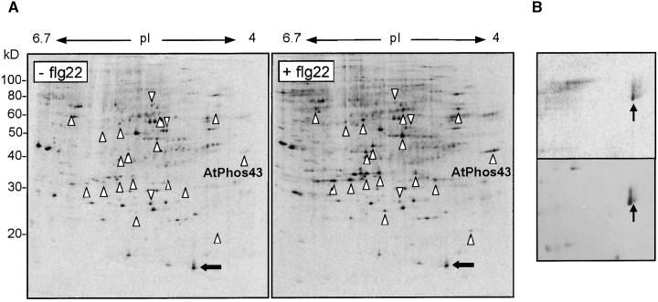
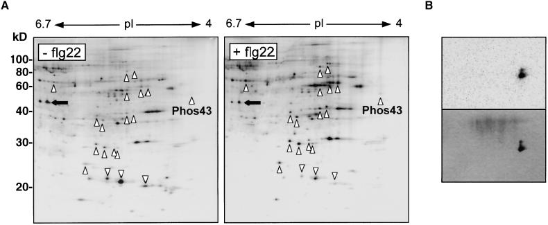
Separation of 33P-labeled soluble proteins by 2D gel electrophoresis allowed visualization of the “phosphoprotein proteome,” that is, the total complement of rapidly phosphorylated proteins in suspension-cultured cells, without any previous purification steps (Figure 1A). The proteins were separated between a pI of 4 and 7 in the first dimension. Although it is possible to use gradients encompassing a wider range (pI 3 to 10), we found that the resolution of these gradients was not sufficient to give a clear image of the changes in phosphorylation. A number of proteins showed increased or decreased incorporation of radioactive phosphate within 4 min of elicitor treatment, as indicated by the triangles and inverted triangles, respectively. Constitutively phosphorylated proteins (Figure 1A, arrows) served as internal controls for loading and phosphate incorporation. The highly reproducible phosphorylation pattern was specific for primary elicitor responses. Compounds generating reactive oxygen species or nitric oxide did not produce the pattern, nor did inhibitors of the formation of these compounds affect the pattern during elicitor responses (data not shown). The acidic pI of one of these phosphoproteins, AtPhos43, placed it in a region of low protein complexity, facilitating identification of the protein. To test the efficacy of this 2D gel-based approach, we selected this protein for further analysis.
Figure 1.
Phosphoprotein Proteome of flg22-Treated Arabidopsis Cell Culture.
(A) Phosphorimage of radiolabeled soluble protein extracted from cells treated for 3 min with water (left) or 10 nM flg22 (right). Proteins that increase in incorporation of 33P after elicitor treatment are indicated by triangles, and those that decrease are indicated by inverted triangles. An example of a constitutively phosphorylated protein is indicated by horizontal arrows. The phosphoprotein examined in this study, AtPhos43, is labeled.
(B) Alignment of the radioactive images of AtPhos43 (top) and its corresponding silver-stained protein (bottom).
AtPhos43 Is a Plant-Specific Protein Containing Ankyrin Repeats
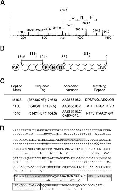
The radioactive image of AtPhos43 (Figure 1B, top) coaligned with a silver-stained protein (Figure 1B, bottom), which was not detectable in the control sample (data not shown). The corresponding protein was isolated from a single undried gel, digested in the gel with trypsin, and prepared for analysis by nanoESI-MS-MS (Shevchenko et al., 1996b). After ionization, a tryptic fragment was bombarded by an inert gas in the second quadrupole during collision-induced dissociation, fragmenting the peptide at the amide bonds. The resulting y-ion series in the spectrum showed the fragmentation of the parent ion (Figure 2A). The mass difference between the peaks was used to determine a “peptide sequence tag” (Mann and Wilm, 1994). This sequence tag consists of the short sequence of amino acid information, the total mass of the unknown amino acids between the phenylalanine residue and the N terminus (Figure 2B, m1), and the total mass of the unknown amino acids between the glutamine residue and the C terminus (Figure 2B, m3).
Figure 2.
Identification and Sequence of AtPhos43.
(A) Spectrum obtained by nanoESI-MS-MS from a single tryptic fragment of the isolated protein. The individual mass peaks represent fragments of the parent ion produced by collision-induced dissociation. m/z, mass-to-charge ratio.
(B) Graphic representation of the data obtained from the spectrum in (A). The circles represent the amino acids of the tryptic peptide, with the three amino acids identified in the shaded circles. The total mass of amino acids between the phenylalanine residue and the N terminus of the peptide is represented by m1. The total mass of amino acids from the glutamine residue to the C terminus is represented by m3.
(C) Peptide sequence tags used to search the Arabidopsis database, and corresponding accession numbers identifying AtPhos43 as the phosphoprotein.
(D) Alignment of AtPhos43 (top sequence) with the highly related protein ARP2 (bottom sequence). Residues of ARP2 identical to those of AtPhos43 are indicated by dashes. Gaps in the alignment are indicated by dots. Peptides corresponding to the peptide sequence tags are shown in the shaded boxes. The putative ankyrin domains are shown in the unshaded boxes, and the glycosyl hydrolase family 1 active site motif is underlined.
A similar analysis of two additional tryptic fragments yielded a total of three peptide sequence tags that were used to search Arabidopsis sequences in GenBank (Figure 2C). All three sequence tags identified AtPhos43 as a protein predicted from the Arabidopsis genome sequencing project (GenBank accession number AAB86516.2; sequences corresponding to the peptide sequence tag are shown in shaded boxes in Figure 2D). One of the sequence tags also identified a second protein (ankyrin repeat–containing protein 2 [ARP2]; GenBank accession number CAB54873.1). ARP2 is highly related to AtPhos43 (77% identity, 84% similarity) and shares an identical sequence within the region of the third sequence tag, but amino acid substitutions in the other two sequence tags unambiguously identified the isolated protein as AtPhos43. We also sequenced the protein immediately to the left of AtPhos43 (Figure 1B, bottom). This protein produced an identical set of sequence tags (data not shown), indicating that this more abundant protein is the unphosphorylated form of AtPhos43. The appearance of the second form of AtPhos43 (Figure 1B, arrows) after elicitor treatment of the cells is consistent with the fact that the pI of a newly phosphorylated protein will become more acidic, thus shifting its position away from the unphosporylated form during isoelectric focusing.
AtPhos43 is a predicted cytosolic protein (PSORT; http://psort.nibb.ac.jp) with two ankyrin motifs at the C terminus, indicating a possible involvement in protein–protein interactions (Sedgwick and Smerdon, 1999). The remainder of the protein shows no significant similarity to any protein of known function. A putative glycosyl hydrolase family 1 active site signature is present within the second ankyrin domain (predicted using PSITE; http://genomic.sanger.ac.uk/pss/pss.shtml), but no other portion of the protein displays similarity to known glycosyl hydrolases. Interestingly, highly related sequences (51 to 69% identity over the first 200 amino acids) are present in the expressed sequence tag databases for a wide range of plant species, including rice, tomato, cotton, alfalfa, and loblolly pine. No related sequence, however, is present in yeast, Caenorhabditis elegans, or human databases, indicating that AtPhos43 is a plant-specific protein.
AtPhos43 Is Phosphorylated in Response to Bacterial and Fungal Elicitors
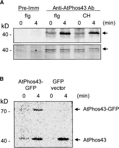
To confirm that AtPhos43 encoded the phosphoprotein and did not merely comigrate with the radioactive spot, we performed immunoprecipitation experiments from radiolabeled cells. Immune serum precipitated a protein of the expected size from cell culture that was not precipitated by preimmune serum (Figure 3A, bottom, arrow). Phosphorimage analysis of the gel showed that the protein is differentially phosphorylated in response to flg22, a bacterial elicitor, and chitin fragments, an elicitor from fungal cell walls (Figure 3A, top).
Figure 3.
AtPhos43 Is Differentially Phosphorylated in Arabidopsis Cell Cultures.
(A) Immunoprecipitation of proteins from suspension cell cultures pulse labeled with 33P after 0 or 4 min of treatment with 10 nM flg22 (flg) or 10 μg/mL chitin fragments (CH). Radioactive image (top) and Coomassie blue stain (bottom) of immunoprecipitated Arabidopsis proteins using preimmune serum (Pre-Imm) or immune serum (Anti-AtPhos43 Ab). Arrows at right indicate the position of AtPhos43.
(B) Radioactive image of proteins immunoprecipitated with anti-AtPhos43 antibody from flg22-treated Arabidopsis cultures transformed with a vector expressing AtPhos43-GFP or GFP alone.
Because it was likely that the antibodies cross-reacted with the highly related protein ARP2, we stably transformed Arabidopsis cell culture with a construct to express an AtPhos43–green fluorescent protein (GFP) fusion protein. A differentially phosphorylated protein of the size expected for the fusion protein was immunoprecipitated from flg22-treated cultures expressing AtPhos43-GFP but not from cultures transformed with the GFP vector alone (Figure 3B). Native AtPhos43 protein was phosphorylated in both cultures after elicitor treatment, confirming that elicitation and phosphorylation occurred in the vector control culture (Figure 3B). These results demonstrate that the protein identified as AtPhos43 is differentially phosphorylated in response to elicitors in Arabidopsis cell culture.
AtPhos43-Related Proteins Are Phosphorylated in Other Plant Species
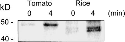
Because related sequences were present in databases for other plant species, we investigated whether these proteins were regulated similarly during elicitor responses. Suspension-cultured cells of tomato were treated with flg22. We found that rice cells did not respond to flg22 (data not shown) but did respond to chitin fragments. In both species, immune serum differentially precipitated phosphorylated proteins with apparent molecular masses slightly larger than that of the Arabidopsis protein, with the protein(s) from rice consistently appearing as a doublet (Figure 4). Thus, the regulation of related proteins is conserved in other dicots and monocots.
Figure 4.
Phos43-Related Proteins Are Phosphorylated in Other Species.
Radioactive image of 32P-labeled proteins immunoprecipitated from tomato or rice suspension cell cultures treated with flg22 or chitin fragments, respectively.
The flg22-Induced Phosphoprotein Proteome Is Not Conserved between Arabidopsis and Tomato
Previously, we reported that AtMPK6, the ortholog of the MAPK activated by elicitors in tomato (Romeis et al., 1999), was activated similarly by microbial elicitors in Arabidopsis (Nühse et al., 2000). In this work, we found that the Phos43 protein was phosphorylated in both Arabidopsis and tomato after treatment with flg22. Thus, it appears that at least some pathways are conserved between the two species. We investigated if it was possible to compare the phosphoprotein proteomes from flg22-treated cells to identify signaling components in parallel systems merely on the basis of position on a 2D gel. As was found for Arabidopsis cells, a number of tomato proteins increased or decreased in their incorporation of radioactive phosphate after treatment with flg22 (Figure 5A). Comparison of the patterns obtained with elicitor-treated Arabidopsis cells (Figure 1A) and tomato cells (Figure 5A), however, showed that there was little conservation between the two phosphoprotein proteomes. A similar lack of conservation was observed when the silver-stained gels were compared (data not shown). In tomato, a weakly phosphorylated spot was located on the acidic end of the gel (Figure 5A, arrows). Using 2D-PAGE to separate radioactively labeled proteins after immunoprecipitation with the anti-Phos43 antibody, we found that this spot was in fact the differentially phosphorylated protein observed in Figure 4 (Figure 5B). No other radioactive proteins were detectable. Thus, although the tomato Phos43 ortholog is differentially phosphorylated after elicitor treatment, the position of the protein on the 2D gel and the relative level of 32P incorporation compared with the other radioactively labeled proteins are quite different between tomato and Arabidopsis. From these results, we conclude that, in general, the identities of related phosphoproteins in plants showing similar responses cannot be assigned by simple comparison of phosphoprotein proteome maps.
Figure 5.
Phosphoprotein Proteome of flg22-Treated Tomato Cell Culture.
(A) Phosphorimage of radiolabeled soluble protein extracted from cells treated for 3 min with water (left) or 10 nM flg22 (right). Proteins that increase in incorporation of 32P after elicitor treatment are indicated by triangles, and those that decrease are indicated by inverted triangles. An example of a constitutively phosphorylated protein is indicated by horizontal arrows. The location of the Phos43 ortholog is labeled.
(B) Confirmation that the spot indicated in (A) is the tomato Phos43 ortholog by 2D separation of immunoprecipitate using anti-AtPhos43 antibody. The top section shows the radioactive image of the silver-stained gel in the bottom section. The spots comigrate to the position indicated in (A).
AtPhos43 Is Phosphorylated in Planta
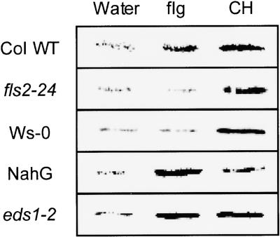
Cell cultures provide an excellent biochemical system to study synchronized elicitor responses, but it is essential to connect these observations to responses in whole plants. To determine if AtPhos43 also is phosphorylated in response to elicitor treatment in planta, 32P-orthophosphate was fed through the roots of 10- to 14-day-old Arabidopsis seedlings before treatment with flg22 or chitin. Changes in the phosphorylation of AtPhos43 were detected by immunoprecipitation within 10 min of treatment of intact seedlings with either flg22 or chitin fragments (Figure 6). These results support previous findings that responses to elicitors in Arabidopsis are similar in intact plants and cell culture (Nühse et al., 2000). To test the specificity of the response to flg22, we examined the phosphorylation of AtPhos43 in the fls2-24 mutant and in the Wassilewskija (Ws-0) ecotype. In the fls2-24 mutant, a single amino acid substitution in the LRR domain of the LRR kinase renders the mutant insensitive to flg22 in all responses tested (Gómez-Gómez and Boller, 2000). The mutant, however, does retain responsiveness to other elicitors. The Ws-0 ecotype also is insensitive to flg22 (Gómez-Gómez et al., 1999), but it cannot be complemented by expression of the FLS2 protein (Gómez-Gómez and Boller, 2000). In both cases, AtPhos43 is not phosphorylated after treatment with flg22, but phosphorylation does occur in response to chitin fragments (Figure 6). Together, these results demonstrate that the phosphorylation of AtPhos43 in response to flg22 is a specific event requiring both a functional FLS2 LRR kinase and a second, as yet unidentified, component.
Figure 6.
AtPhos43 Is Phosphorylated after Treatment of Intact Plants with Microbial Elicitors.
Radioactive image of proteins immunoprecipitated by the anti-AtPhos43 antibody from in planta 32P-labeled Arabidopsis seedlings. Columbia ecotype wild-type (Col WT), fls2-24 mutant, Ws-0 ecotype, eds1-2 mutant, and NahG-expressing seedlings were treated for 10 min with water, 10 μM flg22 (flg), or 100 μg/mL chitin fragments (CH) before immunoprecipitation.
Next, we determined whether SA is required during these rapid changes in phosphorylation. SA is a signaling molecule implicated in mediating a subset of defense responses (Sticher et al., 1997). Plants expressing the NahG gene produce SA hydroxylase, thereby inhibiting the accumulation of SA (Gaffney et al., 1993). In NahG-expressing plants, AtPhos43 is phosphorylated normally in response to both elicitors (Figure 6).
Finally, we examined the response to elicitors in the eds1-2 mutant (Aarts et al., 1998). The EDS1 protein shows sequence similarity to lipases (Falk et al., 1999) and appears to function upstream of SA but downstream of a subset of LRR proteins with domains similar to those found in Drosophila Toll and human IL-1 receptors (Aarts et al., 1998). The role of the EDS1 protein in responses to nonrace-specific elicitors, however, has never been investigated. Treatment of eds1-2 seedlings with flg22 or chitin fragments resulted in normal phosphorylation of AtPhos43 (Figure 6). Together, these results show that AtPhos43 phosphorylation does not depend on EDS1 or SA.
DISCUSSION
Phosphorylation of AtPhos43 and Related Proteins
Although phosphorylation plays a central role in defense signaling, few phosphorylated proteins have been identified in any plant species. In fact, the identification of AtPhos43 as a phosphoprotein in this work makes it only the second protein in Arabidopsis known to be phosphorylated after elicitor treatment, the other being a MAPK, AtMPK6 (Nühse et al., 2000). From analysis of both the soluble and membrane-associated phosphoprotein proteomes, we conservatively estimate that at least 30 proteins are differentially phosphorylated within the first 4 min after elicitor treatment of Arabidopsis cell culture (Figure 1A; S.C. Peck and T.S. Nühse, unpublished results). The large number of phosphoproteins is consistent with the emerging paradigm of complex parallel and overlapping pathways involved in defense signaling (Shah et al., 1999; Durrant et al., 2000). In this regard, the use of defense and hormone response mutants to place the phosphoproteins within discreet signaling pathways will facilitate a more thorough analysis of the points of convergence and divergence of pathways.
We found that flg22-induced, but not chitin-induced, phosphorylation of AtPhos43 in intact plants requires the FLS2 LRR kinase and the component absent from Ws-0. These data provide further evidence that FLS2 is involved in the recognition of the flagellin peptide. In addition, from analysis of the eds1-2 mutants and NahG-expressing seedlings, we conclude that neither EDS1 nor SA is required for flg22- or chitin-induced phosphorylation of AtPhos43. It should be noted that these results do not exclude the possibility that SA may elicit the phosphorylation of AtPhos43 during systemic responses. Currently, there is little information on how much of the early signaling pathways are used during later, cell-to-cell signaling. Our attempts to elicit the phosphorylation of AtPhos43 with SA directly were unsuccessful. However, these results must be interpreted cautiously, because we know that the timing of the elicitor treatment and protein isolation were critical parameters for the success of the in planta assay.
Development of Directed Proteomics as a Tool for Functional Genomics
A growing focus in biology is assigning function to unknown open reading frames generated by genome sequencing projects. Because protein phosphorylation is the major mechanism by which eukaryotic cells respond to extracellular signals, the directed proteomics approach described here is a powerful addition to the growing arsenal of techniques used for functional genomics. With commercially available immobilized pH gradients (IPGs) for first dimension separation, the phosphorylation patterns from the 2D gels are extremely reproducible; therefore, they can serve as a checklist for the systematic identification of differentially phosphorylated proteins. The increased sensitivity and mass accuracy of mass spectrometers allows protein identification from femtomoles of protein (Shevchenko et al., 1996a), provided sufficient database coverage is present, as is the case for Arabidopsis and a growing number of plant species.
Identification of AtPhos43 as a novel plant-specific protein phosphorylated in response to microbial elicitors demonstrates that directed proteomics is an unbiased approach, meaning that it can identify rapidly phosphorylated proteins without previous knowledge of protein function. Previously, biochemical characterization of signaling pathways in plants relied primarily on parallels with mammalian or yeast models, such as the identification of MAPK involved in defense responses (Ligterink et al., 1997; Zhang et al., 1998; Romeis et al., 1999; Nühse et al., 2000). Although the identification of conserved signaling elements is necessary and valuable, alternative approaches such as directed proteomics contribute to a more complete understanding of the events that occur rapidly after elicitor perception. It must be noted, however, that limitations to this approach remain. The major problem involves obtaining reliable alignments of the radioactive image with the protein of interest within regions of the gel containing either a high density of proteins or extremely abundant proteins. When we started these studies, only AtPhos43 and a small number of additional proteins (S.C. Peck, unpublished data) could be identified as putative phosphoproteins using the most expanded gradient available, pI 4 to 7. The proteins that were identified had either highly acidic pIs (<4.5), such as AtPhos43, or high apparent molecular weights, placing them in regions of the gel with a low number of proteins. A significant contribution to resolving this problem comes from the expanded IPGs that are now commercially available. These expanded gradients separate a single pI unit over 18 cm in the first dimension, allowing high-resolution alignments of a significantly greater (>80%) proportion of the putative phosphoproteins.
Another limitation is that very large (>100 kD), extremely hydrophobic, or extremely basic (pI >9) proteins generally are difficult to separate by 2D-PAGE. Applying a low voltage during rehydration of the proteins into the IPG strips increases the number of proteins between 100 and 140 kD resolved on the 2D gel. In the case of the hydrophobic and basic proteins, part of the problem appears to be in resolubilization of the proteins. More soluble cytosolic proteins must be removed to allow solubilization of these more difficult proteins. For instance, carbonate washing of highly enriched plasma membranes prior to resolubilization greatly increases the appearance of integral membrane proteins on 2D gels (T.S. Nühse and S.C. Peck, unpublished results). Thus, although this approach is faster than establishing a purification scheme for each phosphoprotein, it is important to understand that it is not possible to run a single gel to visualize all proteins simultaneously.
In conclusion, we propose that directed proteomics is a systematic and reproducible method for analyzing signal transduction pathways in plants. As more species are sequenced, the applicability of this technique will be extended beyond Arabidopsis. In the future, it will be necessary to develop the in planta phospholabeling assay presented in this work such that the analysis of phosphoproteins can be performed using whole plants and not just cell cultures. There are two main considerations for this development. First is the optimization of the orthophosphate labeling time to maximize the pool of radioactive ATP available to the kinases. The second is to design a sufficiently synchronized stimulus to maximize signal to noise. With these advances, it will be possible to characterize entire phosphorylation cascades involved in a broad range of biological functions.
METHODS
Labeling and 2D Separation of Proteins
Growth and 32P pulse labeling of Arabidopsis thaliana and tomato (Lycopersicon esculentum) suspension cell cultures were as described by Nühse et al. (2000) and Felix et al. (1991), respectively. Approximately 300 μL of 6- to 7-day-old cell culture was used for each experiment. After labeling, cells were mixed with an equal volume of two-dimensional (2D) extraction buffer (100 mM Hepes-KOH, pH 7.5, 5% glycerol, 50 mM sodium pyrophosphate, 1 mM Na2Mo4, 25 mM NaF, 15 mM EGTA, 5 mM EDTA, 0.5% polyvinylpyrrolidone, 3 mM DTT, 10 μM leupeptin, 1 mM PMSF, and 1 nM calyculin A), frozen in liquid nitrogen, and thawed in a sonicating water bath. After centrifugation, the supernatant was mixed with an equal volume of Tris-buffered phenol, pH 8, centrifuged, and back-extracted three times with back-extraction buffer (100 mM Tris, pH 8.4, 20 mM KCl, 10 mM EDTA, and 0.4% β-mercaptoethanol). The phenol phase was precipitated with 5 volumes (v/v) of 100 mM ammonium acetate in methanol at −20°C. After washing the pellet with 80% acetone, the final protein pellet was resuspended in 370 μL of 2D loading buffer (9 M urea, 1% Triton X-100, 0.5% DTT, and 0.5% immobilized pH gradient [IPG] buffer 4-7L). Low voltage rehydration and isoelectric focusing of 18-cm 4-7L IPG strips (Pharmacia Biotechnology) were performed using an IPGPhor first dimension electrophoresis unit (Pharmacia Biotechnology; 12 hr at 100 V, 2 hr at 500 V, 2 hr at 1000 V, 12-hr gradient to 4500 V, and 12 hr at 4500 V). After isoelectric focusing, IPG strips were equilibrated for the second dimension for 15 min in IPG Equilibration Buffer (50 mM Tris, pH 6.8, 6 M urea, 30% glycerol, 1% SDS, and 0.01% bromophenol blue) plus 1% DTT and 15 min in IPG Equilibration Buffer plus 4.5% iodoacetamide. One cm was removed from each end before separation on vertical gels (Protean II; Bio-Rad). Silver staining of proteins was performed as described by Shevchenko et al. (1996b). Flagellin peptide (flg22) was synthesized by F. Fischer (Friedrich Miescher Institut, Basel, Switzerland), and chitin in the form of hexa-N-acetylchitohexose was obtained from Seikagaku Corp. (Tokyo, Japan).
NanoESI-MS-MS Identification of AtPhos43
Proteins separated by SDS-PAGE were excised from the gel, reduced with DTT, alkylated with iodoacetamide, cleaved with sequencing-grade trypsin (Promega), and prepared for nanoelectrospray ionization tandem mass spectrometry (nanoESI-MS-MS) as described (Shevchenko et al., 1996a). NanoESI-MS-MS analysis (Wilm and Mann, 1996) was performed on an API 300 mass spectrometer (Applied Biosystems, Foster City, CA) equipped with a nanoelectrospray source (Protana, Odense, Denmark).
Immunoprecipitation from Cell Culture
Proteins were extracted as described for 2D gel analysis with the addition of 250 mM NaCl in the extraction buffer. Extracts were incubated for 1 hr at 4°C with either anti-AtPhos43 serum (A. Iglesias and F. Meins, unpublished results) or preimmune serum. Protein A–Sepharose was then incubated with the extract for 1 hr at 4°C. Immunoprecipitates were washed three times with 2D extraction buffer plus 1 M NaCl before analysis by SDS-PAGE.
Transgenic Cell Culture
The AtPhos43–green fluorescent protein (GFP) fusion protein was constructed by polymerase chain reaction using the Arabidopsis expressed sequence tag clone 119N10T7 as a template (GenBank accession number T43407). The insert was cloned into pCAMBIA1302 (C.S. Roberts, S. Rajagopal, L. Smith, T. Nguyen, W. Yang, S. Nugroho, K.S. Ravi, M.L. Cao, K. Vijayachandra, V. Patell, R.L. Harcourt, L. Dransfield, N. Desamero, I. Slamet, P. Keese, A. Kilian, and R.A. Jefferson, unpublished data) with the native start methionine intact and as a C-terminal fusion to GFP with an intervening unstructured region (GPGGGG). Transformation of suspension cell culture was performed using the model PDS-1000/He Biolistic Particle Delivery System (Bio-Rad) with subsequent selection of calli by resistance to hygromycin (50 μg/μL).
In Planta Labeling
Arabidopsis seedlings were grown aseptically as described previously (Gómez-Gómez et al., 1999) for 10 to 14 days before transfer to a 24-well microtiter plate (five seedlings per well) containing 500 μL of double-distilled water. Seedlings were placed uncovered in the growth chamber for 2 days. Two hours after adding 10 μL (3.7 MBq) of 32P-orthophosphate, water (control), 10 μM flg22, or 100 μg/mL, chitin fragments was added and mixed gently. After 10 min, seedlings were ground in 1.5-mL Eppendorf tubes containing 600 μL of Immunoprecipitation Buffer (100 mM HEPES-KOH, pH 7.5, 250 mM NaCl, 5% glycerol, 50 mM NaPP, 1 mM Na2Mo4, 25 mM NaF, 15 mM EGTA, 5 mM EDTA, 0.5% polyvinylpyrrolidone, 10 μM leupeptin, 1 mM PMSF, and 1 nM calyculin A,). Immunoprecipitations were performed as described above. Seed of the eds1-2 mutant were obtained from J. Parker (Sainsbury Laboratory, John Innes Centre, Norwich, UK), and seed of the NahG-expressing plants were obtained from U. Neustrander (Crop Protection, Novartis, Basel, Switzerland).
Acknowledgments
We thank R. Matthies for technical assistance and Drs. J. Hofsteenge, M. Collinge, and A. Heese-Peck for reading and commenting on the manuscript. S.C.P. was supported by a European Molecular Biology Organization long-term fellowship.
References
- Aarts, N., Metz, M., Holub, E., Staskawicz, B.J., Daniels, M.J., and Parker, J.E. (1998). Different requirements for ESD1 and NDR1 by disease resistance genes define at least two R gene-mediated signalling pathways in Arabidopsis. Proc. Natl. Acad. Sci. USA 95, 10306–10311. [DOI] [PMC free article] [PubMed] [Google Scholar]
- Boller, T. (1995). Chemoperception of microbial signal in plant cells. Annu. Rev. Plant Physiol. Plant Mol. Biol. 46, 189–214. [Google Scholar]
- Dietrich, A., Mayer, J.E., and Hahlbrock, K. (1990). Fungal elicitor triggers rapid, transient, and specific protein phosphorylation in parsley cell suspension. J. Biol. Chem. 265, 6360–6368. [PubMed] [Google Scholar]
- Droge-Laser, W., Kaiser, A., Lindsay, W.P., Halkier, B.A., Loake, G.J., Doerner, P., Dixon, R.A., and Lamb, C. (1997). Rapid stimulation of a soybean protein-serine kinase that phosphorylates a novel bZIP DNA-binding protein, G/HBF1, during the induction of early transcription-dependent defenses. EMBO J. 16, 726–738. [DOI] [PMC free article] [PubMed] [Google Scholar]
- Durrant, W.E., Rowland, O., Piedras, P., Hammond-Kosack, K.E., and Jones, J.D.G. (2000). cDNA-AFLP reveals a striking overlap in race-specific resistance and wound response gene expression profiles. Plant Cell 12, 963–977. [DOI] [PMC free article] [PubMed] [Google Scholar]
- Falk, A., Feys, B.J., Frost, L.N., Jones, J.D., Daniels, M.J., and Parker, J.E. (1999). EDS1, an essential component of R gene-mediated disease resistance in Arabidopsis has homology to eukaryotic lipases. Proc. Natl. Acad. Sci. USA 96, 3292–3297. [DOI] [PMC free article] [PubMed] [Google Scholar]
- Felix, G., Grosskopf, D.G., Regenass, M., and Boller, T. (1991). Rapid changes of protein phosphorylation are involved in transduction of the elicitor signal in plant cells. Proc. Natl. Acad. Sci. USA 88, 8831–8834. [DOI] [PMC free article] [PubMed] [Google Scholar]
- Felix, G., Duran, J.D., Volko, S., and Boller, T. (1999). Plants have a sensitive perception system for the most conserved domain of bacterial flagellin. Plant J. 18, 265–276. [DOI] [PubMed] [Google Scholar]
- Frye, C.A., Tang, D., and Innes, R.W. (2001). Negative regulation of defense responses in plants by a conserved MAPKK kinase. Proc. Natl. Acad. Sci. USA 98, 373–378. [DOI] [PMC free article] [PubMed] [Google Scholar]
- Gaffney, T., Friedrich, L., Vernoooij, B., Negrotto, D., Nye, G., Uknes, S., Ward, E., Kessmann, H., and Ryals, J. (1993). Requirement of salicylic acid for the induction of systemic acquired resistance. Science 261, 754–756. [DOI] [PubMed] [Google Scholar]
- Glazebrook, J., Rogers, E.E., and Ausubel, F.M. (1997). Use of Arabidopsis for genetic dissection of plant defence responses. Annu. Rev. Genet. 31, 547–569. [DOI] [PubMed] [Google Scholar]
- Gómez-Gómez, L., and Boller, T. (2000). FLS2: A LRR receptor-like kinase involved in recognition of the flagellin elicitor in Arabidopsis. Mol. Cell 5, 1–20. [DOI] [PubMed] [Google Scholar]
- Gómez-Gómez, L., Felix, G., and Boller, T. (1999). A single locus determines sensitivity to bacterial flagellin in Arabidopsis thaliana. Plant J. 18, 277–284. [DOI] [PubMed] [Google Scholar]
- Gu, Y.-Q., Yang, C., Thara, V.K., Zhou, J., and Martin, G.B. (2000). Pti4 is induced by ethylene and salicylic acid, and its product is phosphorylated by the Pto kinase. Plant Cell 12, 771–786. [DOI] [PMC free article] [PubMed] [Google Scholar]
- Innes, R.W. (1998). Genetic dissection of R gene signal transduction pathways. Curr. Opin. Plant Biol. 1, 229–304. [DOI] [PubMed] [Google Scholar]
- Lecourieux-Ouaked, F., Pugin, A., and Lebrun-Garcia, A. (2000). Phosphoproteins involved in the signal transduction of cryptogein, an elicitor of defense reactions in tobacco. Mol. Plant-Microbe Interact. 13, 821–829. [DOI] [PubMed] [Google Scholar]
- Ligterink, W., Kroj, T., zur Nieden, U., Hirt, H., and Scheel, D. (1997). Receptor-mediated activation of a MAP kinase in pathogen defence of plants. Science 276, 2054–2057. [DOI] [PubMed] [Google Scholar]
- Mann, M., and Wilm, M. (1994). Error-tolerant identification of peptides in sequence databases by peptide sequence tags. Anal. Chem. 66, 4390–4399. [DOI] [PubMed] [Google Scholar]
- Martin, G.B., Brommonschenkel, S.H., Chunwongse, J., Frary, A., Ganal, M.W., Spivey, R., Wu, T., Earle, E.D., and Tanksley, S.D. (1993). Map-based cloning of a protein kinase gene conferring disease resistance in tomato. Science 262, 1432–1436. [DOI] [PubMed] [Google Scholar]
- McDowell, J.M., and Dangl, J.L. (2000). Signal transduction in the plant immune response. Trends Biochem. Sci. 25, 79–82. [DOI] [PubMed] [Google Scholar]
- Nühse, T.S., Peck, S.C., Hirt, H., and Boller, T. (2000). Microbial elicitors induce activation and dual phosphorylation of the Arabidopsis thaliana MAPK 6. J. Biol. Chem. 275, 7521–7526. [DOI] [PubMed] [Google Scholar]
- Petersen, M., et al. (2000). Arabidopsis MAP kinase 4 negatively regulates systemic acquired resistance. Cell 103, 1111–1120. [DOI] [PubMed] [Google Scholar]
- Romeis, T., Piedras, P., Zhang, S., Klessig, D.F., Hirt, H., and Jones, J.D. (1999). Rapid Avr9- and Cf-9-dependent activation of MAP kinases in tobacco cell cultures and leaves: Convergence of resistance gene, elicitor, wound, and salicylate responses. Plant Cell 11, 273–287. [DOI] [PMC free article] [PubMed] [Google Scholar]
- Romeis, T., Piedras, P., and Jones, J.D.G. (2000). Resistance gene-dependent activation of a calcium-dependent protein kinase in the plant defense response. Plant Cell 12, 803–815. [DOI] [PMC free article] [PubMed] [Google Scholar]
- Scheel, D. (1998). Resistance response physiology and signal transduction. Curr. Opin. Plant Biol. 1, 305–310. [DOI] [PubMed] [Google Scholar]
- Sedgwick, S.G., and Smerdon, S.J. (1999). The ankyrin repeat: A diversity of interactions on a common structural framework. Trends Biochem. Sci. 24, 313–316. [DOI] [PubMed] [Google Scholar]
- Shah, J., Kachroo, P., and Klessig, D.F. (1999). The Arabidopsis ssi1 mutation restores pathogenesis-related gene expression in npr1 plants and renders defensin gene expression salicylic acid dependent. Plant Cell 11, 191–206. [DOI] [PMC free article] [PubMed] [Google Scholar]
- Shevchenko, A., Jensen, O.N., Podtelejnikov, A.V., Sagliocco, F., Wilm, M., Vorm, O., Mortensen, P., Shevchenko, A., Boucherie, H., and Mann, M. (1996. a). Linking genome and proteome by mass spectrometry: Large-scale identification of yeast proteins from two dimensional gels. Proc. Natl. Acad. Sci. USA 93, 14440–14445. [DOI] [PMC free article] [PubMed] [Google Scholar]
- Shevchenko, A., Wilm, M., Vorm, O., and Mann, M. (1996. b). Mass spectrometric sequencing of proteins from silver stained polyacrylamide gels. Anal. Chem. 68, 850–858. [DOI] [PubMed] [Google Scholar]
- Song, W.Y., Wang, G.L., Chen, L.L., Kim, H.S., Pi, L.Y., Holsten, T., Gardner, J., Wang, B., Zhai, W.X., Zhu, L.H., Fauquet, C., and Ronald, P. (1995). A receptor kinase-like protein encoded by the rice disease resistance gene Xa21. Science 270, 1804–1806. [DOI] [PubMed] [Google Scholar]
- Sticher, L., Mauchi-Mani, B., and Métraux, J.P. (1997). Systemic acquired resistance. Annu. Rev. Phytopathol. 35, 235–270. [DOI] [PubMed] [Google Scholar]
- Viard, M.-P., Martin, F., Pugin, A., Ricci, P., and Blein, J.-P. (1994). Protein phosphorylation is induced in tobacco cells by the elicitor cryptogein. Plant Physiol. 104, 1245–1249. [DOI] [PMC free article] [PubMed] [Google Scholar]
- Wilm, M., and Mann, M. (1996). Analytical properties of the nanoelectrospray ion source. Anal. Chem. 68, 1–8. [DOI] [PubMed] [Google Scholar]
- Xing, T., Higgins, V.J., and Blumwald, E. (1997). Race-specific elicitors of Cladosporium fulvum promote translocation of cytosolic components of NADPH oxidase to the plasma membrane of tomato cells. Plant Cell 9, 249–259. [DOI] [PMC free article] [PubMed] [Google Scholar]
- Zhang, S., Du, H., and Klessig, D. (1998). Activation of the tobacco SIP kinase by both a cell wall-derived carbohydrate elicitor and purified proteinaceous elicitins from Phytophthora spp. Plant Cell 10, 435–449. [DOI] [PMC free article] [PubMed] [Google Scholar]
- Zhou, J., Loh, Y.T., Bressan, R.A., and Martin, G.B. (1995). The tomato gene Pti1 encodes a serine/threonine kinase that is phosphorylated by Pto and is involved in the hypersensitive response. Cell 83, 925–935. [DOI] [PubMed] [Google Scholar]


