

Abstract
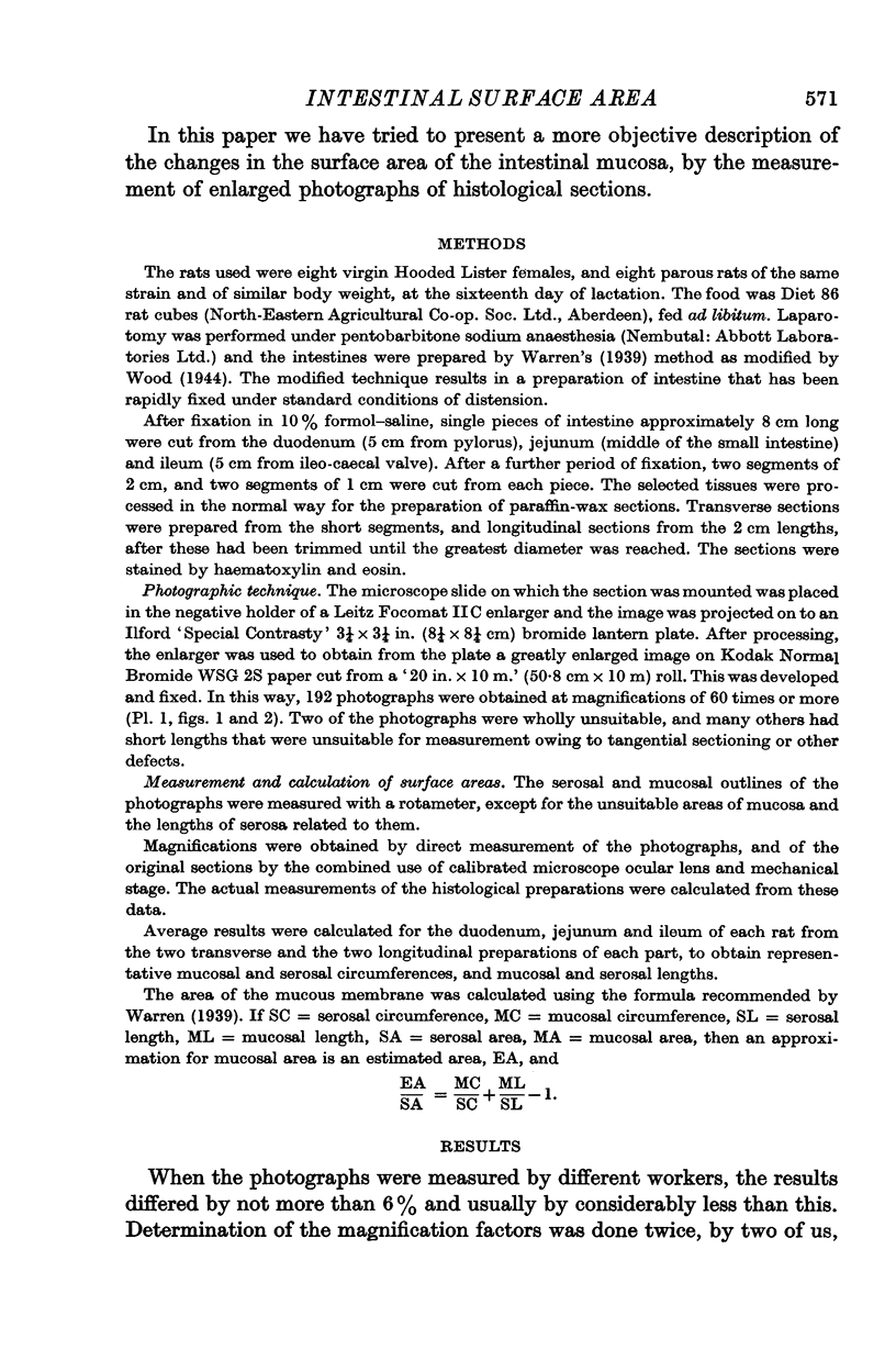
1. The mucosal and serosal surface dimensions of the rat duodenum, jejunum and ileum were obtained by measurement of photographs of histological sections. The surface areas were calculated from these data.
2. Measurements were made on the intestines from virgin rats and from rats on the sixteenth day of lactation. The mucosal area/unit length of serosa was appreciably increased in all parts of the intestine during lactation. The mucosal area/serosal area (MA/SA) ratio was also markedly increased, but to a lesser extent distally, where an important component of the intestinal hypertrophy was dilatation of the intestine.
Full text
PDF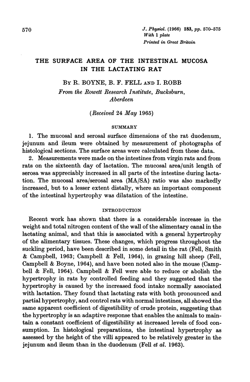


Images in this article

Selected References
These references are in PubMed. This may not be the complete list of references from this article.
- CAMPBELL R. M., FELL B. F. GASTRO-INTESTINAL HYPERTROPHY IN THE LACTATING RAT AND ITS RELATION TO FOOD INTAKE. J Physiol. 1964 May;171:90–97. doi: 10.1113/jphysiol.1964.sp007363. [DOI] [PMC free article] [PubMed] [Google Scholar]
- FELL B. F., SMITH K. A., CAMPBELL R. M. Hypertrophic and hyperplastic changes in the alimentary canal of the lactating rat. J Pathol Bacteriol. 1963 Jan;85:179–188. doi: 10.1002/path.1700850117. [DOI] [PubMed] [Google Scholar]
- FISHER R. B., PARSONS D. S. The gradient of mucosal surface area in the small intestine of the rat. J Anat. 1950 Jul;84(3):272–282. [PMC free article] [PubMed] [Google Scholar]
- PALAY S. L., KARLIN L. J. An electron microscopic study of the intestinal villus. I. The fasting animal. J Biophys Biochem Cytol. 1959 May 25;5(3):363–372. doi: 10.1083/jcb.5.3.363. [DOI] [PMC free article] [PubMed] [Google Scholar]
- Wood H. O. The surface area of the intestinal mucosa in the rat and in the cat. J Anat. 1944 Apr;78(Pt 3):103–105. [PMC free article] [PubMed] [Google Scholar]