

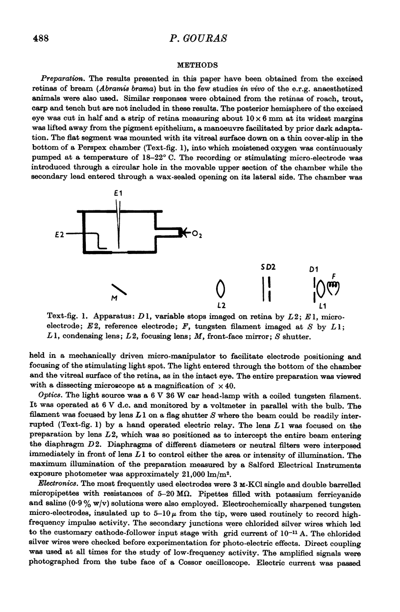
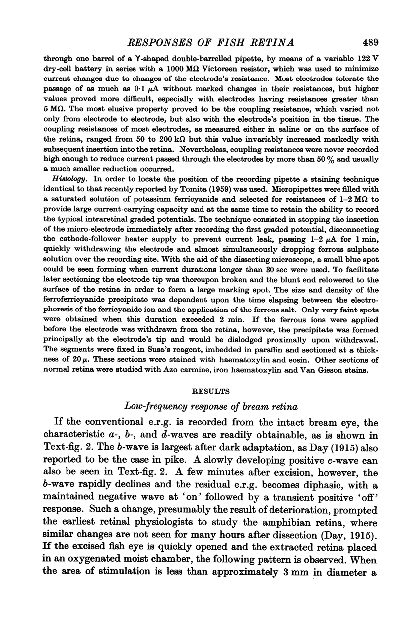
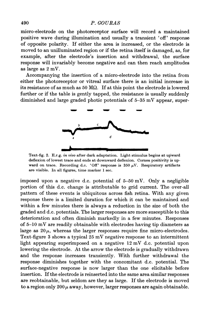
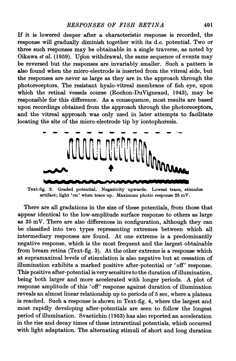
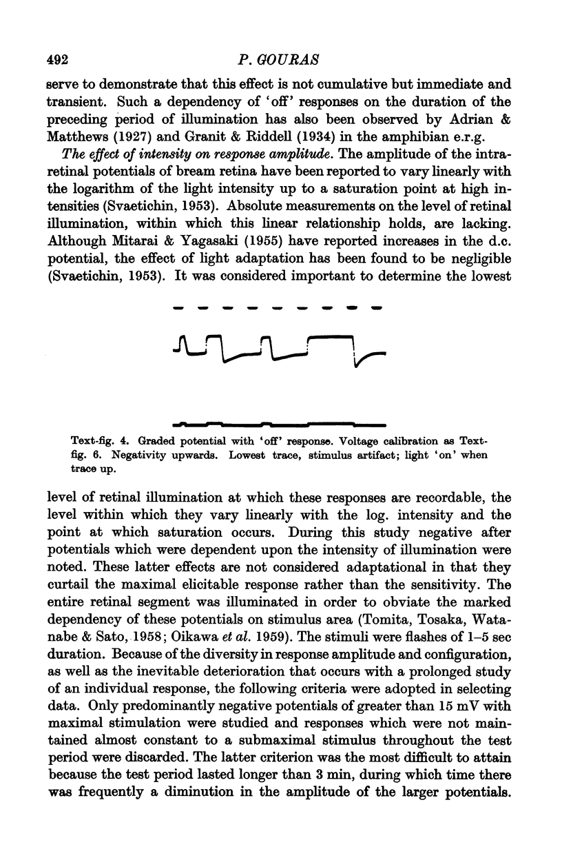
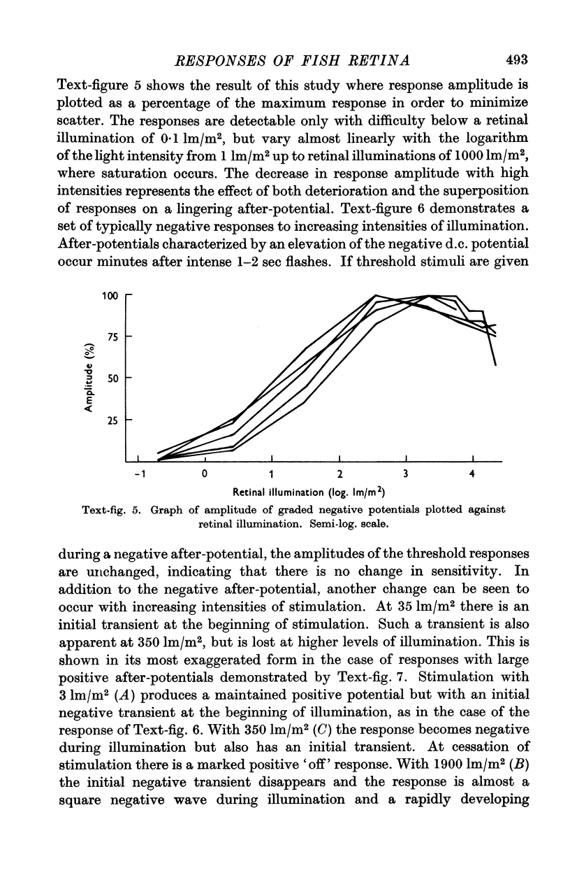
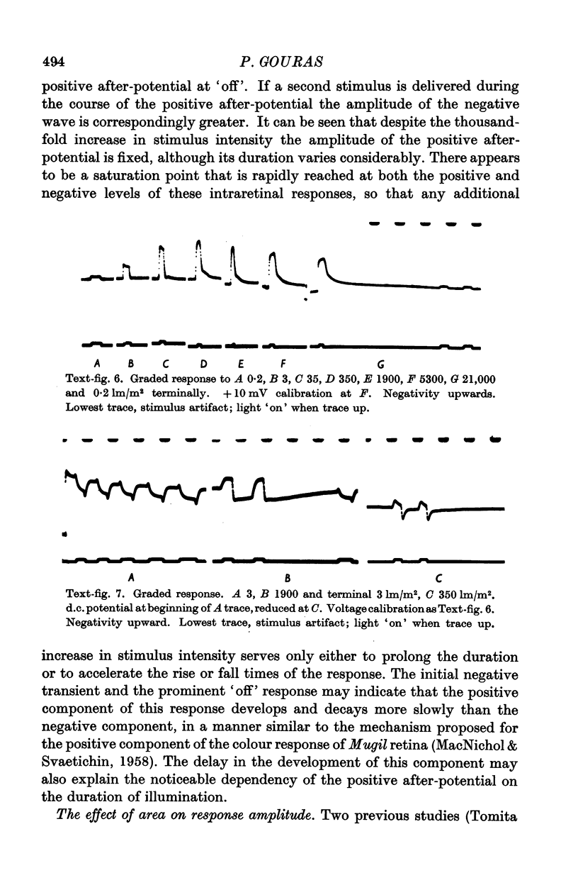
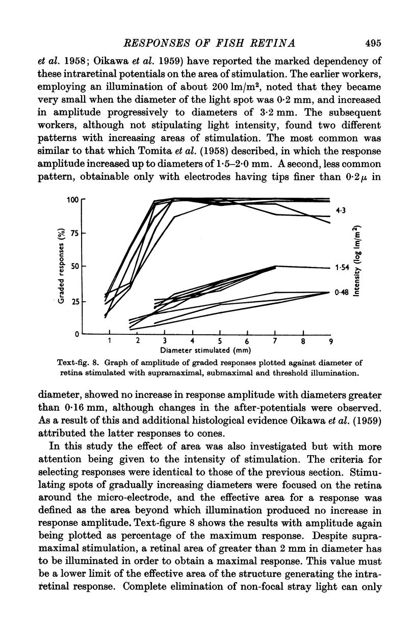
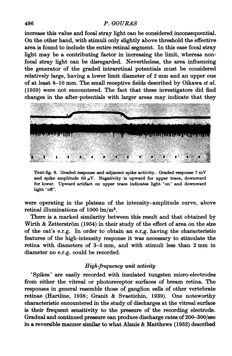
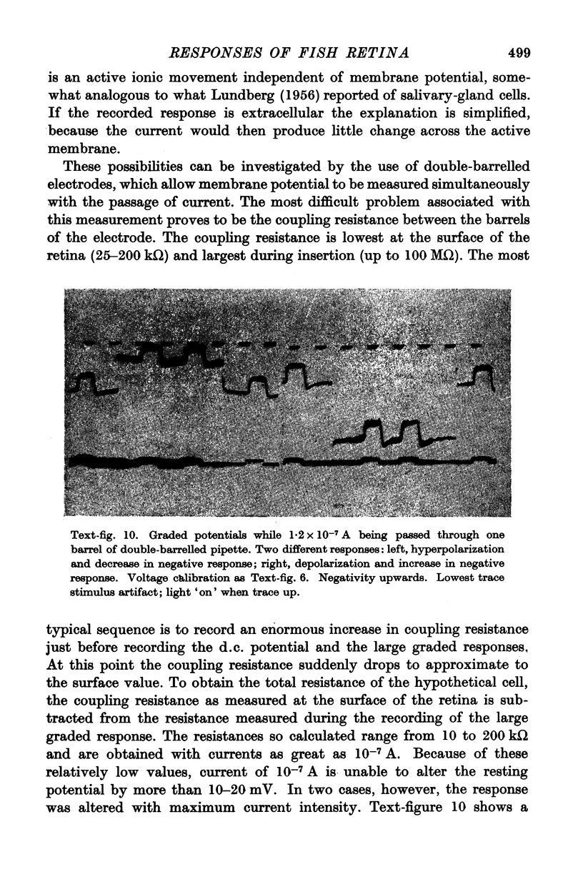
Full text
PDF
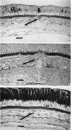
Images in this article
Selected References
These references are in PubMed. This may not be the complete list of references from this article.
- ALANIS J., MATTHEWS B. H. C. The mechano-receptor properties of central neurones. J Physiol. 1952 Aug;117(4):59P–60P. [PubMed] [Google Scholar]
- Adrian E. D., Matthews R. The action of light on the eye: Part I. The discharge of impulses in the optic nerve and its relation to the electric changes in the retina. J Physiol. 1927 Sep 9;63(4):378–414. doi: 10.1113/jphysiol.1927.sp002410. [DOI] [PMC free article] [PubMed] [Google Scholar]
- BRINDLEY G. S. The passive electrical properties of the frog's retina, choroid and sclera for radial fields and currents. J Physiol. 1956 Nov 28;134(2):339–352. doi: 10.1113/jphysiol.1956.sp005647. [DOI] [PMC free article] [PubMed] [Google Scholar]
- BRINDLEY G. S. The sources of slow electrical activity in the frog's retina. J Physiol. 1958 Feb 17;140(2):247–261. doi: 10.1113/jphysiol.1958.sp005931. [DOI] [PMC free article] [PubMed] [Google Scholar]
- BROWN K. T., WIESEL T. N. Intraretinal recording in the unopened cat eye. Am J Ophthalmol. 1958 Sep;46(3 Pt 2):91–98. doi: 10.1016/0002-9394(58)90058-8. [DOI] [PubMed] [Google Scholar]
- Granit R., Riddell L. A. The electrical responses of light- and dark-adapted frogs' eyes to rhythmic and continuous stimuli. J Physiol. 1934 Mar 29;81(1):1–28. doi: 10.1113/jphysiol.1934.sp003112. [DOI] [PMC free article] [PubMed] [Google Scholar]
- LUNDBERG A. Secretory potentials and secretion in the sublingual gland of the cat. Nature. 1956 Jun 9;177(4519):1080–1081. doi: 10.1038/1771080a0. [DOI] [PubMed] [Google Scholar]
- MACNICHOL E. J., SVAETICHIN G. Electric responses from the isolated retinas of fishes. Am J Ophthalmol. 1958 Sep;46(3 Pt 2):26–46. doi: 10.1016/0002-9394(58)90053-9. [DOI] [PubMed] [Google Scholar]
- MOTOKAWA K., OIKAWA T., TASAKI K. Receptor potential of vertebrate retina. J Neurophysiol. 1957 Mar;20(2):186–199. doi: 10.1152/jn.1957.20.2.186. [DOI] [PubMed] [Google Scholar]
- OIKAWA T., OGAWA T., MOTOKAWA K. Origin of so-called cone action potential. J Neurophysiol. 1959 Jan;22(1):102–111. doi: 10.1152/jn.1959.22.1.102. [DOI] [PubMed] [Google Scholar]
- SVAETICHIN G., MACNICHOL E. F., Jr Retinal mechanisms for chromatic and achromatic vision. Ann N Y Acad Sci. 1959 Nov 12;74(2):385–404. doi: 10.1111/j.1749-6632.1958.tb39560.x. [DOI] [PubMed] [Google Scholar]
- TOMITA T. A study on the origin of intraretinal action potential of the cyprinid fish by means of pencil-type microelectrode. Jpn J Physiol. 1957 Mar 15;7(1):80–85. doi: 10.2170/jjphysiol.7.80. [DOI] [PubMed] [Google Scholar]
- TOMITA T., MURAKAMI M., SATO Y., HASHIMOTO Y. Further study on the origin of the so-called cone action potential (s-potential); its histological determination. Jpn J Physiol. 1959 Mar 25;9(1):63–68. doi: 10.2170/jjphysiol.9.63. [DOI] [PubMed] [Google Scholar]
- TOMITA T., TORIHAMA Y. Further study on the intraretinal action potentials and on the site of ERG generation. Jpn J Physiol. 1956 Jun 15;6(2):118–136. doi: 10.2170/jjphysiol.6.118. [DOI] [PubMed] [Google Scholar]
- TOMITA T., TOSAKA T., WATANABE K., SATO Y. The fish EIRG in response to different types of illumination. Jpn J Physiol. 1958 Mar 30;8(1):41–50. doi: 10.2170/jjphysiol.8.41. [DOI] [PubMed] [Google Scholar]
- WIRTH A., ZETTERSTROM B. Effect of area and intensity on the size and shape of the electroretinogram; exclusion of stray light effects. Br J Ophthalmol. 1954 May;38(5):257–265. doi: 10.1136/bjo.38.5.257. [DOI] [PMC free article] [PubMed] [Google Scholar]