Abstract
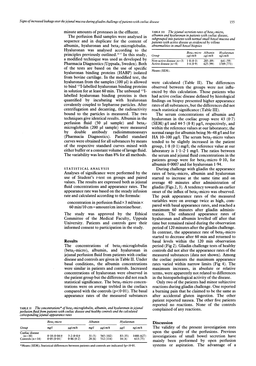
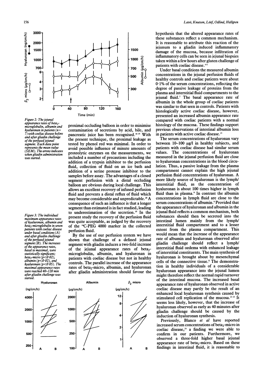
Intestinal secretion rates of albumin, hyaluronan, and beta 2-microglobulin (beta 2-micro) were determined under basal conditions and after gliadin challenge of coeliac patients and healthy controls by the use of a jejunal perfusion technique. A new tube system was used where a jejunal segment is isolated between balloons and then perfused with a balanced salt solution. Under basal conditions the secretion rate of albumin was similar in the patients and controls while the secretion rate of the glycosaminoglycan hyaluronan, a high molecular weight connective tissue component, was increased more than two times in coeliac patients. Beta 2-micro was secreted in on average three-fold rates in coeliacs compared with controls. All three substances were secreted at a higher rate in patients with active disease than in those with inactive disease defined by morphological damage in small bowel biopsies. The concentrations in jejunal perfusion fluids relative to serum levels in the coeliac patients were for albumin 0.0007, beta 2-micro 0.10, and for hyaluronan 1.94. Challenge with a single dose of gliadin into the jejunal segment gave within 60 min a significant, about two-fold, increase of the secretion rates of all three measured substances. The appearance of hyaluronan could reflect a gliadin induced mucosal oedema with an enhanced leakage from the interstitial/lymph fluid, rich in this glycosaminoglycan. The observed parallel increases in the jejunal secretion of albumin and beta 2-micro after gliadin challenge are best explained by a similar mechanism.
Full text
PDF
Selected References
These references are in PubMed. This may not be the complete list of references from this article.
- Anand B. S., Piris J., Jerrome D. W., Offord R. E., Truelove S. C. The timing of histological damage following a single challenge with gluten in treated coeliac disease. Q J Med. 1981;50(197):83–94. [PubMed] [Google Scholar]
- Bernier G. M., Fanger M. W. Synthesis of 2 -microglobulin by stimulated lymphocytes. J Immunol. 1972 Aug;109(2):407–409. [PubMed] [Google Scholar]
- Blanco A., Alonso M., Cilleruelo M. L., Solis P., Calvo C., Sanchez Villares E. Increased serum beta2-microglobulin levels in active celiac disease. J Pediatr Gastroenterol Nutr. 1985 Jun;4(3):388–392. doi: 10.1097/00005176-198506000-00011. [DOI] [PubMed] [Google Scholar]
- Engström-Laurent A., Laurent U. B., Lilja K., Laurent T. C. Concentration of sodium hyaluronate in serum. Scand J Clin Lab Invest. 1985 Oct;45(6):497–504. doi: 10.3109/00365518509155249. [DOI] [PubMed] [Google Scholar]
- Freedman A. R., Macartney J. C., Nelufer J. M., Ciclitira P. J. Timing of infiltration of T lymphocytes induced by gluten into the small intestine in coeliac disease. J Clin Pathol. 1987 Jul;40(7):741–745. doi: 10.1136/jcp.40.7.741. [DOI] [PMC free article] [PubMed] [Google Scholar]
- George J. D. New clinical method for measuring the rate of gastric emptying: the double sampling test meal. Gut. 1968 Apr;9(2):237–242. doi: 10.1136/gut.9.2.237. [DOI] [PMC free article] [PubMed] [Google Scholar]
- Marsh M. N., Haeney M. R. Studies of intestinal lymphoid tissue. VI--Proliferative response of small intestinal epithelial lymphocytes distinguishes gluten- from non-gluten-induced enteropathy. J Clin Pathol. 1983 Feb;36(2):149–160. doi: 10.1136/jcp.36.2.149. [DOI] [PMC free article] [PubMed] [Google Scholar]
- Marsh M. N., Hinde J. Inflammatory component of celiac sprue mucosa. I. Mast cells, basophils, and eosinophils. Gastroenterology. 1985 Jul;89(1):92–101. doi: 10.1016/0016-5085(85)90749-8. [DOI] [PubMed] [Google Scholar]
- Marsh M. N. Immunocytes, enterocytes and the lamina propria: an immunopathological framework of coeliac disease. J R Coll Physicians Lond. 1983 Oct;17(4):205–212. [PMC free article] [PubMed] [Google Scholar]
- Marsh M. N. Studies of intestinal lymphoid tissue. III. Quantitative analyses of epithelial lymphocytes in the small intestine of human control subjects and of patients with celiac sprue. Gastroenterology. 1980 Sep;79(3):481–492. [PubMed] [Google Scholar]
- Modigliani R., Bernier J. J. Absorption of glucose, sodium, and water by the human jejunum studied by intestinal perfusion with a proximal occluding balloon and at variable flow rates. Gut. 1971 Mar;12(3):184–193. doi: 10.1136/gut.12.3.184. [DOI] [PMC free article] [PubMed] [Google Scholar]
- Simpson F. G., Howdle P. D., Robertson D. A., Losowsky M. S. Jejunal biopsy and lymphocyte co-culture in coeliac disease. Scand J Gastroenterol. 1983 Sep;18(6):749–754. doi: 10.3109/00365528309182090. [DOI] [PubMed] [Google Scholar]
- Sladen G. E., Dawson A. M. Further studies on the perfusion method for measuring intestinal absorption in man: the effects of a proximal occlusive balloon and a mixing segment. Gut. 1970 Nov;11(11):947–954. doi: 10.1136/gut.11.11.947. [DOI] [PMC free article] [PubMed] [Google Scholar]
- Soergel K. H. An evaluation of perfusion techniques in the study of water and electrolyte absorption in man: the problem of endogenous secretions. Gut. 1969 Jul;10(7):601–601. doi: 10.1136/gut.10.7.601. [DOI] [PMC free article] [PubMed] [Google Scholar]
- Stern A. I., Hogan D. L., Isenberg J. I. A new method for quantitation of ion fluxes across in vivo human gastric mucosa: effect of aspirin, acetaminophen, ethanol, and hyperosmolar solutions. Gastroenterology. 1984 Jan;86(1):60–70. [PubMed] [Google Scholar]
- Tengblad A., Laurent U. B., Lilja K., Cahill R. N., Engström-Laurent A., Fraser J. R., Hansson H. E., Laurent T. C. Concentration and relative molecular mass of hyaluronate in lymph and blood. Biochem J. 1986 Jun 1;236(2):521–525. doi: 10.1042/bj2360521. [DOI] [PMC free article] [PubMed] [Google Scholar]
- Watson A. J., Wright N. A. Coeliac disease. Morphology and cell kinetics of the jejunal mucosa in untreated patients. Clin Gastroenterol. 1974 Jan;3(1):11–31. [PubMed] [Google Scholar]


