Abstract
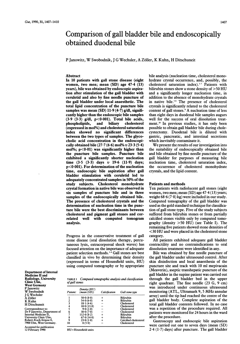
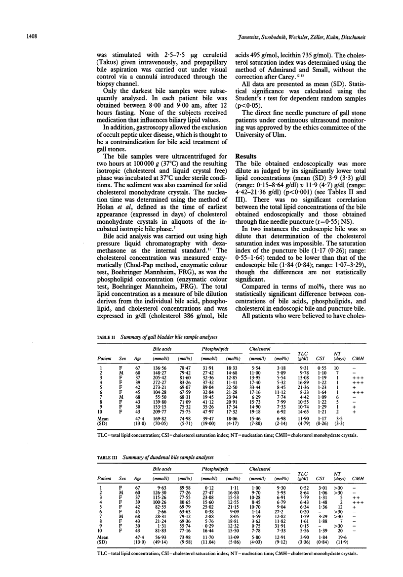
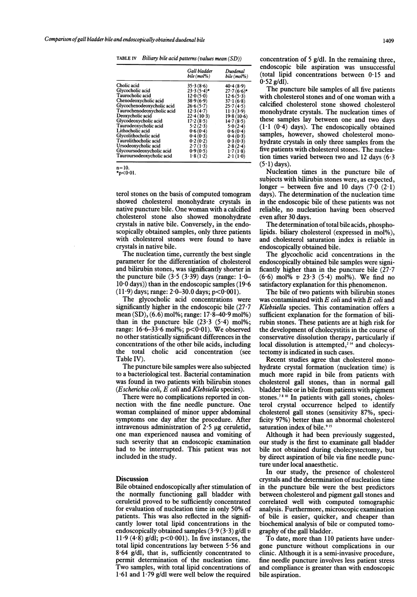
In 10 patients with gall stone disease (eight women, two men; mean (SD) age 47.4 (13) years), bile was obtained by endoscopic aspiration after stimulation of the gall bladder with ceruletid and also by fine needle puncture of the gall bladder under local anaesthetic. The total lipid concentration of the puncture bile samples was mean (SD) 11.9 (4.7) g/dl, significantly higher than the endoscopic bile samples (3.9 (3.3) g/dl, p less than 0.001). Total bile acids, phospholipids, and biliary cholesterol (expressed in mol%) and cholesterol saturation index showed no significant differences between the two types of samples. The glycocholic acid concentration in the endoscopically obtained bile (27.7 (6.6) mol% v 23.3 (5.4) mol%; p less than 0.01) was significantly higher than the puncture bile samples. Puncture bile exhibited a significantly shorter nucleation time (3.5 (3.3) days v 19.6 (11.9) days; p less than 0.001). For determination of the nucleation time, endoscopic bile aspiration after gall bladder stimulation with ceruletid led to adequately concentrated samples in 50% of the study subjects. Cholesterol monohydrate crystal formation in native bile was observed in six samples of puncture bile and in three samples of the endoscopically obtained bile. The presence of cholesterol crystals and the determination of nucleation time in the puncture bile were the best discriminants between cholesterol and pigment gall stones and correlated well with computed tomogram analysis.
Full text
PDF


Selected References
These references are in PubMed. This may not be the complete list of references from this article.
- Admirand W. H., Small D. M. The physicochemical basis of cholesterol gallstone formation in man. J Clin Invest. 1968 May;47(5):1043–1052. doi: 10.1172/JCI105794. [DOI] [PMC free article] [PubMed] [Google Scholar]
- Allen M. J., Borody T. J., Bugliosi T. F., May G. R., LaRusso N. F., Thistle J. L. Cholelitholysis using methyl tertiary butyl ether. Gastroenterology. 1985 Jan;88(1 Pt 1):122–125. doi: 10.1016/s0016-5085(85)80143-8. [DOI] [PubMed] [Google Scholar]
- Carey M. C. Critical tables for calculating the cholesterol saturation of native bile. J Lipid Res. 1978 Nov;19(8):945–955. [PubMed] [Google Scholar]
- Danzinger R. G., Hofmann A. F., Schoenfield L. J., Thistle J. L. Dissolution of cholesterol gallstones by chenodeoxycholic acid. N Engl J Med. 1972 Jan 6;286(1):1–8. doi: 10.1056/NEJM197201062860101. [DOI] [PubMed] [Google Scholar]
- Hickman M. S., Schwesinger W. H., Bova J. D., Kurtin W. E. Computed tomographic analysis of gallstones. An in vitro study. Arch Surg. 1986 Mar;121(3):289–291. doi: 10.1001/archsurg.1986.01400030043007. [DOI] [PubMed] [Google Scholar]
- Holan K. R., Holzbach R. T., Hermann R. E., Cooperman A. M., Claffey W. J. Nucleation time: a key factor in the pathogenesis of cholesterol gallstone disease. Gastroenterology. 1979 Oct;77(4 Pt 1):611–617. [PubMed] [Google Scholar]
- Janowitz P., Swobodnik W., Wechsler J. G., Fischer S., Ditschuneit H. Medikamentöse Cholelitholyse und Nukleationszeit. Dtsch Med Wochenschr. 1989 Jun 23;114(25):983–985. doi: 10.1055/s-2008-1066704. [DOI] [PubMed] [Google Scholar]
- Janowitz P., Zöller A., Swobodnik W., Wechsler J. G., Schumacher K. A., Ditschuneit H. Computed tomography evaluation of radiolucent gallstones in vivo. Gastrointest Radiol. 1990 Winter;15(1):58–60. doi: 10.1007/BF01888736. [DOI] [PubMed] [Google Scholar]
- Podda M., Zuin M., Dioguardi M. L., Festorazzi S., Dioguardi N. A combination of chenodeoxycholic acid and ursodeoxycholic acid is more effective than either alone in reducing biliary cholesterol saturation. Hepatology. 1982 May-Jun;2(3):334–339. doi: 10.1002/hep.1840020308. [DOI] [PubMed] [Google Scholar]
- Ramond M. J., Dumont M., Belghiti J., Erlinger S. Sensitivity and specificity of microscopic examination of gallbladder bile for gallstone recognition and identification. Gastroenterology. 1988 Nov;95(5):1339–1343. doi: 10.1016/0016-5085(88)90370-8. [DOI] [PubMed] [Google Scholar]
- Ros E., Navarro S., Fernández I., Reixach M., Ribó J. M., Rodés J. Utility of biliary microscopy for the prediction of the chemical composition of gallstones and the outcome of dissolution therapy with ursodeoxycholic acid. Gastroenterology. 1986 Sep;91(3):703–712. doi: 10.1016/0016-5085(86)90642-6. [DOI] [PubMed] [Google Scholar]
- Sauerbruch T., Delius M., Paumgartner G., Holl J., Wess O., Weber W., Hepp W., Brendel W. Fragmentation of gallstones by extracorporeal shock waves. N Engl J Med. 1986 Mar 27;314(13):818–822. doi: 10.1056/NEJM198603273141304. [DOI] [PubMed] [Google Scholar]
- Swobodnik W., Baumgaertel H., Janowitz P., Fuchs S., Ditschuneit H. Dissolution of calcified gallbladder stones by treatment with methyl-hexyl ether and urea-EDTA. Lancet. 1988 Jul 23;2(8604):216–216. doi: 10.1016/s0140-6736(88)92314-8. [DOI] [PubMed] [Google Scholar]
- Swobodnik W., Klüppelberg U., Wechsler J. G., Volz M., Normandin G., Ditschuneit H. Rapid and accurate reversed-phase high-performance liquid chromatographic determination of conjugated bile acids in human bile for routine clinical applications. Therapeutic control during gallstone dissolution therapy. J Chromatogr. 1985 May 3;339(2):263–271. doi: 10.1016/s0378-4347(00)84653-8. [DOI] [PubMed] [Google Scholar]
- van Erpecum K. J., van Berge Henegouwen G. P., Stoelwinder B., Stolk M. F., Eggink W. F., Govaert W. H. Cholesterol and pigment gallstone disease: comparison of the reliability of three bile tests for differentiation between the two stone types. Scand J Gastroenterol. 1988 Oct;23(8):948–954. doi: 10.3109/00365528809090152. [DOI] [PubMed] [Google Scholar]


