Abstract
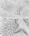
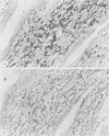
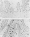
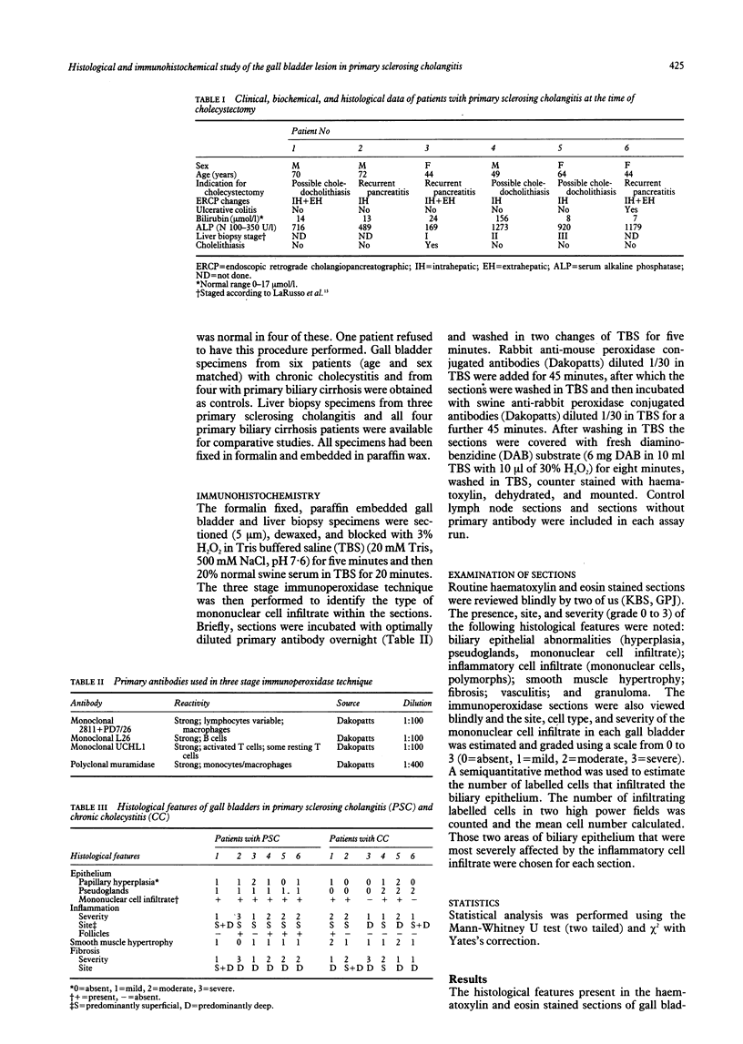
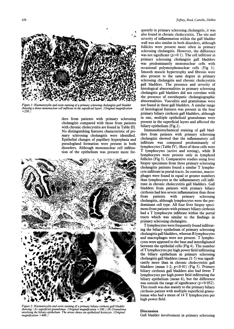
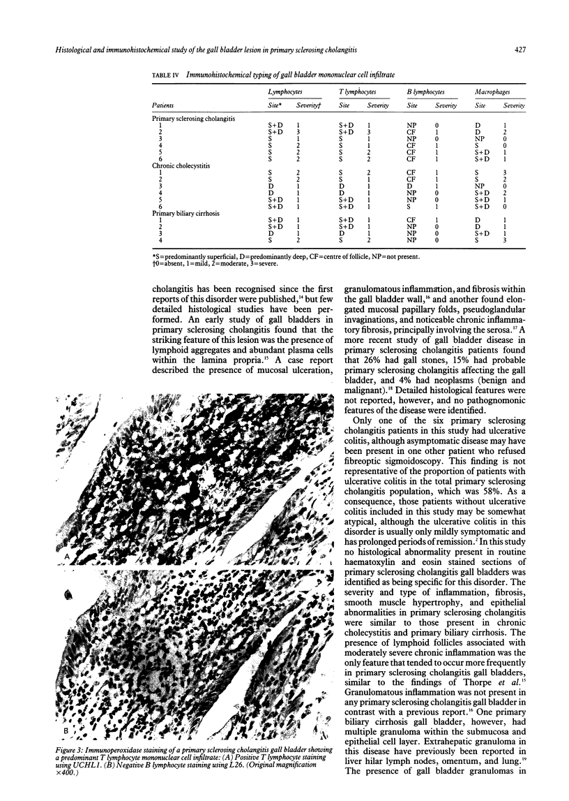
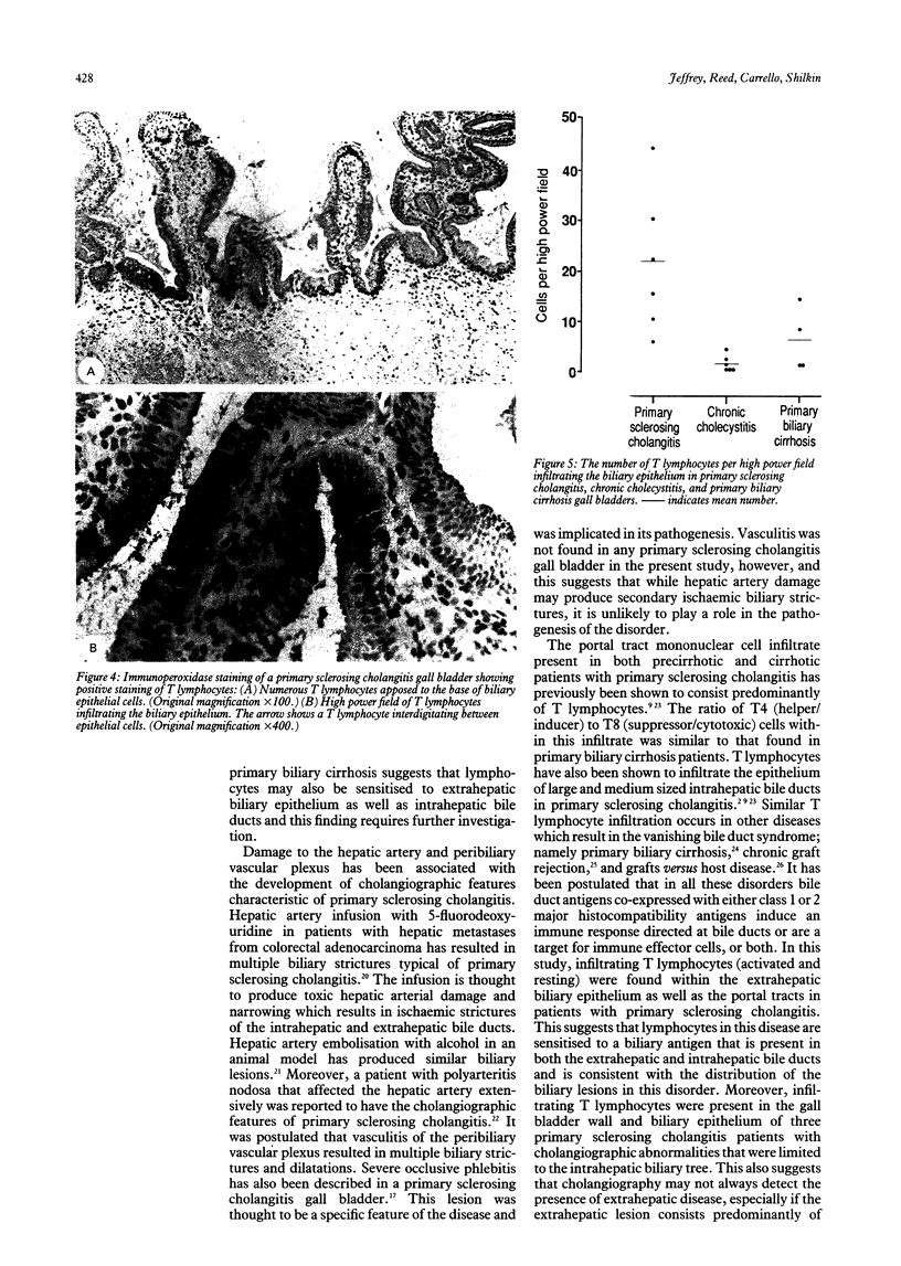
The histological features and type of mononuclear cell infiltrate in gall bladders from six patients with primary sclerosing cholangitis were studied using routine staining techniques and immunohistochemistry. Control studies were performed using the gall bladders from six patients (age and sex matched) with chronic cholecystitis and four with primary biliary cirrhosis. A range of histological abnormalities was present in gall bladders from patients with primary sclerosing cholangitis including a mild to moderate degree of epithelial hyperplasia, pseudogland formation, and mononuclear cell infiltrate of the epithelium; moderate to severe chronic inflammatory cell infiltrate and fibrosis affecting the superficial and deep layers of the gall bladder wall; and minimal smooth muscle hypertrophy. These abnormalities were non-specific and were also present in gall bladders from patients with chronic cholecystitis and primary biliary cirrhosis. Vasculitis and granulomas were not present in the patients with primary sclerosing cholangitis. Immunohistochemistry showed that the superficial and deep mononuclear cell infiltrate in primary sclerosing cholangitis gall bladders was composed predominantly of lymphocytes, in contrast to chronic cholecystitis where macrophages were found in similar or greater numbers. Moreover, T lymphocytes (activated and resting) were present throughout the lymphocytic infiltrate and were apposed to the base and interdigitated between the biliary epithelial cells in significantly greater numbers than in chronic cholecystitis gall bladders. B lymphocytes were present only in lymphoid follicles. Comparative studies using liver biopsy specimens from three of the primary sclerosing cholangitis patients showed a similar T lymphocyte portal tract infiltrate. We conclude that a number of non-specific chronic inflammatory histological abnormalities were present in primary sclerosing cholangitis gall bladders. Immunohistochemistry found other features that were present in this disease - a predominantly lymphocytic mononuclear cell infiltrate of the superficial and deep layers of the gall bladder wall and the presence of T lymphocytes that infiltrated the biliary epithelial cells. These findings support the hypothesis that aberrant cell mediated immune mechanisms may play a role in the pathogenesis of both the intrahepatic and extrahepatic lesions in primary sclerosing cholangitis.
Full text
PDF
Images in this article
Selected References
These references are in PubMed. This may not be the complete list of references from this article.
- Alberti-Flor J. J., de Medina M., Jeffers L., Schultz D. R., Schiff E. R. Elevated levels of immunoglobulins and immune complexes in the bile of patients with primary sclerosing cholangitis. Am J Gastroenterol. 1986 May;81(5):325–328. [PubMed] [Google Scholar]
- Bodenheimer H. C., Jr, LaRusso N. F., Thayer W. R., Jr, Charland C., Staples P. J., Ludwig J. Elevated circulating immune complexes in primary sclerosing cholangitis. Hepatology. 1983 Mar-Apr;3(2):150–154. doi: 10.1002/hep.1840030203. [DOI] [PubMed] [Google Scholar]
- Brandt D. J., MacCarty R. L., Charboneau J. W., LaRusso N. F., Wiesner R. H., Ludwig J. Gallbladder disease in patients with primary sclerosing cholangitis. AJR Am J Roentgenol. 1988 Mar;150(3):571–574. doi: 10.2214/ajr.150.3.571. [DOI] [PubMed] [Google Scholar]
- Brinch L., Teisberg P., Schrumpf E., Akesson I. The in vivo metabolism of C3 in hepatobiliary disease associated with ulcerative colitis. Scand J Gastroenterol. 1982 Jun;17(4):523–527. doi: 10.3109/00365528209182243. [DOI] [PubMed] [Google Scholar]
- Chapman R. W., Arborgh B. A., Rhodes J. M., Summerfield J. A., Dick R., Scheuer P. J., Sherlock S. Primary sclerosing cholangitis: a review of its clinical features, cholangiography, and hepatic histology. Gut. 1980 Oct;21(10):870–877. doi: 10.1136/gut.21.10.870. [DOI] [PMC free article] [PubMed] [Google Scholar]
- Chapman R. W., Kelly P. M., Heryet A., Jewell D. P., Fleming K. A. Expression of HLA-DR antigens on bile duct epithelium in primary sclerosing cholangitis. Gut. 1988 Apr;29(4):422–427. doi: 10.1136/gut.29.4.422. [DOI] [PMC free article] [PubMed] [Google Scholar]
- Doppman J. L., Dunnick N. R., Girton M., Fauci A. S., Popovsky M. A. Bile duct cysts secondary to liver infarcts: report of a case and experimental production by small vessel hepatic artery occlusion. Radiology. 1979 Jan;130(1):1–5. doi: 10.1148/130.1.1. [DOI] [PubMed] [Google Scholar]
- Doppman J. L., Girton M. E. Bile duct scarring following ethanol embolization of the hepatic artery: an experimental study in monkeys. Radiology. 1984 Sep;152(3):621–626. doi: 10.1148/radiology.152.3.6463243. [DOI] [PubMed] [Google Scholar]
- GOLDGRABER M. B., KIRSNER J. B. Chronic granulomatous cholecystitis and chronic fibrosing choledochitis associated with chronic ulcerative cofitis. A case report. Gastroenterology. 1960 May;38:821–828. [PubMed] [Google Scholar]
- Jeffrey G. P., Reed W. D., Laurence B. H., Shilkin K. B. Primary sclerosing cholangitis: clinical and immunopathological review of 21 cases. J Gastroenterol Hepatol. 1990 Mar-Apr;5(2):135–140. doi: 10.1111/j.1440-1746.1990.tb01818.x. [DOI] [PubMed] [Google Scholar]
- Kemeny M. M., Battifora H., Blayney D. W., Cecchi G., Goldberg D. A., Leong L. A., Margolin K. A., Terz J. J. Sclerosing cholangitis after continuous hepatic artery infusion of FUDR. Ann Surg. 1985 Aug;202(2):176–181. doi: 10.1097/00000658-198508000-00007. [DOI] [PMC free article] [PubMed] [Google Scholar]
- LaRusso N. F., Wiesner R. H., Ludwig J., MacCarty R. L. Current concepts. Primary sclerosing cholangitis. N Engl J Med. 1984 Apr 5;310(14):899–903. doi: 10.1056/NEJM198404053101407. [DOI] [PubMed] [Google Scholar]
- Ludwig J., MacCarty R. L., LaRusso N. F., Krom R. A., Wiesner R. H. Intrahepatic cholangiectases and large-duct obliteration in primary sclerosing cholangitis. Hepatology. 1986 Jul-Aug;6(4):560–568. doi: 10.1002/hep.1840060403. [DOI] [PubMed] [Google Scholar]
- Magrin S., Nouri-Aria K. T., Donaldson P. T., Wilkinson M. L., Portmann B. C., Williams R., Eddleston A. L. The relationship between HLA-DR3 and T-cell regulation of immunoglobulin production in primary sclerosing cholangitis. Clin Immunol Immunopathol. 1989 Feb;50(2):205–212. doi: 10.1016/0090-1229(89)90129-3. [DOI] [PubMed] [Google Scholar]
- McFarlane I. G., Wojcicka B. M., Tsantoulas D. C., Portmann B. C., Eddleston A. L., Williams R. Leukocyte migration inhibition in response to biliary antigens in primary biliary cirrhosis, sclerosing cholangitis, and other chronic liver diseases. Gastroenterology. 1979 Jun;76(6):1333–1340. [PubMed] [Google Scholar]
- Minuk G. Y., Angus M., Brickman C. M., Lawley T. J., Frank M. M., Hoofnagle J. H., Jones E. A. Abnormal clearance of immune complexes from the circulation of patients with primary sclerosing cholangitis. Gastroenterology. 1985 Jan;88(1 Pt 1):166–170. doi: 10.1016/s0016-5085(85)80149-9. [DOI] [PubMed] [Google Scholar]
- SCHWARTZ S. I., DALE W. A. Primary sclerosing cholangitis; review and report of six cases. AMA Arch Surg. 1958 Sep;77(3):439–451. [PubMed] [Google Scholar]
- Sherlock S., Fox R. A., James D. G., Scheuer P. J., Sharma O. Impaired delayed hypersensitivity in primary biliary cirrhosis. Lancet. 1969 May 10;1(7602):959–962. doi: 10.1016/s0140-6736(69)91860-1. [DOI] [PubMed] [Google Scholar]
- Sherlock S. The syndrome of disappearing intrahepatic bile ducts. Lancet. 1987 Aug 29;2(8557):493–496. doi: 10.1016/s0140-6736(87)91802-2. [DOI] [PubMed] [Google Scholar]
- Shulman H. M., Sharma P., Amos D., Fenster L. F., McDonald G. B. A coded histologic study of hepatic graft-versus-host disease after human bone marrow transplantation. Hepatology. 1988 May-Jun;8(3):463–470. doi: 10.1002/hep.1840080305. [DOI] [PubMed] [Google Scholar]
- Snook J. A., Chapman R. W., Sachdev G. K., Heryet A., Kelly P. M., Fleming K. A., Jewell D. P. Peripheral blood and portal tract lymphocyte populations in primary sclerosing cholangitis. J Hepatol. 1989 Jul;9(1):36–41. doi: 10.1016/0168-8278(89)90073-1. [DOI] [PubMed] [Google Scholar]
- Vierling J. M., Fennell R. H., Jr Histopathology of early and late human hepatic allograft rejection: evidence of progressive destruction of interlobular bile ducts. Hepatology. 1985 Nov-Dec;5(6):1076–1082. doi: 10.1002/hep.1840050603. [DOI] [PubMed] [Google Scholar]
- Whiteside T. L., Lasky S., Si L., Van Thiel D. H. Immunologic analysis of mononuclear cells in liver tissues and blood of patients with primary sclerosing cholangitis. Hepatology. 1985 May-Jun;5(3):468–474. doi: 10.1002/hep.1840050321. [DOI] [PubMed] [Google Scholar]


