Abstract
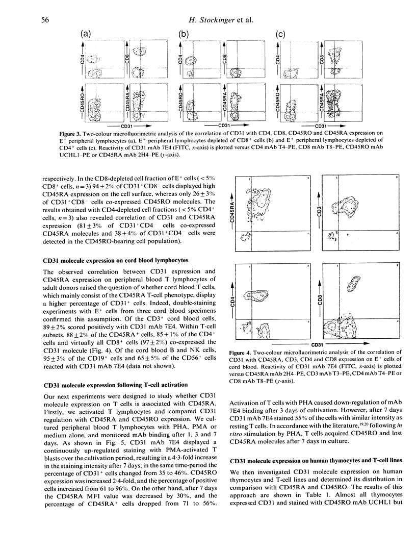
The CD31 molecule is a leucocyte-surface glycoprotein of 130 kDa with homology to the immunoglobulin gene superfamily. In this study we report on the expression of CD31 on human T cells and demonstrate that it subdivides peripheral blood T lymphocytes into two novel subsets. CD31 is expressed by 38% of CD3+ lymphocytes. About 85% of CD31+ T cells display the CD45RA phenotype, 35% the CD45RO phenotype, 24% the CD4 phenotype and 72% the CD8 phenotype. There is also a correlation between CD31 expression and CD45RA expression in cord blood T cells; 89% of CD3+ cord blood cells express CD31, and most of them have the CD45RA phenotype. A discrepancy was found with thymocytes, which are positive for CD31 but negative for CD45RA. Stimulation of human T cells leads to down-regulation of CD45RA, while CD31 continues to be expressed. In functional studies, CD31 antibody binding to T lymphocytes does not lead to mobilization of intracellular calcium, proliferation or modulation of T-cell proliferation.
Full text
PDF


Selected References
These references are in PubMed. This may not be the complete list of references from this article.
- Akbar A. N., Salmon M., Janossy G. The synergy between naive and memory T cells during activation. Immunol Today. 1991 Jun;12(6):184–188. doi: 10.1016/0167-5699(91)90050-4. [DOI] [PubMed] [Google Scholar]
- Akbar A. N., Terry L., Timms A., Beverley P. C., Janossy G. Loss of CD45R and gain of UCHL1 reactivity is a feature of primed T cells. J Immunol. 1988 Apr 1;140(7):2171–2178. [PubMed] [Google Scholar]
- Goyert S. M., Ferrero E. M., Seremetis S. V., Winchester R. J., Silver J., Mattison A. C. Biochemistry and expression of myelomonocytic antigens. J Immunol. 1986 Dec 15;137(12):3909–3914. [PubMed] [Google Scholar]
- Holter W., Majdic O., Stockinger H., Liszka K., Fischer G., Knapp W. Analysis of CD3-antibody-mediated inhibition of T-cell activation. Cell Immunol. 1986 Jun;100(1):140–148. doi: 10.1016/0008-8749(86)90014-6. [DOI] [PubMed] [Google Scholar]
- Muller W. A., Ratti C. M., McDonnell S. L., Cohn Z. A. A human endothelial cell-restricted, externally disposed plasmalemmal protein enriched in intercellular junctions. J Exp Med. 1989 Aug 1;170(2):399–414. doi: 10.1084/jem.170.2.399. [DOI] [PMC free article] [PubMed] [Google Scholar]
- Newman P. J., Berndt M. C., Gorski J., White G. C., 2nd, Lyman S., Paddock C., Muller W. A. PECAM-1 (CD31) cloning and relation to adhesion molecules of the immunoglobulin gene superfamily. Science. 1990 Mar 9;247(4947):1219–1222. doi: 10.1126/science.1690453. [DOI] [PubMed] [Google Scholar]
- Novel G., Novel M. Mutants d'Escherichia coli K 12 affectés pour leur croissance sur méthyl-beta-D-glucuronide: localisation of gène de structure de la beta-D-glucuronidase (uid A. Mol Gen Genet. 1973;120(4):319–335. [PubMed] [Google Scholar]
- Ohto H., Maeda H., Shibata Y., Chen R. F., Ozaki Y., Higashihara M., Takeuchi A., Tohyama H. A novel leukocyte differentiation antigen: two monoclonal antibodies TM2 and TM3 define a 120-kd molecule present on neutrophils, monocytes, platelets, and activated lymphoblasts. Blood. 1985 Oct;66(4):873–881. [PubMed] [Google Scholar]
- Rabinovitch P. S., June C. H., Grossmann A., Ledbetter J. A. Heterogeneity among T cells in intracellular free calcium responses after mitogen stimulation with PHA or anti-CD3. Simultaneous use of indo-1 and immunofluorescence with flow cytometry. J Immunol. 1986 Aug 1;137(3):952–961. [PubMed] [Google Scholar]
- Sanders M. E., Makgoba M. W., Shaw S. Human naive and memory T cells: reinterpretation of helper-inducer and suppressor-inducer subsets. Immunol Today. 1988 Jul-Aug;9(7-8):195–199. doi: 10.1016/0167-5699(88)91212-1. [DOI] [PubMed] [Google Scholar]
- Simmons D. L., Walker C., Power C., Pigott R. Molecular cloning of CD31, a putative intercellular adhesion molecule closely related to carcinoembryonic antigen. J Exp Med. 1990 Jun 1;171(6):2147–2152. doi: 10.1084/jem.171.6.2147. [DOI] [PMC free article] [PubMed] [Google Scholar]
- Stockinger H., Gadd S. J., Eher R., Majdic O., Schreiber W., Kasinrerk W., Strass B., Schnabl E., Knapp W. Molecular characterization and functional analysis of the leukocyte surface protein CD31. J Immunol. 1990 Dec 1;145(11):3889–3897. [PubMed] [Google Scholar]
- Takeuchi A., Shimizu A., Ohto H., Hashimoto T. Inhibition of neutrophil and monocyte functions by the monoclonal antibody TM2. Clin Immunol Immunopathol. 1988 Dec;49(3):439–449. doi: 10.1016/0090-1229(88)90131-6. [DOI] [PubMed] [Google Scholar]
- Wallace D. L., Beverley P. C. Phenotypic changes associated with activation of CD45RA+ and CD45RO+ T cells. Immunology. 1990 Mar;69(3):460–467. [PMC free article] [PubMed] [Google Scholar]
- Zocchi M. R., Poggi A., Mariani S., Gianazza E., Rugarli C. Identification of a new surface molecule expressed by human LGL and LAK cells production of a specific monoclonal antibody and comparison with other NK/LAK markers. Cell Immunol. 1989 Nov;124(1):144–157. doi: 10.1016/0008-8749(89)90118-4. [DOI] [PubMed] [Google Scholar]
- van Mourik J. A., Leeksma O. C., Reinders J. H., de Groot P. G., Zandbergen-Spaargaren J. Vascular endothelial cells synthesize a plasma membrane protein indistinguishable from the platelet membrane glycoprotein IIa. J Biol Chem. 1985 Sep 15;260(20):11300–11306. [PubMed] [Google Scholar]


