Abstract
Francisella tularensis is the intracellular pathogen that causes human tularemia. It is recognized as a potential agent of bioterrorism due to its low infectious dose and multiple routes of entry. We report the development of a Himar1-based random mutagenesis system for F. tularensis (HimarFT). In vivo mutagenesis of F. tularensis live vaccine strain (LVS) with HimarFT occurs at high efficiency. Approximately 12 to 15% of cells transformed with the delivery plasmid result in transposon insertion into the genome. Results from Southern blot analysis of 33 random isolates suggest that single insertions occurred, accompanied by the loss of the plasmid vehicle in most cases. Nucleotide sequence analysis of rescued genomic DNA with HimarFT indicates that the orientation of integration was unbiased and that insertions occurred in open reading frames and intergenic and repetitive regions of the chromosome. To determine the utility of the system, transposon mutagenesis was performed, followed by a screen for growth on Chamberlain's chemically defined medium (CDM) to isolate auxotrophic mutants. Several mutants were isolated that grew on complex but not on the CDM. We genetically complemented two of the mutants for growth on CDM with a newly constructed plasmid containing a nourseothricin resistance marker. In addition, uracil or aromatic amino acid supplementation of CDM supported growth of isolates with insertions in pyrD, carA, or aroE1 supporting the functional assignment of genes within each biosynthetic pathway. A mutant containing an insertion in aroE1 demonstrated delayed replication in macrophages and was restored to the parental growth phenotype when provided with the appropriate plasmid in trans. Our results suggest that a comprehensive library of mutants can be generated in F. tularensis LVS, providing an additional genetic tool to identify virulence determinants required for survival within the host.
Francisella tularensis is the etiologic agent of human tularemia. Four subspecies of F. tularensis have been recognized, including (i) the virulent type A F. tularensis subsp. tularensis, (ii) the less virulent type B F. tularensis subsp. holarctica, (iii) F. tularensis subsp. mediasiatica, and (iv) F. tularensis subsp. novicida. The F. tularensis LVS (live vaccine strain) is derived from F. tularensis subsp. holarctica. This strain demonstrates an attenuated phenotype in humans but remains virulent for mice, making it a potential model system to identify virulence factors (13, 23). Although Francisella replicates in several cell types including macrophages (4, 22, 25), hepatocytes (17), and amoebae (1), the virulence determinants that contribute to its intracellular lifestyle remain an active area of investigation. Approaches to identify and functionally characterize specific genes involved in intracellular maintenance and replication will contribute toward a general understanding of Francisella and host biology.
There has been steady progress toward the development and use of genetic methods to manipulate Francisella. Chemically induced or spontaneous mutants have been reported for both Francisella novicida (40) and F. tularensis LVS (10, 50). Tn10- or Tn1721-based transposon shuttle mutagenesis has been performed in F. tularensis subsp. novicida (5, 7-9, 18, 27, 41), but the observed instability of the transposons is problematic (35). Recently, transposon-transposase complexes were used to construct stable Tn5-derived insertion mutants in F. tularensis LVS in vivo (31). The functional aspects of candidate virulence genes have been tested using allelic replacement strategies (26, 35), and genetic strategies to complement attenuated mutants to wild-type have been developed (7, 36, 37, 42). The recent availability of genomic sequence information and chip arrays provides additional opportunities to identify genomic and transcript expression differences between subspecies and strains. As the mechanisms of pathogenesis remain poorly understood, particularly for the highly virulent type A strains of F. tularensis, random mutagenesis strategies may facilitate the discovery of new genes involved in maintaining an intracellular lifestyle. The identification of virulence factors should, in turn, facilitate the rational design of therapeutics or vaccines.
In previous work we constructed shuttle plasmids with expanded capabilities for use in Francisella (37). In this study, we report the utilization of a conditionally replicating derivative for delivery of a modified Himar1 (HimarFT) into F. tularensis LVS in vivo. Our results demonstrate that HimarFT-based mutagenesis of F. tularensis LVS results in random, single, stable insertions at high efficiency, providing a new genetic tool for the potential identification of coding regions important for pathogenesis.
MATERIALS AND METHODS
Bacterial strains, plasmids, and growth conditions.
All bacterial strains and plasmids used in this study are listed in Table 1. F. tularensis LVS was routinely grown at 37°C in modified Mueller-Hinton (MH) broth or on agar (Difco Laboratories) as previously described (37). In some experiments, cysteine heart agar (Difco) with 5% defibrinated horse blood (Becton Dickinson) was used. Screening for auxotrophic mutants was performed with Chamberlain's chemically defined medium (CDM) (15). Strains containing temperature-sensitive plasmid derivatives were grown at 30°C (permissive temperature) or 40°C (nonpermissive temperature). When required, medium was supplemented with kanamycin (10 μg ml−1) or nourseothricin (5 μg ml−1). Selection for nourseothricin resistance was performed at 30°C. Escherichia coli was grown aerobically in Luria-Bertani (LB) medium (Difco) at 37°C, supplemented with kanamycin (50 μg ml−1), ampicillin (100 μg ml−1), or nourseothricin (50 μg ml−1). Kanamycin and ampicillin were purchased from Sigma-Aldrich (St. Louis, Mo.) or United States Biochemical Corporation (Cleveland, Ohio). Nourseothricin was purchased from WERNER BioAgents (Jena, Germany).
TABLE 1.
Bacterial strains, plasmids, and primers used in this study
| Strain, plasmid, or primer | Descriptiona | Reference or source |
|---|---|---|
| Strains | ||
| F. tularensis | ||
| LVS | F. tularensis subsp. holartica live vaccine strain | K. L. Elkins |
| LVS carA::HimarFT | LVS containing a HimarFT insertion within carA | This study |
| LVS pyrD::HimarFT | LVS containing a HimarFT insertion within pyrD | This study |
| LVS aroE1::HimarFT | LVS containing a HimarFT insertion within aroE1 | This study |
| E. coli | ||
| DH5α | F−φ80lacZΔM15 endA1 recA1 hsdR17 supE44 thi-1 gyrA96 relA1 Δ(lacZYA-argF)U169 | Invitrogen |
| DH5αλpir | as DH5α, lysogenized with λpir phage; used as host for Himar1 rescue | A. Camilli |
| TOP10 | F−mcrA Δ(mrr-hsdRMS-mcrBC) φ80lacZΔM15 ΔlacX74 deoR recA1 araD139 Δ(ara-leu)7697 galU galK rpsL (Smr) endA1 nupG; used as host for plasmids derived from pCR2.1-TOPO | Invitrogen |
| Plasmids | ||
| pCR2.1-TOPO | 3.9-kb plasmid for cloning PCR products; Kmr Apr | Invitrogen |
| pAG36 | 6.1-kb plasmid used as the source of the nourseothricin resistance marker; Apr Ntr | Euroscarf |
| pMiniHimar | 4.0-kb plasmid containing the magellan4 transposon constructed with a neomycin phosphotransferase (npt) promoter upstream of npt and a mycobacterial-specific promoter upstream of the transposase (tnp); Kmr | 49 |
| pFNLTP1 | 6.9-kb plasmid obtained by spontaneous deletion of pTOPO/FNL10; Kmr Apr | 37 |
| pFNLTP8 | 6.9-kb pFNLTP1 derivative with EcoRI, NdeI, NotI, NheI, SmaI, and SalI restriction enzyme sites (MCS4) cloned between the KpnI and BamHI sites; Kmr Apr | 37 |
| pFNLTP9 | 6.9-kb temperature-sensitive derivative of pFNLTP1 with a mutation in amino acid 120 (M120I) of RepA; Kmr Apr | 37 |
| pFNLTP11 | 6.9-kb derivative of pFNLTP9 with NdeI, EcoRI, SmaI, NotI, NheI, and XhoI restriction enzymes sites (MCS2) cloned between the KpnI and BamHI sites; Kmr Apr | This study |
| pFNLTP16 | 5.7-kb derivative of pFNLTP11 lacking kanamycin resistance used for delivery of Himar1 into Francisella; Apr | This study |
| pFNLTP16 H1 | 9.7-kb derivative of pFNLTP16 combined with pMiniHimar using NotI; Kmr Apr | This study |
| pFNLTP16 H2 | 9.9-kb derivative of pFNLTP16 containing Himar1 modified with the F. tularensis LVS acpA promoter in the PvuI site upstream of tnp; Kmr Apr | This study |
| pFNLTP16 H3 | 10-kb derivative of pFNLTP16 containing HimarFT (Himar1 modified with the F. tularensis LVS groEL promoter in the BclI site upstream of npt); Kmr Apr | This study |
| pFNLTP16 H4 | 10.2-kb derivative of pFNLTP16 containing Himar1 modified with the F. tularensis LVS groEL and acpA promoters upstream of npt and tnp, respectively; Kmr Apr | This study |
| pFNLTP25 H3 | 11.3-kb derivative of pFNLTP16 H3 containing nourseothricin acetyltransferase at the SmaI site outside of the transposon; Kmr Apr Ntr | This study |
| pFNLTP23 | 7.0-kb derivative of pFNLTP8 lacking kanamycin resistance and containing nourseothricin acetyltransferase flanked with XbaI at NheI; Apr Ntr | This study |
| pFNLTP23 pyrDF | 1.8-kb fragment containing pyrDF and 344 bp upstream sequence cloned into pFNLTP23 at NotI for complementation of pyrD::HimarFT; Apr Ntr | This study |
| pFNLTP23 aroE1 | 1.2-kb fragment containing aroE1 and 319 bp upstream sequence cloned into pFNLTP23 at NotI for complementation of aroE1::HimarFT; Apr Ntr | This study |
| Primersb | ||
| A1 | 5′-ATTTCCGTGTCGCCCTTATTC-3′; forward primer for β-lactamase (bla) probe | |
| A2 | 5′-TTATCCGCCTCCATCCAGTC-3′; reverse primer for β-lactamase (bla) probe | |
| A3 | 5′-ATAAGAATGCGGCCGCTTGGTGTAGCATTCATTGCC-3′; forward primer for aroE1 | |
| A4 | 5′-ATAAGAATGCGGCCGCAACTAGCCTAGCAACTATGC-3′; reverse primer for aroE1 | |
| K1 | 5′-GCTATTCGGCTATGACTG-3′; forward primer for kanamycin marker (npt) probe | |
| K2 | 5′-CAGCAATATCACGGGTAG-3′; reverse primer for kanamycin marker (npt) probe | |
| K3 | 5′-GCTTCCTCGTGCTTTACGG-3′; npt primer for Himar1 insertion sequencing | |
| K4 | 5′-GAGGATCTCGTCGTGACC-3′; forward primer for npt removal from pFNLTP11 by inverse PCR | |
| K5 | 5′-TAACCAATAGGCCGAAATCG-3′; reverse primer for npt removal from pFNLTP11 by inverse PCR | |
| N1 | 5′-TCTAGACCAGCTGAAGCTTCGTACGC-3′; nourseothricin acetyltransferase forward primer | |
| N2 | 5′-TCTAGAGCATAGGCCACTAGTGGATCTGAT-3′; nourseothricin acetyltransferase reverse primer | |
| R1 | 5′-TGCCACCTAAATTGTAAGCG-3′; R6K primer for Himar1 insertion sequencing | |
| P1 | 5′-ATAAGAATGCGGCCGCGGTCTTAATCTTATTATATGCGG-3′; forward primer for pyrDF | |
| P2 | 5′-ATAAGAATGCGGCCGCGTATGTCGCTAAAACTTACGC-3′; reverse primer for pyrDF |
Kmr, Apr, and Ntr indicate resistance to kanamycin, ampicillin, and nourseothricin, respectively.
The primers for the promoters groEL and acpA are as described (37) except BglII and PvuI restriction enzyme sites were added in this study, respectively. Underlined regions are restriction enzyme sites for NotI (A3, A4, P1 and P2) or XbaI (N1 and N2).
DNA manipulation and transformation.
Purification and manipulation of plasmid or genomic DNA, electroporation of F. tularensis LVS, and chemical transformation of E. coli were performed as described previously (37). Custom oligonucleotide primers (Table 1) were synthesized by Operon (Huntsville, AL). DNA maps were constructed using MacPlasmap Pro (CGC Scientific, Inc., Ballwin, Mo.).
To determine insertion locations in Francisella, genomic DNA was isolated from each mutant strain, digested with SpeI, and treated with T4 DNA ligase. HimarFT-containing fragments were recovered as plasmid DNA in E. coli DH5αλpir (14). Analysis of genomic DNA adjacent to insertions was performed as previously described (37). The HimarFT insertion site sequences were compared against the F. tularensis LVS genome at http://bbrp.llnl.gov/bbrp/bin/f.tularensis_blast and the F. tularensis Schu S4 genome available at http://artedi.ebc.uu.se/Projects/Francisella/blast/.
Southern blot analysis.
Genomic DNA (0.5 μg) was digested with SpeI overnight, resolved on a 0.7% agarose gel, and transferred to a positively charged nylon membrane (Roche Diagnostics Corporation, Indianapolis, IN) using an LKB 2016 VacuGene Vacuum Blotting System (Pharmacia). Blots were probed with DNA fragments randomly labeled using a DIG High Prime DNA Labeling and Detection Starter Kit II (Roche); probes detected either the HimarFT transposon with a 634-bp fragment recognizing the neomycin phosphotransferase (npt) gene responsible for kanamycin resistance or the pFNLTP16 vehicle with a 624-bp fragment recognizing the β-lactamase (bla) gene responsible for ampicillin resistance. Gel processing, probe labeling, and detection were performed as recommended by the manufacturer.
Construction and transposition of Himar1 derivatives.
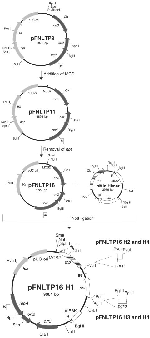
All Himar1 derivatives are listed in Table 1. The vehicle pFNLTP16 is a derivative of pFNLTP9 (37) in which npt was removed by inverse PCR and multiple cloning sites were added (Fig. 1). The pMiniHimar plasmid containing Himar1 was linearized and ligated to pFNLTP16 to generate pFNLTP16 H1 (Fig. 1). Derivatives of pFNLTP16 H1 were generated by cloning promoters for groEL (pgro) and/or acpA (pacp) (21, 48) upstream of npt and/or tnp, respectively (Fig. 1). A derivative of pFNLTP16 H3 containing a 1.3-kb fragment encoding nourseothricin resistance, including the translation elongation factor promoter and terminator (24), was amplified from pAG36 (Euroscarf) to construct pFNLTP25 H3.
FIG. 1.
Construction of pFNLTP16 derivatives containing modified and unmodified pMiniHimar to optimize transposase and kanamycin resistance. The pFNLTP16 delivery vehicle was derived from pFNLTP9 (37), a temperature-sensitive derivative of pFNLTP1. Unique restriction sites (multiple cloning site 2 [MCS2]; NdeI, EcoRI, SmaI, NotI, NheI, and XhoI between the KpnI and BamHI sites) were added to pFNLTP9 to generate pFNLTP11. Inverse PCR was performed on pFNLTP11 to generate pFNLTP16, deleting npt. The pUC ori and β-lactamase gene (bla) encoding ampicillin resistance are from pCR2.1-TOPO (light gray arrow or box) (Invitrogen), while repA, orf2, and orf3 are from pFNL10 (dark gray arrows) (46). The pFNLTP16 vehicle was combined with unmodified pMiniHimar using NotI to generate pFNLTP16 H1. The transposase (tnp; striped arrow) is provided in cis with (pFNLTP16 H2 and H4) or without (pFNLTP16 H1 and H3) the addition of the acpA promoter (pacp; striped small box) upstream at PvuI. Himar1, shown in white between the inverted repeats (IR), contains npt and the R6K origin. Expression of npt in pFNLTP16 H3 and H4 is under the control of the F. tularensis LVS groEL promoter (pgro).
Several transposition conditions were tested to determine the optimal growth medium and length of time for outgrowth. For optimal transposition, 100 ng of each transposon-containing plasmid was electroporated into newly prepared electrocompetent F. tularensis LVS. After outgrowth at 30°C on a shaker for 5 h, dilutions were plated on MH medium containing kanamycin and incubated at 40°C to select for Himar1 integration and loss of plasmid (one-step protocol) or incubated at 30°C to obtain replicating plasmid before secondary selection for plasmid loss at 40°C (two-step protocol). In addition, dilutions were plated onto MH medium with or without kanamycin to determine total CFU transformed or total potential recipients, respectively. Colonies grown for 3 or 4 days at 40°C were picked and struck onto MH medium containing kanamycin and grown at 37°C to recover individual clones containing Himar1 insertions in the genome.
Complementation of HimarFT auxotrophic mutants.
The transposon insertion was mapped in each auxotrophic mutant identified. Wild-type aroE1 or pyrDF and predicted promoter regions were amplified from F. tularensis LVS genomic DNA and cloned into pFNLTP23 (see Fig. 4), a derivative of pFNLTP8 (37) containing the gene encoding nourseothricin acetyltransferase. After electroporation into the respective F. tularensis LVS HimarFT mutant strains, the ability to restore growth on CDM was determined. To functionally complement carA, pyrD, or aroE1 mutant strains, uracil (50 μg ml−1) or a mixture of phenylalanine and tryptophan (100 μg ml−1 each) was added to CDM. The intracellular growth phenotype was determined by measuring bacterial replication over 3 days in the murine BALB/c macrophage cell line J774A.1 as described previously (37).
FIG. 4.
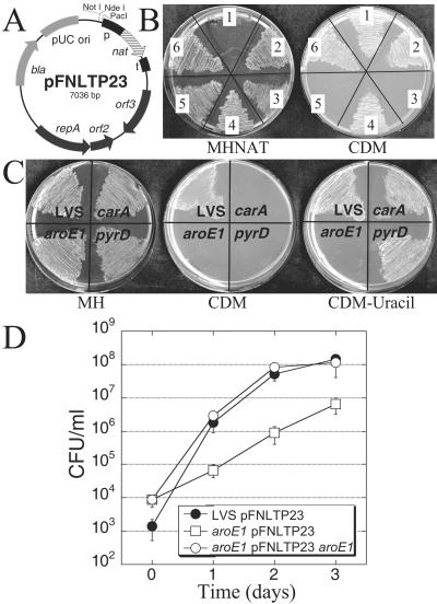
(A) Map of complementation plasmid pFNLTP23 containing nourseothricin acetyltransferase (nat; striped arrow) with the promoter (p; black box) and terminator (t; black box) for the translation elongation factor. The pUC ori and β-lactamase gene (bla) are from pCR2.1-TOPO (light gray arrow or box) (Invitrogen), while repA, orf2, and orf3 are from pFNL10 (dark gray arrows) (46). (B) Genetic complementation of two auxotrophic mutants. Growth is illustrated on medium containing nourseothricin (MHNAT) or on CDM. Culture designations 1 to 6 are LVS, LVS pFNLTP23, pyrD::HimarFT pFNLTP23, pyrD::HimarFT pFNLTP23 pyrDF, aroE1::HimarFT pFNLTP23, and aroE1::HimarFT pFNLTP23 aroE1, respectively. (C) Functional complementation of two auxotrophic mutants using CDM supplemented with uracil. Growth is illustrated on MH medium, CDM, and CDM containing uracil. In panels C and D the insertionally inactive gene name is used for space considerations (i.e., aroE1 is aroE1::HimarFT). (D) Growth of F. tularensis LVS pFNLTP23, aroE1 pFNLTP23, and aroE1 pFNLTP23 aroE1 in the murine BALB/c macrophage cell line J774A.1. Macrophage monolayers were infected at a multiplicity of infection of 1 for 2 h, washed, and incubated for a total of 3 days. Cells were lysed at different times, and bacteria were enumerated. Experiments were performed in triplicate (results are means ± standard deviations). The growth of aroE1::HimarFT pFNLTP23 on days 1 to 3 was significantly different from LVS pFNLTP23 to P < 0.03.
Nucleotide sequence accession numbers.
Sequence information for pFNLTP16 H3 and pFNLTP23 is available from the GenBank database under accession numbers DQ236098 and DQ266433, respectively.
RESULTS
Construction of a Himar1 transposon for use in Francisella.
Initial attempts to isolate Himar1 insertions in Francisella using pMiniHimar (Table 1), a plasmid unable to replicate in Francisella, were unsuccessful. This result could be due to failures in (i) transposon delivery, (ii) expression of the transposase, (iii) expression of the selection marker, or (iv) a combination of these factors. To address delivery of the transposon, Himar1 derivatives were cloned into the temperature-sensitive shuttle plasmid pFNLTP16 (Fig. 1), a derivative of pFNLTP9 in which npt had been removed. This plasmid can be efficiently electroporated into Francisella species and maintained at a permissive (30°C) but not at a nonpermissive (40°C) temperature (37). To ensure transcriptional initiation of the selectable marker and transposase, the Francisella promoter for groEL (pgro) (21) was cloned upstream of npt (pFNLTP16 H3 and H4), and/or the promoter for acpA (pacp) (48) was cloned upstream of tnp (pFNLTP16 H2 and H4) (Fig. 1). Previous experiments demonstrated that both pgro and pacp drive the expression of gfp cloned into pFNLTP6 in F. tularensis LVS (37).
Optimization of Himar1 transposition in F. tularensis LVS.
One-step and two-step transposition protocols were tested using various pFNLTP16 Himar1 derivatives, including those that had been modified by the addition of one or both Francisella promoters. All derivatives were electroporated into F. tularensis LVS to determine if Kmr colonies could be recovered at 40°C in a one-step procedure. Transposition was not detected with pFNLTP16, pMiniHimar, pFNLTP16 H1, or pFNLTP16 H2. However, Kmr isolates were recovered at high efficiency using the pFNLTP16 H3 or H4 derivatives (Table 2). The efficiency of plating at the nonpermissive temperature was approximately 12 to 15% of that observed at the permissive temperature (Table 2). Loss of plasmid DNA was investigated using a derivative of pFNLTP16 H3 that contains the nourseothricin resistance cassette as a selectable marker (pFNLTP25 H3) (Table 1). After electroporation and growth at the nonpermissive temperature, 100 kanamycin-resistant isolates were picked onto MH medium containing either nourseothricin or kanamycin. All isolates retained kanamycin resistance, but none was able to grow on medium with nourseothricin, consistent with Himar1 transposition and loss of the plasmid vehicle. Isolates from an electroporation plated at permissive temperature grew on medium containing kanamycin or nourseothricin, a result consistent with plasmid maintenance (data not shown).
TABLE 2.
Himar1 derivatives and frequency of transposition into F. tularensis LVSa
| Himar1 derivative | No. of transformantsb | No. of insertionsc | Avg no. of insertions/avg no. of transformants | Avg no. of insertions/avg total no. of cells |
|---|---|---|---|---|
| pFNLTP16 H1 | 0 | 0 | 0 | 0 |
| pFNLTP16 H2 | 0 | 0 | 0 | 0 |
| pFNLTP16 H3 | 8.73 × 10⁵ ± 5.03 × 10⁵ | 1.04 × 10⁵ ± 5.03 × 10⁴ | 0.12 | 1.82 × 10⁻⁵ |
| pFNLTP16 H4 | 7.35 × 10⁵ ± 6.14 × 10⁴ | 1.13 × 10⁵ ± 1.55 × 10⁴ | 0.15 | 1.97 × 10⁻⁵ |
Results shown are averages from four separate experiments. The average total number of cells recovered after electroporation and 5 h of outgrowth was 5.73 × 10⁹ ± 2.38 × 10⁹ CFU ml−1. Both plasmids pFNLTP16 and pMiniHimar resulted in no viable CFU ml−1 recovered as transformants or insertions.
Transformants are the average number of viable CFU ml−1 recovered after growth on selective medium at the permissive temperature.
Insertions are the average number of viable CFU ml−1 recovered after growth on selective medium at the nonpermissive temperature.
To determine if the frequency of transposition could be further improved, the procedure was repeated for all transposon derivatives using a two-step method. Strains were first selected for inheritance and maintenance of plasmid DNA at 30°C. Cultures were then shifted to the restrictive temperature (40°C) to inhibit subsequent plasmid replication. As in the one-step protocol, Kmr colonies were obtained only with the pFNLTP16 H3 and H4 derivatives after incubation at 40°C. Plasmid DNA, however, remained detectable in these isolates. The presence of plasmid DNA may be due to a residual plasmid-containing subpopulation that should be lost by subsequent replication cycles at 40°C. Since the retention of plasmid-encoded Kmr could be problematic in subsequent screening steps, we concluded that a one-step protocol using pFNLTP16 H3 was optimal for the delivery and transposition of HimarFT into F. tularensis LVS.
Verification of HimarFT transposition in F. tularensis LVS.
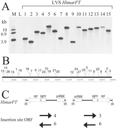
Southern blot analysis was performed on genomic DNA from 33 random Kmr colonies recovered after a one-step transposition protocol with pFNLTP16 H3. Hybridization of SpeI-digested genomic DNA with a probe specific to npt present on HimarFT resulted in single bands of various sizes, as shown for 15 representative isolates (Fig. 2A). No signal was obtained with a probe specific to the β-lactamase gene (bla) present on pFNLTP16 for 31 of these random isolates (data not shown). DNA from two isolates hybridized to the plasmid-specific probe, suggesting that plasmid DNA was incompletely resolved during subsequent replication.
FIG. 2.
Transposition of HimarFT into F. tularensis LVS. (A) Southern blot analysis. Genomic DNA from Kmr colonies selected at the nonpermissive temperature was prepared, digested with SpeI, separated on an agarose gel, and transferred to a nylon membrane. The membrane was hybridized to a probe specific to the kanamycin resistance marker present on the HimarFT transposon. Isolates 1 to 15 are representative of the 33 random isolates screened for HimarFT insertion (numbers 13 to 15 were recovered as auxotrophic strains). Wild-type F. tularensis LVS (lane L) was included as a negative control. Plasmid markers (lane M) included pFNLTP1 (6.9 kb), pFNLTP16 H1 (10 kb), and pCR2.1 TOPO (3.9 kb). (B) Map of the F. tularensis LVS genome with identification of the HimarFT insertion site locations. (C) Graphical representation of the orientation of HimarFT insertion. HimarFT was found in both orientations in the F. tularensis LVS genome with no bias to the orientation of the ORF (arrows) at the insertion site based on the F. tularensis Schu S4 genome annotation. The number of isolates containing insertions into ORFs in the indicated orientation is shown adjacent to each arrow.
Rescue of HimarFT and DNA sequence analysis of insertions.
HimarFT insertion sites were mapped by ligation of SpeI-digested genomic fragments and recovery of plasmid DNA in E. coli DH5αλpir. Nucleotide sequence analysis was performed with primers annealing to HimarFT and reading into flanking genomic DNA. When insertion locations were mapped to the F. tularensis LVS genome (Fig. 2B), a random distribution of HimarFT insertion was observed with no apparent regional bias. A variety of insertions occurred with no observed preference for the open reading frame (ORF) or transposon orientation (Fig. 2C). Of the 31 rescued genomic insertions, 19 possessed HimarFT insertions in a predicted ORF (Table 3). HimarFT inserted into genes involved in secretion, transport, energy production, metabolism, cell division, and protein turnover. Eight insertions mapped to intergenic regions, and two were located in repetitive regions of the F. tularensis LVS genome. The remaining two isolates likely represent aberrant transposition events. We detected one deletion and one duplication of flanking genomic sequence. The typical TA insertion site for Himar1 transposons was present in all but one isolate.
TABLE 3.
Compilation of HimarFT insertions within open reading framesa
| Location of insertion | Putative function |
|---|---|
| FTT1495c | Hypothetical membrane protein |
| FTT0194c | Conserved hypothetical membrane protein |
| FTT1738c | kdpB, K-transporting ATPase B chain |
| FTT1425c | naoX, NADH oxidase |
| FTT0432 | speA, putative arginine decarboxylase |
| FTT1591 | Lipoprotein |
| FTT0088 | pilT, type IV pilus nucleotide-binding protein |
| FTT1171c | hsdM, DNA methyltransferase subunit M |
| FTT0491c | gph, phosphoglycolate phosphatase |
| FTT0948c | Aldo/keto reductase |
| FTT1133 | pilB, type IV pilus binding protein |
| FTT0679c | GTP-binding protein |
| FTT0745c | Hypothetical protein |
| FTT1339c | Sulfate permease family protein |
| FTT1769c | clpB, ClpB protein |
| FTT0742 | Hypothetical lipoprotein |
| FTT1663 | carA, carbamoyl-P synthase small chain |
| FTT1647c | pyrD, dihydroorotate dehydrogenase |
| FTT0238 | aroE1, shikimate 5-dehydrogenase |
Location of HimarFT insertion and putative functions according to the F. tularensis subsp. tularensis (FTT) designation of the Schu S4 genome.
Stability of HimarFT insertions in F. tularensis LVS.
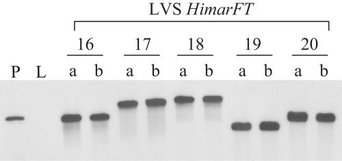
Five HimarFT mutants were serially passaged in MH broth without kanamycin for 5 days. After ∼40 generations, cultures passaged in the absence of selective pressure maintained the kanamycin marker. A Southern blot comparing the genomic DNA isolated from cultures grown under kanamycin selection and those isolated after 5 days without kanamycin confirmed that the insertion site was identical (Fig. 3). These results indicate that once transposition and plasmid loss has occurred, the kanamycin marker remains stably integrated within the genome.
FIG. 3.
Southern blot analysis of five HimarFT insertion mutants (isolates 16 to 20) and wild-type F. tularensis LVS (L) with a probe specific to the kanamycin resistance marker on HimarFT. Genomic DNA was prepared from the mutants after initial isolation (a) or after approximately 40 generations in the absence of kanamycin selection (b).
Phenotypic screen for auxotrophic mutants after HimarFT transposition.
To test the utility of HimarFT, a one-step mutagenesis protocol was performed, and Kmr colonies were screened for growth by replica plating onto Chamberlain's CDM. From approximately 6,500 clones, three isolates were obtained that failed to grow on CDM. Each strain possessed a single insertion of HimarFT, as shown by Southern blot analysis with the npt probe (Fig. 2A, lanes 13 to 15). A hybridization signal was undetectable with the bla probe (data not shown). HimarFT insertions resulting in auxotrophy mapped to carA (required for carbamoyl phosphate synthesis, an intermediate in arginine and pyrimidine synthesis), pyrD (required for pyrimidine synthesis), and aroE1 (required for the shikimate pathway involved in aromatic amino acid, ubiquinone/menaquinone, and folate synthesis, respectively) based on the annotated Schu S4 genome (Table 3).
Complementation of the auxotrophic HimarFT mutants.
To genetically complement HimarFT insertion strains, we constructed a plasmid expressing nourseothricin resistance as the selective marker (pFNLTP23). Nourseothricin is an aminoglycoside that inhibits a broad spectrum of organisms, including bacteria, protozoa, yeasts, viruses, and plants. Treatment with nourseothricin is postulated to inhibit protein synthesis by inducing miscoding events. It is an attractive marker for research because it is not used in human or veterinary medicine and has not been shown to display cross-resistance (24). The MIC of nourseothricin for F. tularensis LVS is approximately 2 to 5 μg ml−1 when it is incorporated in complex medium (data not shown). No cross-resistance to kanamycin was observed in strain LVS.
DNA fragments containing the coding sequences and putative promoter regions for the HimarFT-interrupted ORFs were amplified from the genome and cloned into pFNLTP23 for complementation (Fig. 4A). F. tularensis LVS containing pFNLTP23 grew on defined medium (Fig. 4B, sector 2), while the auxotrophic mutants containing pFNLTP23 were unable to grow (Fig. 4B, sectors 3 and 5). When pyrDF and aroE1 (including ∼300 bp upstream) were provided in trans, they restored the ability of pyrD::HimarFT and aroE1::HimarFT, respectively, to grow on CDM (Fig. 4B, sectors 4 and 6). We were unable to genetically test complementation of carAB::HimarFT as several attempts to clone this fragment in pFNLTP23 were unsuccessful. However, the addition of uracil to CDM functionally complemented both carAB and pyrD insertion strains (Fig. 4C), consistent with the predicted function of these genes in pyrimidine biosynthesis (12). Similarly, aroE1::HimarFT grew on CDM supplemented with phenylalanine and tryptophan (data not shown).
To determine if any of the auxotrophic mutants possessed intracellular replication defects, a growth analysis was performed in J774A.1 macrophages. Strains with insertions in pyrD or carA were indistinguishable from LVS when replication in macrophages was assessed (data not shown). In contrast, aroEI::HimarFT pFNLTP23 exhibited a distinct delay in replication compared to LVS pFNLTP23 (Fig. 4D). This delay is specific to intracellular growth since no growth difference was seen at 37°C in MH broth between LVS and aroEI::HimarFT with or without pFNLTP23 (data not shown). The replication defect of aroEI::HimarFT pFNLTP23 in J774A.1 macrophages was complemented with the cloned gene in trans (Fig. 4D).
DISCUSSION
Himar1 has been useful for in vitro (2, 3, 45) and in vivo (6, 49, 51, 54) transposon mutagenesis of a variety of bacteria. Moreover, since Himar1 does not require host-specific factors for transposition and displays a lack of site specificity, it seemed ideal for mutagenesis of the AT-rich Francisella genome (32-34). The modified Himar1 transposon developed in this study (HimarFT) allows efficient in vivo random mutagenesis of F. tularensis LVS. Analysis of 31 insertions from a single transposition reaction suggested that HimarFT could transpose randomly within the F. tularensis LVS genome with no known sequence specificity apart from the TA dinucleotide previously reported to be required for all mariner transposition events (19, 33). The frequency of insertion for HimarFT, either 15% of transformed cells or 2 × 10⁻⁵ of potential recipient organisms, is comparable to other reported in vivo random mutagenesis systems in Francisella (31) or in other bacteria (49, 51, 54). This frequency allows saturation mutagenesis to be conducted in Francisella from a single electroporation, an advantage over transposon-transposase complexes (31).
Implementing a transposon mutagenesis strategy in diverse organisms may require optimization of several parameters, including expression of the transposase and antibiotic resistance marker for selection. Alteration or substitution of promoter sequences has been used in prior studies to eliminate host restriction (49, 51, 54). Recent studies indicate that this could also be a limitation encountered in Francisella. Interestingly, detection of EZ::TN insertions in the Francisella genome appears to be limited to transcriptionally active regions, perhaps due to poor expression of the resistance marker in single copy. In HimarFT, the orientation of the groEL promoter relative to npt and tnp may result in sufficient expression of both genes, as the addition of the acpA promoter upstream of tnp does not affect the frequency of transposition. This configuration may actually limit tnp expression to a single event per plasmid.
Although single, stable genomic insertions were identified in most cases, spurious events were also detected using HimarFT as constructed. Isolates were obtained that possessed rearrangements adjacent to the insertion or parts of the delivery vehicle. This is not specific to Francisella or HimarFT but is common for transposon mutagenesis (6, 11, 16, 20, 38, 53). Additionally, two isolates appeared to maintain plasmid sequences, due possibly to reversion of the temperature-sensitive mutation within RepA. We anticipate that spurious events represent only a minority of the HimarFT-containing clones in a genomic library, some of which may not generate a phenotype during subsequent screening steps.
Auxotrophy is one phenotype that has been considered for the development of a suitable attenuated vaccine candidate. Both the shikimate and the purine biosynthetic pathways have been proposed as targets to generate vaccine strains in Francisella (30, 47). The attenuation of growth in vivo while maintaining the expression of protective antigens is important in considering immunization strategies (28, 43). Mutants of the aro pathway in Listeria monocytogenes are attenuated in virulence in epithelial cell culture and in mice (52), while Mycobacterium tuberculosis requires this pathway for viability (44). Disruption of carA or pyrD did not result in any phenotypic change for entry or replication within macrophages in F. tularensis LVS. In contrast, disruption of aroE1 results in delayed or reduced replication in F. tularensis LVS, a characteristic fully reversible when aroE1 is provided in trans. Further analysis of LVS aroE::HimarFT in mice will be necessary to better delineate the in vivo phenotype and its potential as a live vaccine.
Isolation of auxotrophs in other bacteria often results in 1 to 2% recovery (6, 29). We had expected to obtain more auxotrophs in our screen, consistent with the F. tularensis Schu S4 genomic analysis that identified approximately 350 enzymes postulated to participate in metabolism (34). Although ∼6,500 clones were analyzed on CDM, replica plating resulted in crowding on the assay medium, reducing the ability to detect loss of growth. We arrayed the library in a 96-well format and repeated the screen for growth on CDM. From 5,467 clones, eight isolates grew on complex medium and reproducibly failed to grow on CDM (0.15%) (data not shown). All insertions mapped within genes annotated as components of biosynthetic pathways. Additionally, different insertions in carB and aroE1 were identified in the second screen. A similar frequency of auxotrophy (0.5%) was reported in a screen of Xenorhabdus nematophila transposon mutants on a defined medium supplemented with all 20 l-amino acids (39). The complexity of the CDM and the exclusion of essential genes from our screen may influence the total number of auxotrophs that could be isolated by insertional mutagenesis.
The in vivo HimarFT mutagenesis system utilized in this study for Francisella expands the array of newly developed tools to analyze gene function in this intracellular pathogen. Continued development of random mutagenesis strategies should accelerate the discovery of determinants required for virulence and replication and perhaps guide future work toward a defined vaccine strain.
Acknowledgments
This work was supported by the Center for Biopreparedness and Infectious Disease at the Medical College of Wisconsin and the Great Lakes Regional Center of Excellence (GLRCE) for Biodefense and Emerging Infectious Disease Research (DP5, to T.C.Z. and D.W.F.). T.M.M. is supported by a GLRCE Career Development Fellowship (CDP9).
We thank Rachel H. Becker and Renee Penoske for their technical assistance and Eric J. Rubin for the pMiniHimar used in this study.
REFERENCES
- 1.Abd, H., T. Johansson, I. Golovliov, G. Sandström, and M. Forsman. 2003. Survival and growth of Francisella tularensis in Acanthamoeba castellanii. Appl. Environ. Microbiol. 69:600-606. [DOI] [PMC free article] [PubMed] [Google Scholar]
- 2.Akerley, B. J., E. J. Rubin, A. Camilli, D. J. Lampe, H. M. Robertson, and J. J. Mekalanos. 1998. Systematic identification of essential genes by in vitro mariner mutagenesis. Proc. Natl. Acad. Sci. USA 95:8927-8932. [DOI] [PMC free article] [PubMed] [Google Scholar]
- 3.Akerley, B. J., E. J. Rubin, V. N. Novik, K. Amaya, N. Judson, and J. J. Mekalanos. 2002. A genome-scale analysis for identification of genes required for growth or survival of Haemophilus influenzae. Proc. Natl. Acad. Sci. USA 99:966-971. [DOI] [PMC free article] [PubMed] [Google Scholar]
- 4.Anthony, L. D., R. D. Burke, and F. E. Nano. 1991. Growth of Francisella spp. in rodent macrophages. Infect. Immun. 59:3291-3296. [DOI] [PMC free article] [PubMed] [Google Scholar]
- 5.Anthony, L. S. D., M. Z. Gu, S. C. Cowley, W. W. S. Leung, and F. E. Nano. 1991. Transformation and allelic replacement in Francisella spp. J. Gen. Microbiol. 137:2697-2703. [DOI] [PubMed] [Google Scholar]
- 6.Ashour, J., and M. K. Hondalus. 2003. Phenotypic mutants of the intracellular actinomycete Rhodococcus equi created by in vivo Himar1 transposon mutagenesis. J. Bacteriol. 185:2644-2652. [DOI] [PMC free article] [PubMed] [Google Scholar]
- 7.Baron, G. S., and F. E. Nano. 1998. MglA and MglB are required for the intramacrophage growth of Francisella novicida. Mol. Microbiol. 29:247-259. [DOI] [PubMed] [Google Scholar]
- 8.Baron, G. S., T. J. Reilly, and F. E. Nano. 1999. The respiratory burst-inhibiting acid phosphatase AcpA is not essential for the intramacrophage growth or virulence of Francisella novicida. FEMS Microbiol. Lett. 176:85-90. [DOI] [PubMed] [Google Scholar]
- 9.Berg, J. M., K. E. Mdluli, and F. E. Nano. 1992. Molecular cloning of the recA gene and construction of a recA strain of Francisella novicida. Infect. Immun. 60:690-693. [DOI] [PMC free article] [PubMed] [Google Scholar]
- 10.Bhatnagar, N., E. Getachew, S. Straley, J. Williams, M. Meltzer, and A. Fortier. 1994. Reduced virulence of rifampicin-resistant mutants of Francisella tularensis. J. Infect. Dis. 170:841-847. [DOI] [PubMed] [Google Scholar]
- 11.Biery, M. C., F. J. Stewart, A. E. Stellwagen, E. A. Raleigh, and N. L. Craig. 2000. A simple in vitro Tn7-based transposition system with low target site selectivity for genome and gene analysis. Nucleic Acids Res. 28:1067-1077. [DOI] [PMC free article] [PubMed] [Google Scholar]
- 12.Bringel, F., and J.-C. Hubert. 2003. Extent of genetic lesions of the arginine and pyrimidine biosynthetic pathways in Lactobacillus plantarum, L. paraplantarum, L. pentosus, and L. casei: prevalence of CO2-dependent auxotrophs and characterization of deficient arg genes in L. plantarum. Appl. Environ. Microbiol. 69:2674-2683. [DOI] [PMC free article] [PubMed] [Google Scholar]
- 13.Burke, D. S. 1977. Immunization against tularemia: analysis of the effectiveness of live Francisella tularensis vaccine in prevention of laboratory-acquired tularemia. J. Infect. Dis. 135:55-60. [DOI] [PubMed] [Google Scholar]
- 14.Camilli, A., D. A. Portnoy, and P. Youngman. 1990. Insertional mutagenesis of Listeria monocytogenes with a novel Tn917 derivative that allows direct cloning of DNA flanking transposon insertions. J. Bacteriol. 172:3738-3744. [DOI] [PMC free article] [PubMed] [Google Scholar]
- 15.Chamberlain, R. E. 1965. Evaluation of live tularemia vaccine prepared in a chemically defined medium. Appl. Microbiol. 13:232-235. [DOI] [PMC free article] [PubMed] [Google Scholar]
- 16.Chow, W.-Y. W., J. B. Peterson, and A. G. Atherly. 1989. Unusual genetic phenomena associated with Tn5 mutagenesis in Alcaligenes eutrophus strain H1. Arch. Microbiol. 152:289-295. [Google Scholar]
- 17.Conlan, J. W., and R. J. North. 1992. Early pathogenesis of infection in the liver with the facultative intracellular bacteria Listeria monocytogenes, Francisella tularensis, and Salmonella typhimurium involves lysis of infected hepatocytes by leukocytes. Infect. Immun. 60:5164-5171. [DOI] [PMC free article] [PubMed] [Google Scholar]
- 18.Cowley, S. C., C. J. Gray, and F. E. Nano. 2000. Isolation and characterization of Francisella novicida mutants defective in lipopolysaccharide biosynthesis. FEMS Microbiol. Lett. 182:63-67. [DOI] [PubMed] [Google Scholar]
- 19.Craig, N. L. 1997. Target site selection in transposition. Annu. Rev. Biochem. 66:437-474. [DOI] [PubMed] [Google Scholar]
- 20.Engel, P., and A. R. Lax. 1997. Partial deletion of transposon Tn4560 integrated into the genome of Streptomyces tendae. Lett. Appl. Microbiol. 25:225-228. [DOI] [PubMed] [Google Scholar]
- 21.Ericsson, M., I. Golovliov, G. Sandström, A. Tärnvik, and A. Sjöstedt. 1997. Characterization of the nucleotide sequence of the groE operon encoding heat shock proteins chaperone-60 and -10 of Francisella tularensis and determination of the T-cell response to the proteins in individuals vaccinated with F. tularensis. Infect. Immun. 65:1824-1829. [DOI] [PMC free article] [PubMed] [Google Scholar]
- 22.Fortier, A. H., S. J. Green, T. Polsinelli, T. R. Jones, R. M. Crawford, D. A. Leiby, K. L. Elkins, M. S. Meltzer, and C. A. Nacy. 1994. Life and death of an intracellular pathogen: Francisella tularensis and the macrophage. Immunol. Ser. 60:349-361. [PubMed] [Google Scholar]
- 23.Fortier, A. H., M. V. Slayter, R. Ziemba, M. S. Meltzer, and C. A. Nacy. 1991. Live vaccine strain of Francisella tularensis: infection and immunity in mice. Infect. Immun. 59:2922-2928. [DOI] [PMC free article] [PubMed] [Google Scholar]
- 24.Goldstein, A. L., and J. H. McCusker. 1999. Three new dominant drug resistance cassettes for gene disruption in Saccharomyces cerevisiae. Yeast 15:1541-1553. [DOI] [PubMed] [Google Scholar]
- 25.Golovliov, I., V. Baranov, Z. Krocova, H. Kovarova, and A. Sjöstedt. 2003. An attenuated strain of facultative intracellular bacterium Francisella tularensis can escape the phagosome of monocytic cells. Infect. Immun. 71:5940-5950. [DOI] [PMC free article] [PubMed] [Google Scholar]
- 26.Golovliov, I., A. Sjöstedt, A. N. Mokrievich, and V. M. Pavlov. 2003. A method for allelic replacement in Francisella tularensis. FEMS Microbiol. Lett. 222:273-280. [DOI] [PubMed] [Google Scholar]
- 27.Gray, C. G., S. C. Cowley, K. K. Cheung, and F. E. Nano. 2002. The identification of five genetic loci of Francisella novicida associated with intracellular growth. FEMS Microbiol. Lett. 215:53-56. [DOI] [PubMed] [Google Scholar]
- 28.Isherwood, K. E., R. W. Titball, D. H. Davies, P. L. Felgner, and J. W. Morrow. 2005. Vaccination strategies for Francisella tularensis. Adv. Drug Deliv. Rev. 57:1403-1414. [DOI] [PubMed] [Google Scholar]
- 29.Jacobs, M. A., A. Alwood, I. Thaipisuttikul, D. Spencer, E. Haugen, S. Ernst, O. Will, R. Kaul, C. Raymond, R. Levy, L. Chun-Rong, D. Guenthner, D. Bovee, M. V. Olson, and C. Manoil. 2003. Comprehensive transposon mutant library of Pseudomonas aeruginosa. Proc. Natl. Acad. Sci. USA 100:14339-14344. [DOI] [PMC free article] [PubMed] [Google Scholar]
- 30.Karlsson, J., R. G. Prior, K. Williams, L. Lindler, K. A. Brown, N. Chatwell, K. Hjalmarsson, N. Loman, K. A. Mack, M. Pallen, M. Popek, G. Sandström, A. Sjöstedt, T. Svensson, I. Tamas, S. G. Andersson, B. W. Wren, P. C. Oyston, and R. W. Titball. 2000. Sequencing of the Francisella tularensis strain Schu 4 genome reveals the shikimate and purine metabolic pathways, targets for the construction of a rationally attenuated auxotrophic vaccine. Microb. Comp. Genomics 5:25-39. [DOI] [PubMed] [Google Scholar]
- 31.Kawula, T. H., J. D. Hall, J. R. Fuller, and R. R. Craven. 2004. Use of transposon-transposase complexes to create stable insertion mutant strains of Francisella tularensis LVS. Appl. Environ. Microbiol. 70:6901-6904. [DOI] [PMC free article] [PubMed] [Google Scholar]
- 32.Lampe, D. J., M. E. A. Churchill, and H. M. Robertson. 1996. A purified mariner transposase is sufficient to mediate transposition in vitro. EMBO J. 15:5470-5479. [PMC free article] [PubMed] [Google Scholar]
- 33.Lampe, D. J., T. E. Grant, and H. M. Robertson. 1998. Factors affecting transposition of the Himar1 mariner transposon in vitro. Genetics 149:179-187. [DOI] [PMC free article] [PubMed] [Google Scholar]
- 34.Larsson, P., P. C. F. Oyston, P. Chain, M. C. Chu, M. Duffield, H.-H. Fuxelius, E. Garcia, G. Halltorp, D. Johansson, K. E. Isherwood, P. D. Karp, E. Larsson, Y. Liu, S. Michell, J. Prior, R. Prior, S. Malfatti, A. Sjostedt, K. Svensson, N. Thompson, L. Vergez, J. K. Wagg, B. W. Wren, L. Lindler, S. G. E. Andersson, M. Forsman, and R. W. Titball. 2005. The complete genome sequence of Francisella tularensis, the causative agent of tularemia. Nat. Genet. 37:153-159. [DOI] [PubMed] [Google Scholar]
- 35.Lauriano, C. M., J. R. Barker, F. E. Nano, B. P. Arulanandam, and K. E. Klose. 2003. Allelic exchange in Francisella tularensis using PCR products. FEMS Microbiol. Lett. 229:195-202. [DOI] [PubMed] [Google Scholar]
- 36.Lindgren, H., I. Golovliov, V. Baranov, R. K. Ernst, M. Telepnev, and A. Sjöstedt. 2004. Factors affecting the escape of Francisella tularensis from the phagolysosome. J. Med. Microbiol. 53:953-958. [DOI] [PubMed] [Google Scholar]
- 37.Maier, T. M., A. Havig, M. Casey, F. E. Nano, D. W. Frank, and T. C. Zahrt. 2004. Construction and characterization of a highly efficient Francisella shuttle plasmid. Appl. Environ. Microbiol. 70:7511-7519. [DOI] [PMC free article] [PubMed] [Google Scholar]
- 38.Maier, T. M., J. M. Myers, and C. R. Myers. 2003. Identification of the gene encoding the sole physiological fumarate reductase in Shewanella oneidensis MR-1. J. Basic Microbiol. 43:312-327. [DOI] [PubMed] [Google Scholar]
- 39.Martens, E. C., F. M. Russell, and H. Goodrich-Blair. 2005. Analysis of Xenorhabdus nematophila metabolic mutants yields insight into stages of Steinernema carpocapsae nematode intestinal colonization. Mol. Microbiol. 58:28-45. [DOI] [PubMed] [Google Scholar]
- 40.McDonald, M. K., S. C. Cowley, and F. E. Nano. 1997. Temperature-sensitive lesions in the Francisella novicida valA gene cloned into an Escherichia coli msbA lpxK mutant affecting deoxycholate resistance and lipopolysaccharide assembly at the restrictive temperature. J. Bacteriol. 179:7638-7643. [DOI] [PMC free article] [PubMed] [Google Scholar]
- 41.Mdluli, K. E., L. S. Anthony, G. S. Baron, M. K. McDonald, S. V. Myltseva, and F. E. Nano. 1994. Serum-sensitive mutation of Francisella novicida: association with an ABC transporter gene. Microbiology 140:3309-3318. [DOI] [PubMed] [Google Scholar]
- 42.Nano, F. E., N. Zhang, S. C. Cowley, K. E. Klose, K. K. M. Cheung, M. J. Roberts, J. S. Ludu, G. W. Letendre, A. I. Meierovics, G. Stephens, and K. L. Elkins. 2004. A Francisella tularensis pathogenicity island required for intramacrophage growth. J. Bacteriol. 186:6430-6436. [DOI] [PMC free article] [PubMed] [Google Scholar]
- 43.Oyston, P. C. F., and J. E. Quarry. 2005. Tularemia vaccine: past, present and future. Antonie Leeuwenhoek 87:277-281. [DOI] [PubMed] [Google Scholar]
- 44.Parish, T., and N. G. Stoker. 2002. The common aromatic amino acid biosynthesis pathway is essential in Mycobacterium tuberculosis. Microbiology 148:3069-3077. [DOI] [PubMed] [Google Scholar]
- 45.Pelicic, V., S. Morelle, D. Lampe, and X. Nassif. 2000. Mutagenesis of Neisseria meningitidis by in vitro transposon of Himar1 mariner. J. Bacteriol. 182: 5391-5398. [DOI] [PMC free article] [PubMed] [Google Scholar]
- 46.Pomerantsev, A. P., I. R. Golovliov, Y. Ohara, A. N. Mokrievich, M. Obuchi, A. Norqvist, K. Kuoppa, and V. M. Pavlov. 2001. Genetic organization of the Francisella plasmid pFNL10. Plasmid 46:210-222. [DOI] [PubMed] [Google Scholar]
- 47.Prior, R. G., L. Klasson, P. Larsson, K. Williams, L. Lindler, A. Sjöstedt, T. Svensson, I. Tamas, B. W. Wren, P. C. Oyston, S. G. Andersson, and R. W. Titball. 2001. Preliminary analysis and annotation of the partial genome sequence of Francisella tularensis strain Schu 4. J. Appl. Microbiol. 91:614-620. [DOI] [PubMed] [Google Scholar]
- 48.Reilly, T. J., G. S. Baron, F. E. Nano, and M. S. Kuhlenschmidt. 1996. Characterization and sequencing of a respiratory burst-inhibiting acid phosphatase from Francisella tularensis. J. Biol. Chem. 271:10973-10983. [DOI] [PubMed] [Google Scholar]
- 49.Rubin, E. J., B. J. Akerley, V. N. Novik, D. J. Lampe, R. N. Husson, and J. J. Mekalanos. 1999. In vivo transposition of mariner-based elements in enteric bacteria and mycobacteria. Proc. Natl. Acad. Sci. USA 96:1645-1650. [DOI] [PMC free article] [PubMed] [Google Scholar]
- 50.Sandström, G., S. Löfgren, and A. Tärnvik. 1988. A capsule-deficient mutant of Francisella tularensis LVS exhibits enhanced sensitivity to killing by serum but diminished sensitivity to killing by polymorphonuclear leukocytes. Infect. Immun. 56:1194-1202. [DOI] [PMC free article] [PubMed] [Google Scholar]
- 51.Stewart, P. E., J. Hoff, E. Fischer, J. G. Krum, and P. A. Rosa. 2004. Genome-wide transposon mutagenesis of Borrelia burgdorferi for identification of phenotypic mutants. Appl. Environ. Microbiol. 70:5973-5979. [DOI] [PMC free article] [PubMed] [Google Scholar]
- 52.Stritzker, J., J. Janda, C. Schoen, M. Taupp, S. Pilgrim, I. Gentschev, P. Schreier, G. Geginat, and W. Goebel. 2004. Growth, virulence, and immunogenicity of Listeria monocytogenes aro mutants. Infect. Immun. 72:5622- 5629. [DOI] [PMC free article] [PubMed] [Google Scholar]
- 53.Vandeyar, M. A., and S. A. Zahler. 1986. Chromosomal insertions of Tn917 in Bacillus subtilis. J. Bacteriol. 167:530-534. [DOI] [PMC free article] [PubMed] [Google Scholar]
- 54.Zhang, J. K., M. A. Pritchett, D. J. Lampe, H. M. Robertson, and W. W. Metcalf. 2000. In vivo transposon mutagenesis of the methanogenic archaeon Methanosarcina acetivorans C2A using a modified version of the insect mariner-family transposable element Himar1. Proc. Natl. Acad. Sci. USA 97:9665-9670. [DOI] [PMC free article] [PubMed] [Google Scholar]


