Abstract
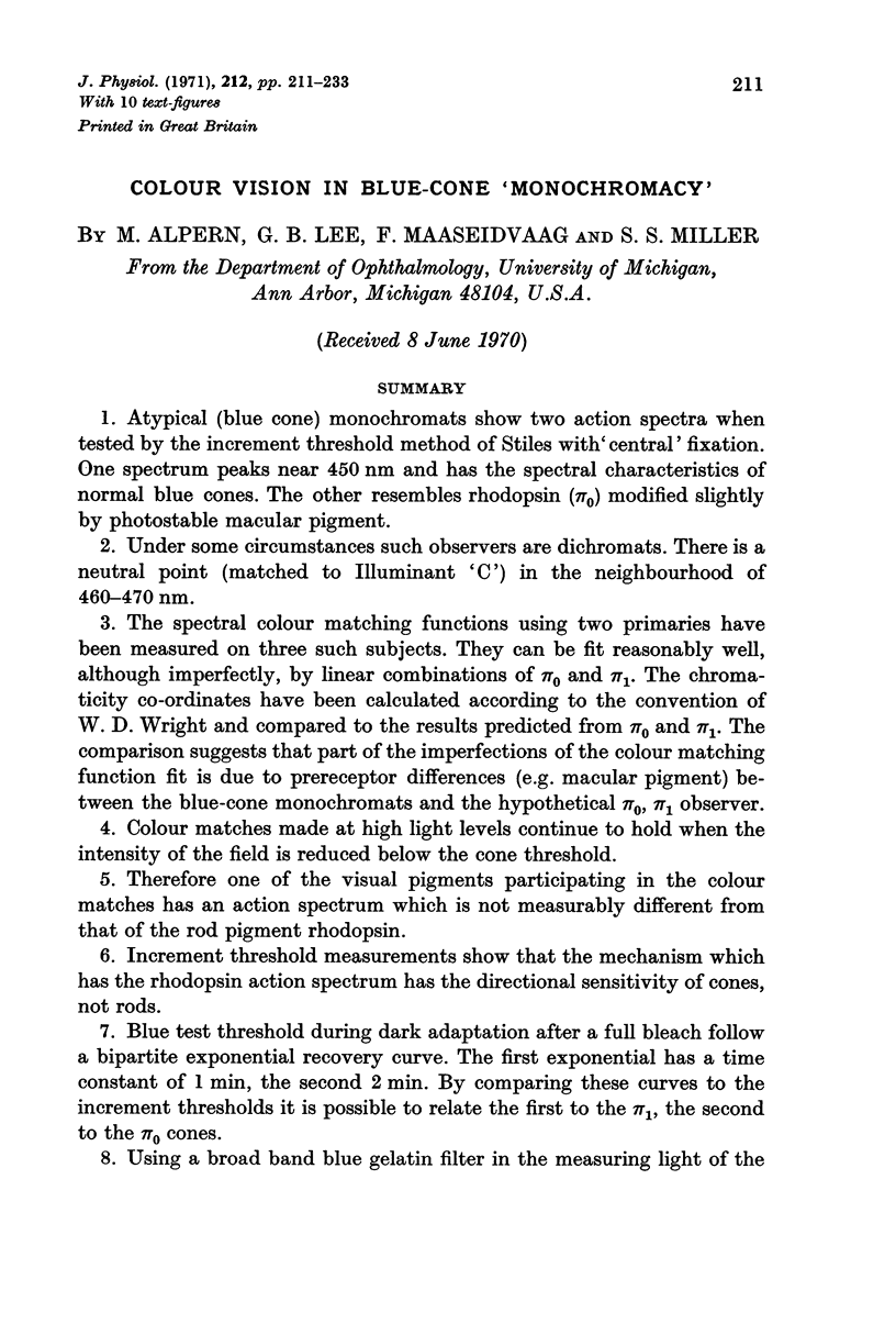
1. Atypical (blue cone) monochromats show two action spectra when tested by the increment threshold method of Stiles with `central' fixation. One spectrum peaks near 450 nm and has the spectral characteristics of normal blue cones. The other resembles rhodopsin (π0) modified slightly by photostable macular pigment.
2. Under some circumstances such observers are dichromats. There is a neutral point (matched to Illuminant `C') in the neighbourhood of 460-470 nm.
3. The spectral colour matching functions using two primaries have been measured on three such subjects. They can be fit reasonably well, although imperfectly, by linear combinations of π0 and π1. The chromaticity co-ordinates have been calculated according to the convention of W. D. Wright and compared to the results predicted from π0 and π1. The comparison suggests that part of the imperfections of the colour matching function fit is due to prereceptor differences (e.g. macular pigment) between the blue-cone monochromats and the hypothetical π0, π1 observer.
4. Colour matches made at high light levels continue to hold when the intensity of the field is reduced below the cone threshold.
5. Therefore one of the visual pigments participating in the colour matches has an action spectrum which is not measurably different from that of the rod pigment rhodopsin.
6. Increment threshold measurements show that the mechanism which has the rhodopsin action spectrum has the directional sensitivity of cones, not rods.
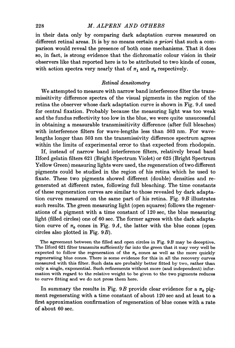
7. Blue test threshold during dark adaptation after a full bleach follow a bipartite exponential recovery curve. The first exponential has a time constant of 1 min, the second 2 min. By comparing these curves to the increment thresholds it is possible to relate the first to the π1, the second to the π0 cones.
8. Using a broad band blue gelatin filter in the measuring light of the retinal densitometer and studying the same retinal region tested in 7 it is possible to follow the regeneration of a pigment after a full bleach which has an exponential recovery with a time constant of 1·0 min. With a yellow green filter in the measuring light the exponential recovery observed after a full bleach has a time constant of 2·0 min.
9. One of the two visual pigments participating in the colour matches resides in receptors which have the action spectrum, the directional sensitivity and probably the dark adaptation curve of normal blue cones.
10. The other resides in receptors which have the action spectrum of normal rods but the directional sensitivity and the dark adaptation curve of normal red and green cones.
Full text
PDF


Selected References
These references are in PubMed. This may not be the complete list of references from this article.
- ALPERN M., FALLS H. F., LEE G. B. The enigma of typical total monochromacy. Am J Ophthalmol. 1960 Nov;50:996–1012. doi: 10.1016/0002-9394(60)90353-6. [DOI] [PubMed] [Google Scholar]
- ALPERN M., LEE G. B., SPIVEY B. E. PI-1 CONE MONOCHROMATISM. Arch Ophthalmol. 1965 Sep;74:334–337. doi: 10.1001/archopht.1965.00970040336008. [DOI] [PubMed] [Google Scholar]
- Alpern M., Torii S. Prereceptor colour vision distortions in protanomalous trichromacy. J Physiol. 1968 Oct;198(3):549–560. doi: 10.1113/jphysiol.1968.sp008625. [DOI] [PMC free article] [PubMed] [Google Scholar]
- Alpern M., Torii S. The luminosity curve of the protanomalous fovea. J Gen Physiol. 1968 Nov;52(5):717–737. doi: 10.1085/jgp.52.5.717. [DOI] [PMC free article] [PubMed] [Google Scholar]
- Blaurock A. E., Wilkins M. H. Structure of frog photoreceptor membranes. Nature. 1969 Aug 30;223(5209):906–909. doi: 10.1038/223906a0. [DOI] [PubMed] [Google Scholar]
- Cohen A. I. New evidence supporting the linkage to extracellular space of outer segment saccules of frog cones but not rods. J Cell Biol. 1968 May;37(2):424–444. doi: 10.1083/jcb.37.2.424. [DOI] [PMC free article] [PubMed] [Google Scholar]
- DOWLING J. E. Chemistry of visual adaptation in the rat. Nature. 1960 Oct 8;188:114–118. doi: 10.1038/188114a0. [DOI] [PubMed] [Google Scholar]
- Daw N. W., Pearlman A. L. Cat colour vision: one cone process or several? J Physiol. 1969 May;201(3):745–764. doi: 10.1113/jphysiol.1969.sp008785. [DOI] [PMC free article] [PubMed] [Google Scholar]
- Du Croz J. J., Rushton W. A. The separation of cone mechanisms in dark adaptation. J Physiol. 1966 Mar;183(2):481–496. doi: 10.1113/jphysiol.1966.sp007878. [DOI] [PMC free article] [PubMed] [Google Scholar]
- Falls H. F., Wolter J. R., Alpern M. Typical total monochromacy. A histological and psychophysical study. Arch Ophthalmol. 1965 Nov;74(5):610–616. doi: 10.1001/archopht.1965.00970040612005. [DOI] [PubMed] [Google Scholar]
- Flamant F., Stiles W. S. The directional and spectral sensitivities of the retinal rods to adapting fields of different wave-lengths. J Physiol. 1948 Mar 15;107(2):187–202. doi: 10.1113/jphysiol.1948.sp004262. [DOI] [PMC free article] [PubMed] [Google Scholar]
- Gras W. J., Worthington C. R. X-ray analysis of retinal photoreceptors. Proc Natl Acad Sci U S A. 1969 Jun;63(2):233–238. doi: 10.1073/pnas.63.2.233. [DOI] [PMC free article] [PubMed] [Google Scholar]
- Liebman P. A., Entine G. Visual pigments of frog and tadpole (Rana pipiens). Vision Res. 1968 Jul;8(7):761–775. doi: 10.1016/0042-6989(68)90128-4. [DOI] [PubMed] [Google Scholar]
- Pearlman A. L., Daw N. W. Opponent color cells in the cat lateral geniculate nucleus. Science. 1970 Jan 2;167(3914):84–86. doi: 10.1126/science.167.3914.84. [DOI] [PubMed] [Google Scholar]
- RUSHTON W. A. CONE PIGMENT KINETICS IN THE DEUTERANOPE. J Physiol. 1965 Jan;176:38–45. doi: 10.1113/jphysiol.1965.sp007533. [DOI] [PMC free article] [PubMed] [Google Scholar]
- RUSHTON W. A. CONE PIGMENT KINETICS IN THE PROTANOPE. J Physiol. 1963 Sep;168:374–388. doi: 10.1113/jphysiol.1963.sp007198. [DOI] [PMC free article] [PubMed] [Google Scholar]
- Rushton W. A. Rod/cone rivalry in pigment regeneration. J Physiol. 1968 Sep;198(1):219–236. doi: 10.1113/jphysiol.1968.sp008603. [DOI] [PMC free article] [PubMed] [Google Scholar]
- STILES W. S. Increment thresholds and the mechanisms of colour vision. Doc Ophthalmol. 1949;3:138–165. doi: 10.1007/BF00162601. [DOI] [PubMed] [Google Scholar]
- WALD G., BROWN P. K., KENNEDY D. The visual system of the alligator. J Gen Physiol. 1957 May 20;40(5):703–713. doi: 10.1085/jgp.40.5.703. [DOI] [PMC free article] [PubMed] [Google Scholar]
- WALD G., BROWN P. K., SMITH P. H. Iodopsin. J Gen Physiol. 1955 May 20;38(5):623–681. doi: 10.1085/jgp.38.5.623. [DOI] [PMC free article] [PubMed] [Google Scholar]
- WALLS G. L., HEATH G. G. Typical total color blindness reinterpreted. Acta Ophthalmol (Copenh) 1954;32(3):253–contd. [PubMed] [Google Scholar]
- Watkins R. D. Foveal increment thresholds in normal and deutan observers. Vision Res. 1969 Oct;9(10):1185–1196. doi: 10.1016/0042-6989(69)90108-4. [DOI] [PubMed] [Google Scholar]


