Abstract
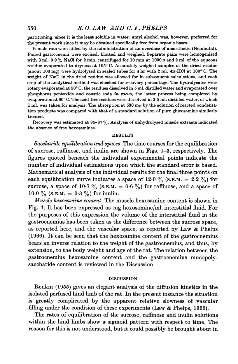
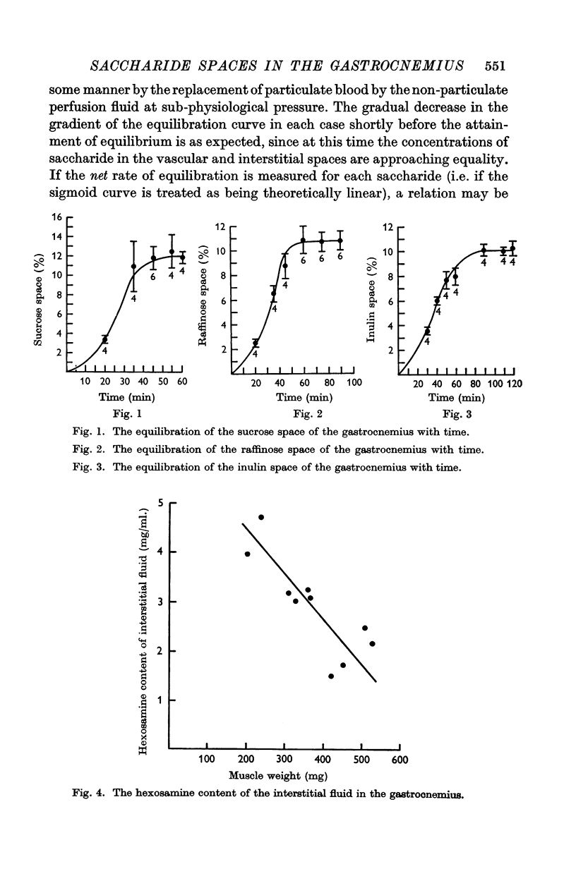
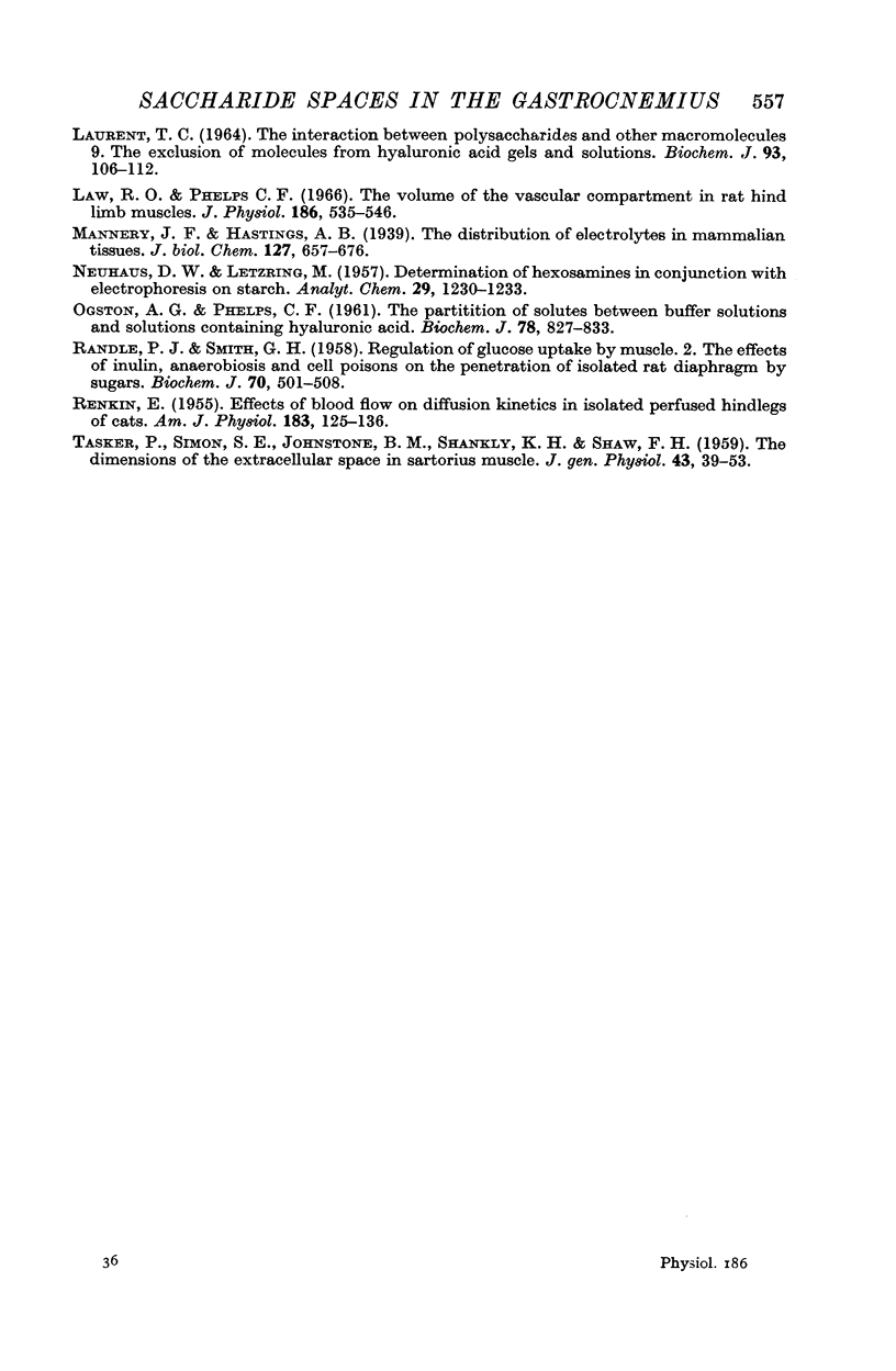
1. The sucrose, raffinose and inulin spaces within the gastrocnemius of the female rat have been estimated at 12·0 ± 2·2%, 10·7 ± 0·6%, and 10·0 ± 0·3% respectively, expressed as a percentage of the total muscle volume.
2. The mucopolysaccharide and hexosamine contents of the gastrocnemius have been found to be inversely dependent upon the weight of the muscle.
3. The possibility that a relation exists between the saccharide spaces and the muscle musopolysaccharide content has been discussed in terms of saccharide molecular radii.
Full text
PDF


Selected References
These references are in PubMed. This may not be the complete list of references from this article.
- BOAS N. F. Method for the determination of hexosamines in tissues. J Biol Chem. 1953 Oct;204(2):553–563. [PubMed] [Google Scholar]
- Boyle P. J., Conway E. J., Kane F., O'reilly H. L. Volume of interfibre spaces in frog muscle and the calculation of concentrations in the fibre water. J Physiol. 1941 Jun 30;99(4):401–414. doi: 10.1113/jphysiol.1941.sp003911. [DOI] [PMC free article] [PubMed] [Google Scholar]
- COTLOVE E. Mechanism and extent of distribution of inulin and sucrose in chloride space of tissues. Am J Physiol. 1954 Mar;176(3):396–410. doi: 10.1152/ajplegacy.1954.176.3.396. [DOI] [PubMed] [Google Scholar]
- Conway E. J., Fitzgerald O. Diffusion relations of urea, inulin and chloride in some mammalian tissues. J Physiol. 1942 Jun 2;101(1):86–105. doi: 10.1113/jphysiol.1942.sp003968. [DOI] [PMC free article] [PubMed] [Google Scholar]
- EDWARDS C., HARRIS E. J. Factors influencing the sodium movement in frog muscle with a discussion of the mechanism of sodium movement. J Physiol. 1957 Mar 11;135(3):567–580. doi: 10.1113/jphysiol.1957.sp005731. [DOI] [PMC free article] [PubMed] [Google Scholar]
- Elson L. A., Morgan W. T. A colorimetric method for the determination of glucosamine and chondrosamine. Biochem J. 1933;27(6):1824–1828. doi: 10.1042/bj0271824. [DOI] [PMC free article] [PubMed] [Google Scholar]
- KULKA R. G. Colorimetric estimation of ketopentoses and ketohexoses. Biochem J. 1956 Aug;63(4):542–548. doi: 10.1042/bj0630542. [DOI] [PMC free article] [PubMed] [Google Scholar]
- Laurent T. C. The interaction between polysaccharides and other macromolecules. 9. The exclusion of molecules from hyaluronic acid gels and solutions. Biochem J. 1964 Oct;93(1):106–112. doi: 10.1042/bj0930106. [DOI] [PMC free article] [PubMed] [Google Scholar]
- Law R. O., Phelps C. F. The volume of vascular compartment in rat hind limb muscles. J Physiol. 1966 Oct;186(3):535–546. doi: 10.1113/jphysiol.1966.sp008054. [DOI] [PMC free article] [PubMed] [Google Scholar]
- OGSTON A. G., PHELPS C. F. The partition of solutes between buffer solutions and solutions containing hyaluronic acid. Biochem J. 1961 Apr;78:827–833. doi: 10.1042/bj0780827. [DOI] [PMC free article] [PubMed] [Google Scholar]
- RANDLE P. J., SMITH G. H. Regulation of glucose uptake by muscle. 2. The effects of insulin, anaerobiosis and cell poisons on the penetration of isolated rat diaphragm by sugars. Biochem J. 1958 Nov;70(3):501–508. doi: 10.1042/bj0700501. [DOI] [PMC free article] [PubMed] [Google Scholar]
- RENKIN E. M. Effects of blood flow on diffusion kinetics in isolated, perfused hindlegs of cats; a double circulation hypothesis. Am J Physiol. 1955 Oct;183(1):125–136. doi: 10.1152/ajplegacy.1955.183.1.125. [DOI] [PubMed] [Google Scholar]
- TASKER P., SIMON S. E., JOHNSTONE B. M., SHANKLY K. H., SHAW F. H. The dimensions of the extracellular space in sartorius muscle. J Gen Physiol. 1959 Sep;43:39–53. doi: 10.1085/jgp.43.1.39. [DOI] [PMC free article] [PubMed] [Google Scholar]


