Abstract
1. Spatial and temporal summation have been measured in perimetrically impaired regions of the visual field. Two classes of impairment have been studied: that resulting from lesions in the pre-geniculate visual pathways, and that resulting from post-geniculate lesions (optic radiation and/or striate cortex).
2. Control measurements were made in the perimetrically normal visual fields of subjects without visual pathway damage.
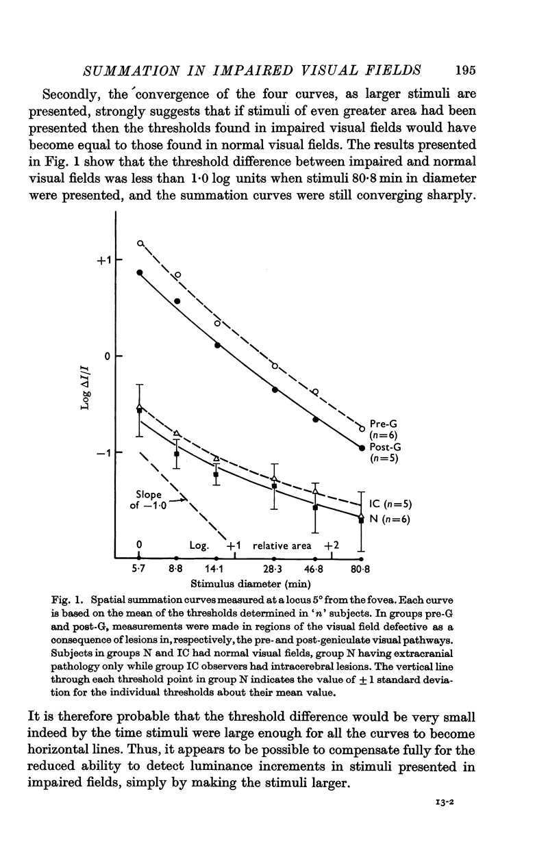
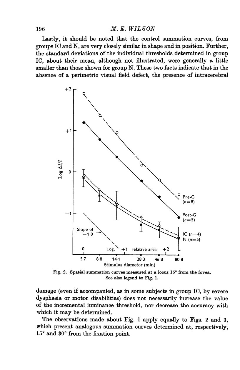
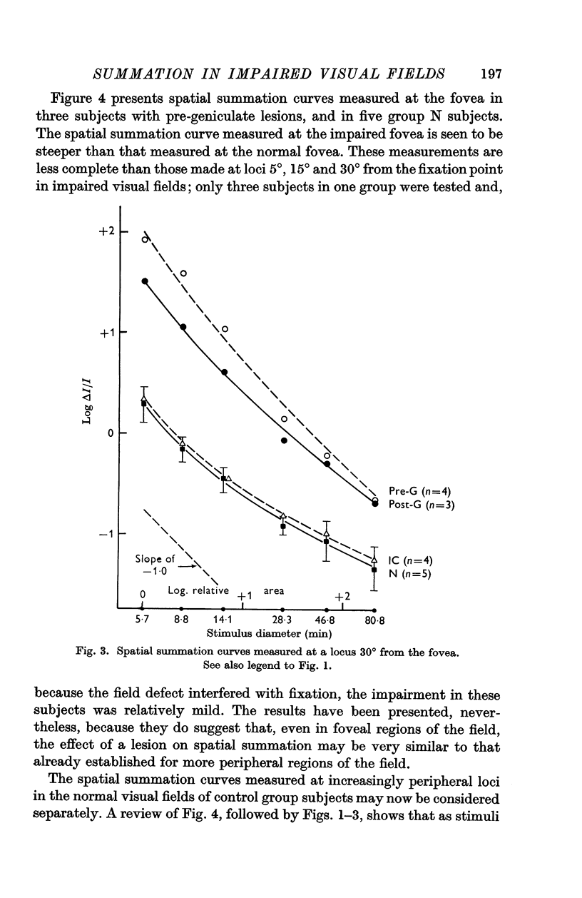
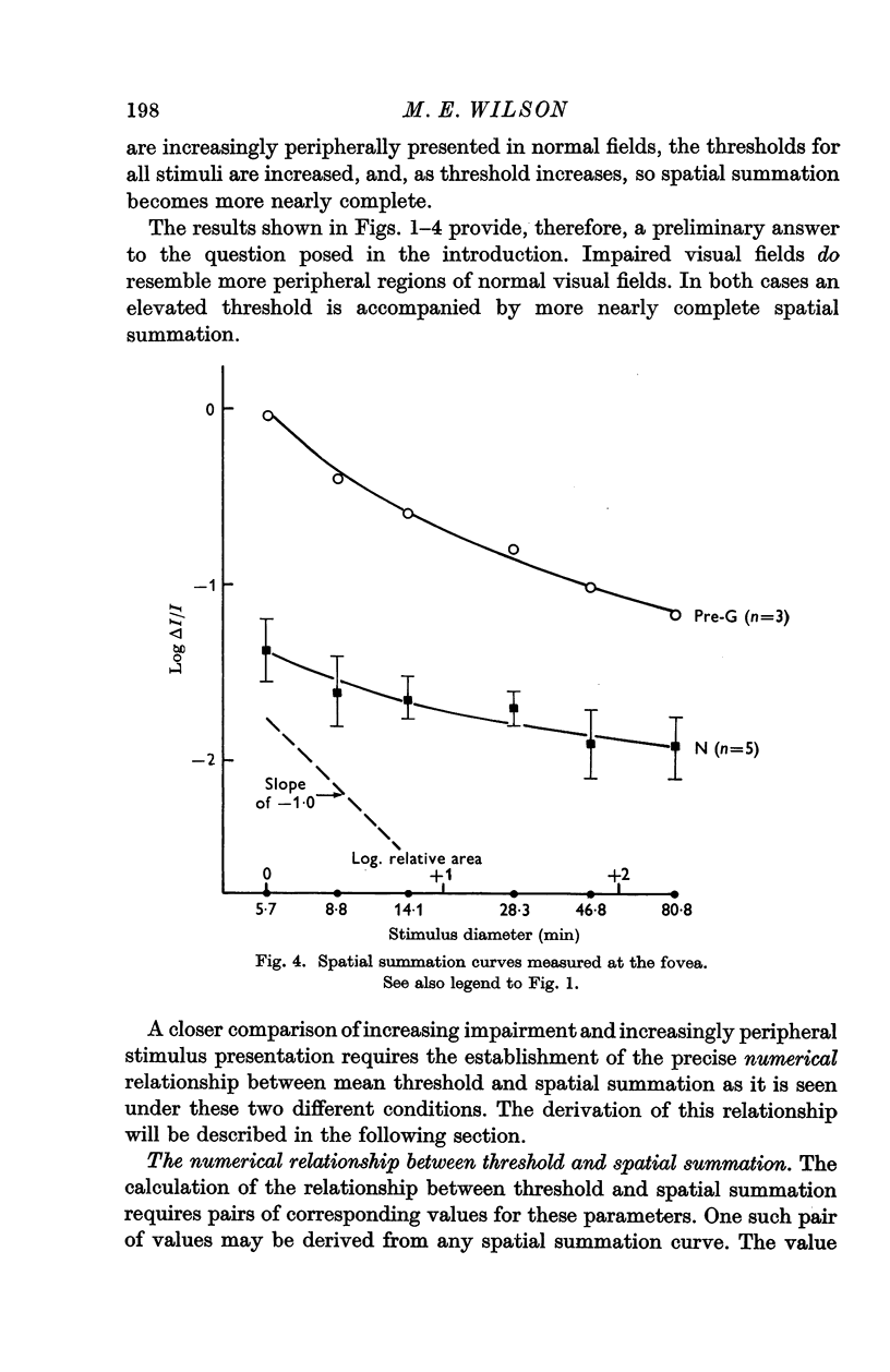
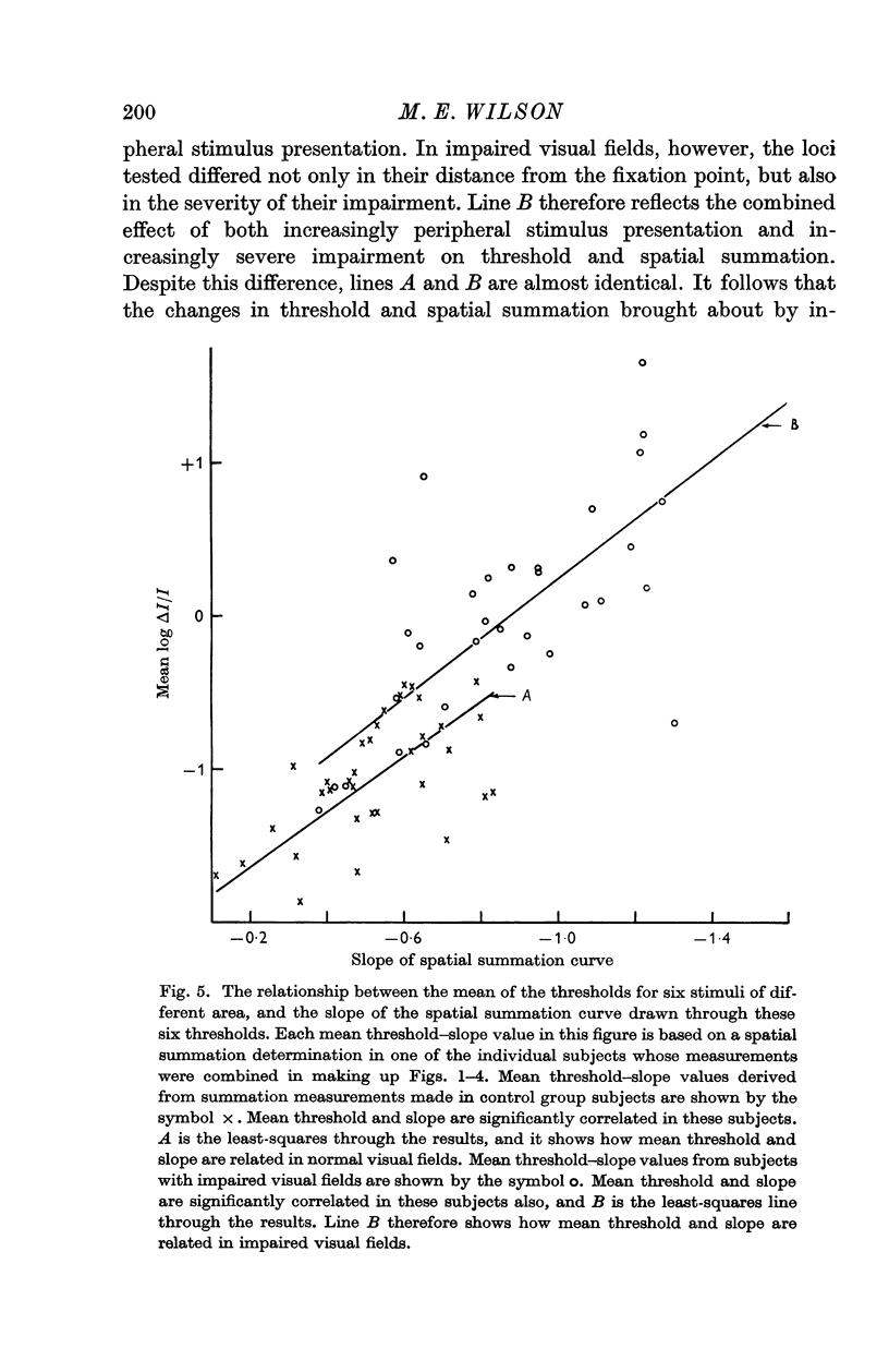
3. Spatial summation was found altered in all impaired visual fields: the greater the threshold elevation produced by the lesion, the more nearly complete was spatial summation.
4. The above relation between threshold and spatial summation has also been given numerical form. This has been shown to be very nearly identical to the threshold—spatial summation relation which is seen as stimuli are increasingly peripherally presented in normal visual fields.
5. It has been shown that the alterations of spatial summation brought about by a lesion are found only in those parts of the visual field which are perimetrically impaired: spatial summation is always normal in perimetrically normal regions of a visual field, even if other parts of the same field show impairment.
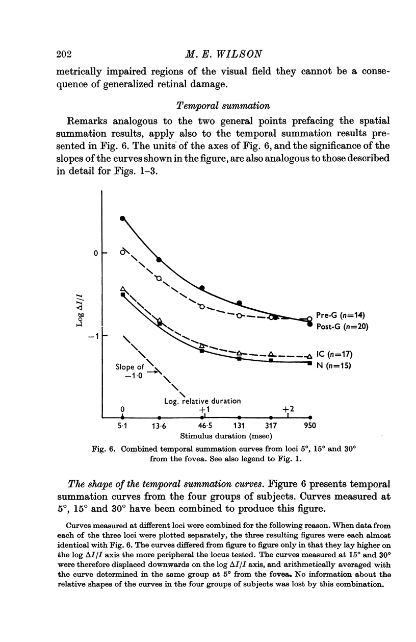
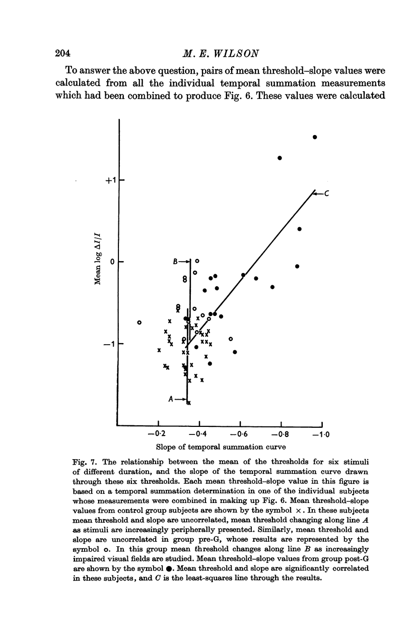
6. Temporal summation has been found altered in visual fields impaired by post-geniculate lesions: the greater the threshold elevation produced by the lesion, the more nearly complete was temporal summation. These changes in temporal summation were found only in perimetrically impaired regions of the field.
7. Temporal summation was normal in visual fields impaired by pregeniculate lesions.
Full text
PDF
Selected References
These references are in PubMed. This may not be the complete list of references from this article.
- Adrian E. D., Matthews R. The action of light on the eye: Part I. The discharge of impulses in the optic nerve and its relation to the electric changes in the retina. J Physiol. 1927 Sep 9;63(4):378–414. doi: 10.1113/jphysiol.1927.sp002410. [DOI] [PMC free article] [PubMed] [Google Scholar]
- BARLOW H. B., FITZHUGH R., KUFFLER S. W. Dark adaptation, absolute threshold and Purkinje shift in single units of the cat's retina. J Physiol. 1957 Aug 6;137(3):327–337. doi: 10.1113/jphysiol.1957.sp005816. [DOI] [PMC free article] [PubMed] [Google Scholar]
- BARLOW H. B. Temporal and spatial summation in human vision at different background intensities. J Physiol. 1958 Apr 30;141(2):337–350. doi: 10.1113/jphysiol.1958.sp005978. [DOI] [PMC free article] [PubMed] [Google Scholar]
- BAUMGARDT E. Seuils visuels et quanta de lumière; précisions. Annee Psychol. 1953;53(2):431–441. [PubMed] [Google Scholar]
- DUBOIS-POULSEN A., MAGIS C. La notion de sommation spatiale en physio-pathologie oculaire. Bibl Ophthalmol. 1957;12(47):218–238. [PubMed] [Google Scholar]
- FUORTES M. G., GUNKEL R. D., RUSHTON W. A. Increment thresholds in a subject deficient in cone vision. J Physiol. 1961 Apr;156:179–192. doi: 10.1113/jphysiol.1961.sp006667. [DOI] [PMC free article] [PubMed] [Google Scholar]
- GREGORY R. L., CANE V. A statistical information theory of visual thresholds. Nature. 1955 Dec 31;176(4496):1272–1272. doi: 10.1038/1761272a0. [DOI] [PubMed] [Google Scholar]
- WEYMOUTH F. W. Visual sensory units and the minimal angle of resolution. Am J Ophthalmol. 1958 Jul;46(1 Pt 2):102–113. doi: 10.1016/0002-9394(58)90042-4. [DOI] [PubMed] [Google Scholar]


