

Abstract
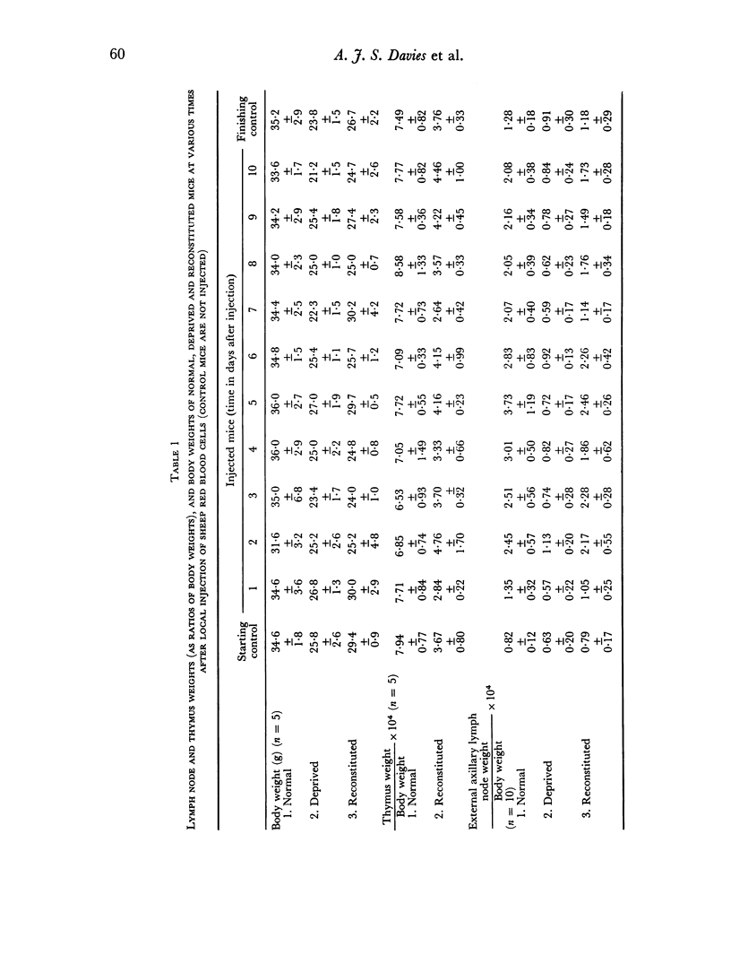
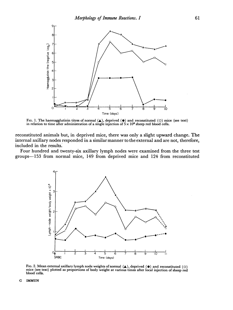
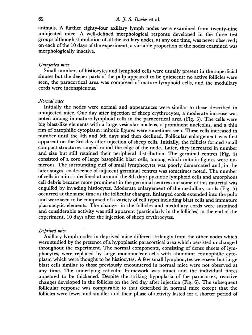
The response to sheep red blood cells has been studied in the lymph nodes draining their site of injection in normal mice, and in thymectomized, irradiated, bone-marrow injected mice with and without a reconstituting thymus graft. By using a chromosome marker to differentiate between cells derived from the bone-marrow and thymus graft it has proved possible to show that the immune response should be thought of in terms of at least two cell populations. Cells of thymic origin are stimulated to mitotic activity in the interfollicular cortex, and their activity precedes both antibody production and morphological signs of activity in the follicular regions. Mitotic divisions of cells of bone-marrow origin reached a peak a day later than did the thymic cells and their activity was sustained. Follicular enlargement and germinal centre production were coincident in time both with antibody production and bone-marrow cell mitotic activity. Lymph nodes of animals lacking a thymic influence showed only minor changes after antigenic stimulation and these were restricted to the follicular regions. There appeared to be only a small quantitative difference between the responses of normal and of reconstituted animals.
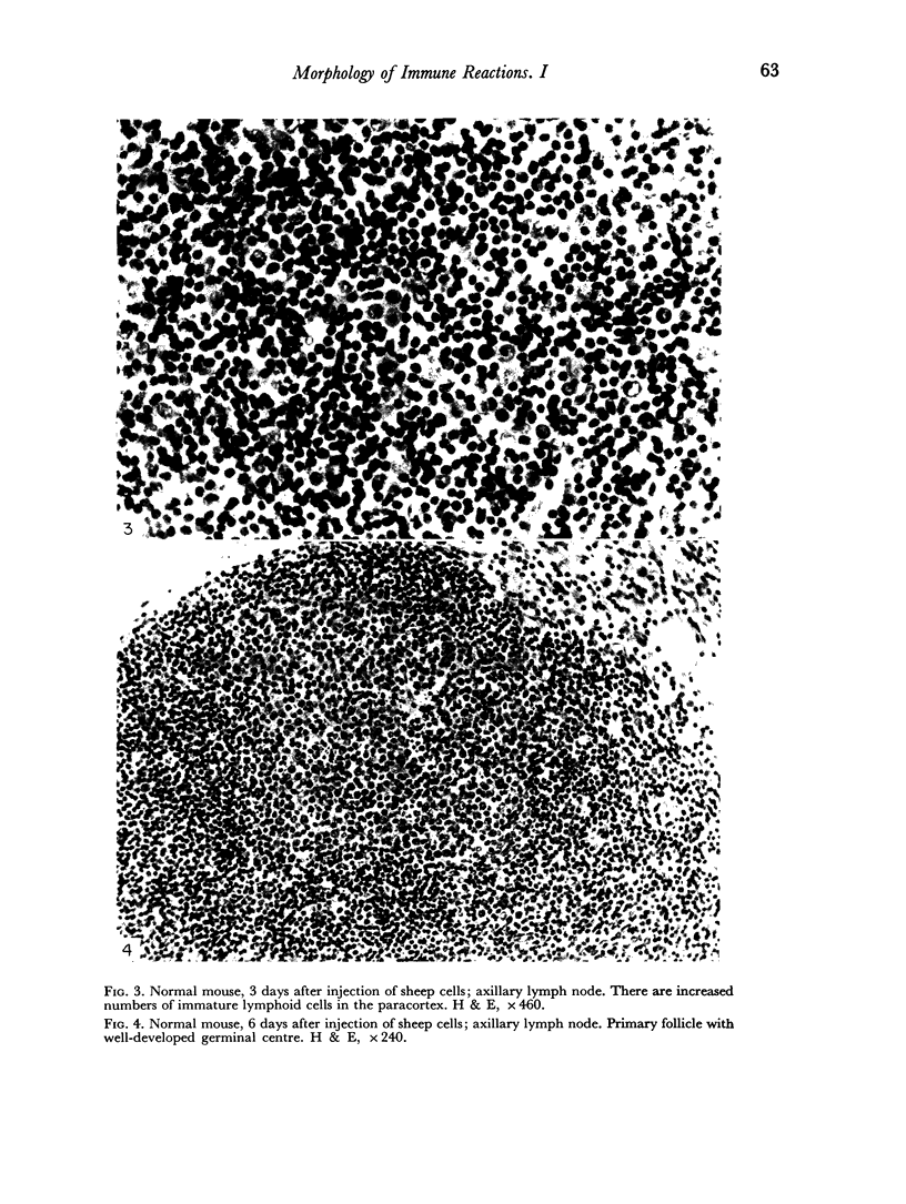
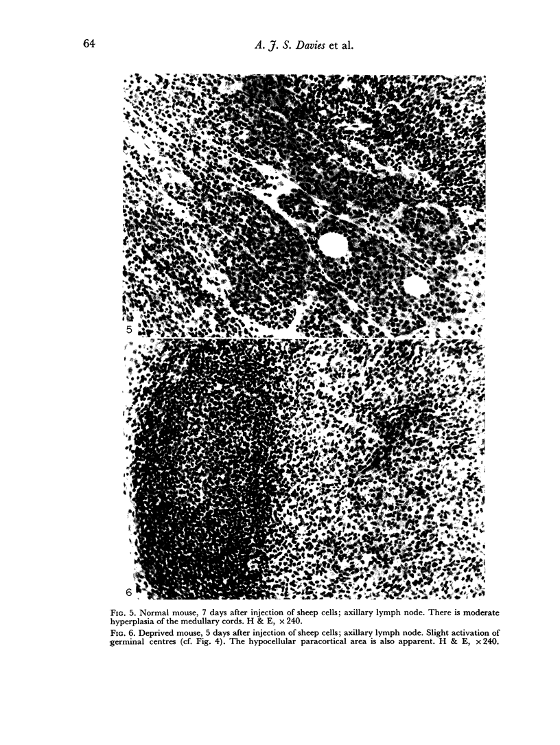
Full text
PDF
Images in this article
Selected References
These references are in PubMed. This may not be the complete list of references from this article.
- Davies A. J., Leuchars E., Wallis V., Koller P. C. The mitotic response of thymus-derived cells to antigenic stimulus. Transplantation. 1966 Jul;4(4):438–451. doi: 10.1097/00007890-196607000-00008. [DOI] [PubMed] [Google Scholar]
- Gershon R. K., Wallis V., Davies A. J., Leuchars E. Inactivation of thymus cells after multiple injections of antigen. Nature. 1968 Apr 27;218(5139):380–381. doi: 10.1038/218380a0. [DOI] [PubMed] [Google Scholar]
- LEUCHARS E., CROSS A. M., DUKOR P. THE RESTORATION OF IMMUNOLOGICAL FUNCTION BY THYMUS GRAFTING IN THYMECTOMIZED IRRADIATED MICE. Transplantation. 1965 Jan;3:28–38. doi: 10.1097/00007890-196501000-00004. [DOI] [PubMed] [Google Scholar]
- Miller J. F. The thymus. Yesterday, today, and tomorrow. Lancet. 1967 Dec 16;2(7529):1299–1302. doi: 10.1016/s0140-6736(67)90407-2. [DOI] [PubMed] [Google Scholar]
- OORT J., TURK J. L. A HISTOLOGICAL AND AUTORADIOGRAPHIC STUDY OF LYMPH NODES DURING THE DEVELOPMENT OF CONTACT SENSITIVITY IN THE GUINEA-PIG. Br J Exp Pathol. 1965 Apr;46:147–154. [PMC free article] [PubMed] [Google Scholar]
- Parrott D. M., de Sousa M. A. Changes in the thymus-dependent areas of lymph nodes after immunological stimulation. Nature. 1966 Dec 17;212(5068):1316–1317. doi: 10.1038/2121316a0. [DOI] [PubMed] [Google Scholar]
- Parrott D. V., De Sousa M. A., East J. Thymus-dependent areas in the lymphoid organs of neonatally thymectomized mice. J Exp Med. 1966 Jan 1;123(1):191–204. doi: 10.1084/jem.123.1.191. [DOI] [PMC free article] [PubMed] [Google Scholar]
- Turk J. L. Lymphocyte response to antigens. A. Afferent side of sensitization arc. Response of lymphocytes to antigens. Transplantation. 1967 Jul;5(4 Suppl):952–961. doi: 10.1097/00007890-196707001-00025. [DOI] [PubMed] [Google Scholar]