Abstract
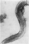
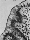
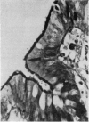
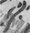
An abnormal condition of the large intestine is described in which the surface epithelium is infested by short spirochaetes. Diagnosis can be made by light microscopy. A review of 14 cases diagnosed by rectal biopsy and 62 cases involving the appendix shows no consistent symptom complex. The possible significance is discussed.
Full text
PDF
Images in this article
Selected References
These references are in PubMed. This may not be the complete list of references from this article.
- Gear E. V., Jr Dobbins WO 3d,+DOBBINS WO III: Rectal biopsy. A review of its diagnostic usefulness. Gastroenterology. 1968 Oct;55(4):522–544. [PubMed] [Google Scholar]
- Harland W. A., Lee F. D. Intestinal spirochaetosis. Br Med J. 1967 Sep 16;3(5567):718–719. doi: 10.1136/bmj.3.5567.718. [DOI] [PMC free article] [PubMed] [Google Scholar]
- SHERA A. G. Specific granular lesions associated with intestinal spirochaetosis. Br J Surg. 1962 Jul;50:68–77. doi: 10.1002/bjs.18005021917. [DOI] [PubMed] [Google Scholar]
- SWAIN R. H. Electron microscopic studies of the morphology of pathogenic spirochaetes. J Pathol Bacteriol. 1955 Jan-Apr;69(1-2):117–128. doi: 10.1002/path.1700690117. [DOI] [PubMed] [Google Scholar]
- THOMAS G. K. Strawberry lesion of the recto-sigmoid. Br Med J. 1956 Jul 28;2(4986):209–210. doi: 10.1136/bmj.2.4986.209. [DOI] [PMC free article] [PubMed] [Google Scholar]


