Abstract
The Arabidopsis Ca2+/H+ transporter CAX1 (Cation Exchanger1) may be an important regulator of intracellular Ca2+ levels. Here, we describe the preliminary localization of CAX1 to the tonoplast and the molecular and biochemical characterization of cax1 mutants. We show that these mutants exhibit a 50% reduction in tonoplast Ca2+/H+ antiport activity, a 40% reduction in tonoplast V-type H+-translocating ATPase activity, a 36% increase in tonoplast Ca2+-ATPase activity, and increased expression of the putative vacuolar Ca2+/H+ antiporters CAX3 and CAX4. Enhanced growth was displayed by the cax1 lines under Mn2+ and Mg2+ stress conditions. The mutants exhibited altered plant development, perturbed hormone sensitivities, and altered expression of an auxin-regulated promoter-reporter gene fusion. We propose that CAX1 regulates myriad plant processes and discuss the observed phenotypes with regard to the compensatory alterations in other transporters.
INTRODUCTION
As in all eukaryotic systems, plant Ca2+ signaling depends on the regulation of cytosolic Ca2+ levels. For secondary transporters, animals predominately use Na+ as the coupling ion to circulate Ca2+ across biological membranes, whereas plants use protons as the coupling ion almost exclusively (Sze et al., 1999; Gaxiola et al., 2002). Proton gradients are generated by primary H+-translocating pumps that hydrolyze either ATP (plasma membrane P-type H+-translocating ATPase and tonoplast V-type H+-translocating ATPase [V-ATPase]) or PPi (tonoplast H+-translocating pyrophosphatase [V-PPase]) as the energy source to pump protons, generating a proton motive force that energizes the membrane (Drozdowicz and Rea, 2001; Palmgren, 2001; Sze et al., 2002). Thus, plants use the proton motive force to directly and indirectly regulate the transport of ions such as Ca2+ across membranes.
The design and architecture of the plant cell contribute spatial features to the Ca2+ spike not seen in mammalian systems, particularly the Ca2+ spikes around the vacuole. The plant vacuole can occupy up to 99% of a plant cell's volume (Marty, 1999) and contains various Ca2+ channels, including Ca2+-permeable inositol 1,4,5-trisphosphate– and cyclic ADP-ribose–activated channels (Schumaker and Sze, 1987; Allen et al., 1995). These types of channels on the tonoplast suggest that localized Ca2+ spikes around the plant vacuole play a pivotal role in determining signal specificity. Furthermore, these findings imply that vacuolar Ca2+/H+ antiporters driven by the V-ATPase or V-PPase and Ca2+-ATPases help reset cytosolic Ca2+ levels after signal transduction. However, there is a paucity of mutants in plant vacuolar Ca2+ transporters that can be used to assess the biological impact of these transporters in plant signaling (Wu et al., 2002).
Initially, plant Ca2+/H+ antiporter genes were cloned by their ability to suppress the Ca2+-hypersensitive phenotype of a Saccharomyces cerevisiae mutant (Hirschi et al., 1996; Ueoka-Nakanishi et al., 2000). These genes are termed cation exchangers (CAX). CAX1 from Arabidopsis is a high-capacity Ca2+ transporter, whereas CAX2 has a lower capacity for Ca2+ transport (Hirschi et al., 1996) and also can transport other metals (Hirschi et al., 2000). Arabidopsis appears to have up to 10 other putative cation/H+ antiporters (CAX3 to CAX11 and MHX) (Mäser et al., 2001). Some of these, such as CAX2, CAX4, and MHX, have been shown to localize to the plant vacuole (Shaul et al., 1999; Hirschi et al., 2000; Cheng et al., 2002). Understanding the intracellular localization and function of these individual CAX transporters is an important component of understanding the specificity of Ca2+ signals.
The activity of CAX1 appears to be regulated by an N-terminal regulatory region (NRR) that was absent from the initial clone characterized by heterologous expression in yeast (Pittman and Hirschi, 2001; Pittman et al., 2002). Ectopic expression of deregulated CAX1 (termed sCAX1, missing the N-terminal autoinhibitor) in tobacco increases Ca2+ levels in the plants and causes numerous stress-sensitive phenotypes often associated with Ca2+ deficiencies (Hirschi, 1999, 2001). Thus, a wide range of environmental responses appear to require the judicious control of CAX1 transport activity; however, these studies have not addressed the phenotypic consequences of diminishing Ca2+/H+ transport around the plant vacuole.
In this study, we tentatively localize CAX1 in Arabidopsis and demonstrate that in planta CAX1 contains the N-terminal autoinhibitory domain. We report the isolation of CAX1 knockout mutants and describe the phenotypes of these plants at the whole-plant, molecular, and biochemical levels. Characterization of the mutant phenotypes indicates that the cax1 disruption alters the expression and/or activity of other vacuolar Ca2+ transporters and the V-ATPase. Despite these compensatory changes, cax1 mutants exhibit alterations in growth, stress responses, and hormone perception. These findings offer a clue to the elaborate regulatory interplay among transporters and suggest that CAX1 transport mediates numerous biological responses.
RESULTS
CAX1 Localizes to the Vacuolar Membrane in Arabidopsis
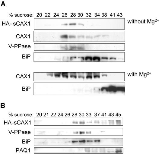
The localization of CAX1 has not been reported. Previous findings suggest that CAX1 localizes to the vacuolar membrane. First, deregulated N-terminal truncations of CAX1 can suppress yeast mutants defective in vacuolar Ca2+ transport (Hirschi et al., 1996); second, when expressed heterologously in yeast, epitope-tagged, full-length, and truncated CAX1 localize to the vacuolar membrane (Pittman and Hirschi, 2001); and third, ectopic expression of deregulated CAX1 increases Ca2+/H+ transport in tobacco tonoplast-enriched fractions (Hirschi, 1999). To further establish the subcellular localization of CAX1 in plants, microsomal membranes from wild-type and transgenic lines harboring the hemagglutinin (HA)-tagged truncated CAX1 fusion protein (HA-sCAX1) were fractionated. Centrifugation through a con-tinuous Suc gradient was first used to compare the distribution of the epitope-tagged transporter in both Arabidopsis (Figure 1A) and tobacco BY-2 (Figure 1B) suspension cells and the native full-length CAX1 (Figure 1A) with that of markers for the vacuole, plasma membrane, and endoplasmic reticulum. As shown in Figure 1, when membrane fractions were assayed for CAX1 and HA-sCAX1 accumulation, proteins of ∼50 kD increased at ∼30% Suc (most abundant in fractions corresponding to 28 and 37% Suc, respectively). Both endogenous CAX1 and HA-sCAX1 accumulated in fractions enriched in vacuolar membranes, as indicated by the sedimentation profiles, which overlap with that of a resident protein (V-PPase) from this membrane. Furthermore, the profile of the protein detected by anti-CAX1 was unchanged in gradients prepared in the presence of Mg2+, whereas the endoplasmic reticulum, as detected by anti-BiP, showed a large characteristic Mg2+ shift to heavier density fractions (Figure 1A).
Figure 1.
Subcellular Localization of CAX1 in Arabidopsis.
Immunoblot analysis of CAX1 in Arabidopsis (A) and tobacco BY-2 cells (B). Equal amounts of protein (10 μg) isolated from HA-sCAX1–expressing transgenic Arabidopsis, wild-type Arabidopsis, and HA-sCAX1–expressing tobacco BY-2 cells were separated by SDS-PAGE, blotted, and subjected to protein gel blot analysis using antibodies against HA (HA-sCAX1), an N-terminal peptide from full-length CAX1 (CAX1), and plant membrane markers: the plant endoplasmic reticulum luminal protein (BiP), mung bean vacuolar pyrophosphatase (V-PPase), and radish plasma membrane aquaporin (PAQ1). Wild-type Arabidopsis membranes were prepared and fractionated in the absence or presence of 5 mM MgCl2, as indicated in (A), and then assayed for changes in CAX1 and BiP expression. Numbers above the gels indicate the Suc concentration of each fraction.
Isolation of CAX1 Knockout Mutants
Insertional mutagenesis is a means of disrupting gene function based on the insertion of foreign DNA, either transposable elements or T-DNA, into the gene of interest (McKinney et al., 1995; Krysan et al., 1996). Although the insertions are random in nature, it is possible to generate a large number of these insertions; thus, there is a high probability of obtaining an insert in the gene of interest. Collections of insertional mutant lines are now available for which the insertion site has been sequenced and the information deposited into databases (Barbier-Brygoo et al., 2001). This information allows the identification of mutants and helps compensate for the inability to perform efficient targeted disruptions in Arabidopsis. To investigate the physiological function of CAX1 in plants, we used PCR to screen a large population of transposon insertion lines from two different sources (Parinov et al., 1999; Tissier et al., 1999).
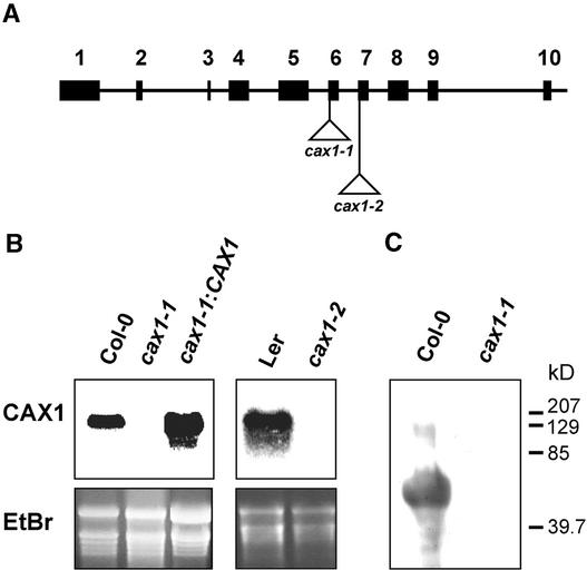
We screened a large population of dSpm insertion lines from the SLAT pools (Sainsbury Laboratory Arabidopsis thaliana Transposants) (Tissier et al., 1999). As shown in Figure 2A, we isolated an Arabidopsis plant of the Columbia ecotype (Col-0) carrying a dSpm insertion within the CAX1 open reading frame located in the acidic motif of CAX1, between Gln-266 and Glu-267 (cax1-1). The dSpm insertion confers BASTA resistance. A homozygous line was isolated by PCR analysis combined with BASTA selection. We also used a SGT (Singapore Gene Trap) Ds transposon insertion pool (Parinov et al., 1999) to isolate another allele, cax1-2, in the Landsberg erecta (Ler) ecotype. This Ds insertion conferred kanamycin resistance; 15 plants from this line contained the proper diagnostic PCR products. Homozygous lines were identified by PCR combined with kanamycin selection. Sequence analysis of the junction of the Ds insert determined that it was located between Ala-313 and Ser-314, which is between putative transmembrane spans 7 and 8 in the CAX1 coding sequence (Figure 2A). These disruptions in CAX1 did not produce detectable levels of CAX1 RNA (Figure 2B) even when high levels (10 mM) of exogenous Ca2+ were added, an environmental factor that increases the levels of CAX1 RNA significantly (Hirschi, 1999). In addition, neither allele produced detectable levels of CAX1 protein (Figure 2C and data not shown), whereas both alleles of cax1 were fertile.
Figure 2.
Identification of Transposon Insertion Lines of CAX1.
(A) Diagram of the genomic CAX1 DNA depicting the sites of transposon insertions. The 4.1 kb of genomic CAX1 DNA is represented by 9 introns (lines) and 10 exons (boxes). Triangles depict the sites of the transposon insertions. The lines harboring these insertions are termed cax1-1 and cax1-2.
(B) RNA gel blot analysis of CAX1 gene expression in wild-type and cax1 plants. Total RNA was extracted from 3-week-old wild-type, cax1 mutant, and CAX1-expressing cax1 plants pretreated with 10 mM CaCl2 overnight. Ten micrograms of total RNA of each sample was loaded, blotted, and hybridized with a 32P-labeled CAX1 cDNA probe. Ethidium bromide (EtBr) staining of the agarose gel is shown as a loading control.
(C) Immunoblot analysis of CAX1 protein expression in wild-type and cax1-1 mutant plants pretreated with 100 mM CaCl2 overnight. Twenty micrograms of vacuole-enriched protein sample was separated by SDS-PAGE, blotted, and subjected to protein gel blot analysis using a CAX1-specific antibody.
Both the cax1-1 and cax1-2 alleles were backcrossed to the wild type, and the F1 and F2 progeny were analyzed. The F2 progeny segregated 3:1 for the antibiotic resistance (378 BASTA-resistant seedlings:128 BASTA-sensitive seedlings for cax1-1 and 205 kanamycin-resistant seedlings:71 kanamycin-sensitive seedlings for cax1-2), indicating that both alleles contain a single transposon insertion with a functional selection marker.
To remove any possibility that the cax1 phenotypes were caused by an unknown mutation, it was important to express CAX1 in the mutants to restore CAX1-mediated Ca2+/H+ antiport activity. For these comparisons, we generated 15 transgenic lines (cax1-1:CAX1) harboring the cDNA encoding deregulated CAX1 (sCAX1) under the control of the 35S promoter of Cauliflower mosaic virus (Hirschi, 1999). The cax1-1 lines expressing deregulated CAX1 contained high levels of CAX1 RNA and CAX1 protein (Figure 2B and data not shown).
V-ATPase Activity Is Altered in the cax1 Mutants
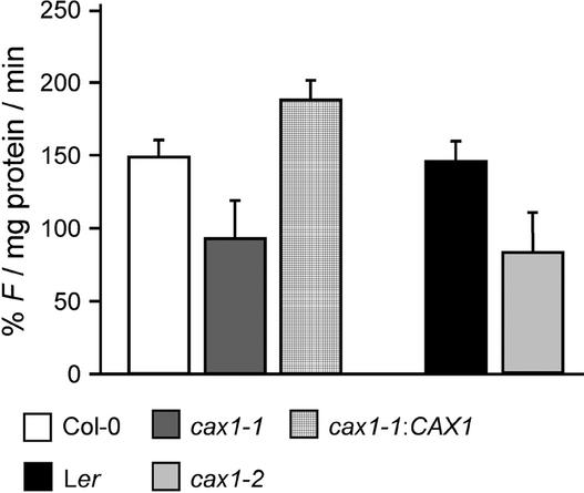
The H+ driving force for CAX1 Ca2+/H+ antiport activity on the tonoplast is provided by primary H+ pumps that co-reside on this membrane and acidify the vacuolar lumen. ATP- and PPi-dependent H+ transport, as determinants of V-ATPase and V-PPase activities, respectively, were measured in tonoplast vesicles from Col-0, cax1-1, cax1-1:CAX1, Ler, and cax1-2. V-ATPase H+ transport activity, determined in the absence of Ca2+ in the reaction buffer, was reduced significantly in the cax1 mutant plants: by 38% in cax1-1 and by 45% in cax1-2 compared with the wild type (Figure 3). This reduction in V-ATPase activity in cax1 was independent of pretreatment of the plants with Ca2+ before harvest (data not shown). V-ATPase activity also was measured in cax1-1 expressing deregulated CAX1. These plants showed a 26% increase in H+ transport activity compared with Col-0 plants (Figure 3). By contrast, V-PPase activity was negligible in roots of Col-0 plants, and no change was detected in cax1 mutants or cax1-1 expressing deregulated CAX1 (data not shown). Furthermore, a preliminary examination of cax1-1 vacuolar morphology suggested no alterations in vacuolar size or shape compared with wild-type plants (data not shown).
Figure 3.
Initial Rates of V-ATPase H+ Transport Activity in Purified Tonoplast Vesicles from the Wild Type, cax1, and cax1-1 Expressing CAX1.
ATP-dependent H+ transport into tonoplast vesicles (vesicle acidification) was monitored by the quenching of quinacrine fluorescence. Initial rates were calculated from the rates of quinacrine fluorescence quenching during the first 40 s after the addition of ATP. Results are means ± se of three independent membrane preparations. F, fluorescence intensity.
cax1-1 Mutants Display Altered Vacuolar Ca2+ Transport
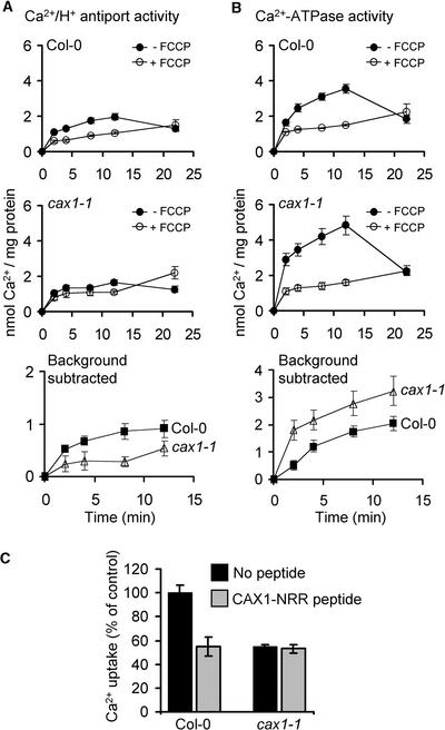
We demonstrated previously that Ca2+/H+ antiport activity can be measured directly in vacuole-enriched membrane vesicles isolated from Arabidopsis after pretreatment of the plants with 100 mM CaCl2, whereas without high Ca2+ induction, Ca2+/H+ antiport activity is difficult to measure (Pittman et al., 2002). Increased expression of CAX1 mRNA is observed in the presence of exogenous Ca2+, and we believe that the Ca2+/H+ antiport activity observed in the Ca2+-treated plants is predominantly the result of CAX1, because this activity is inhibited significantly by a CAX1-specific synthetic peptide (Pittman et al., 2002). To confirm that the loss of the CAX1 transcript in cax1-1 mutants caused the disruption of Ca2+/H+ antiport activity, root tissue was harvested from 4-week-old plants that were pretreated with 100 mM Ca2+ 18 h before harvest. Although significant Ca2+/H+ antiport activity was observed in vesicles from wild-type Col-0 plants, antiport activity in vesicles from cax1-1 plants was reduced by >50% (Figure 4A).
Figure 4.
Ca2+ Uptake Activity into Vacuole-Enriched Membrane Vesicles from Ca2+-Treated Col-0 and cax1-1 Root Tissue.
Time courses of Mg2+-ATP–energized 10 μM 45Ca2+ uptake were determined in the presence of 0.1 mM NaN3, 10 mM KCl, 1 mM ATP, and 1 mM MgSO4. ΔpH-dependent Ca2+/H+ antiport activity (A) was determined in the presence of 0.2 mM orthovanadate (Ca2+-ATPase inhibitor) as the difference between Ca2+ uptake in the absence and presence of 5 μM carbonyl cyanide p-(trifluoromethoxy)phenylhydrazone (FCCP) (protonophore). Calmodulin-stimulated Ca2+-ATPase activity (B) was determined in the absence of orthovanadate and the presence of 0.5 μM calmodulin and 5 μM FCCP. The Ca2+ ionophore A23187 (5 μM) was added at the 12-min time point and significantly dissipated Ca2+ accumulation mediated by Ca2+/H+ antiport or Ca2+-ATPase when measured at the 22-min time point. The results of net Ca2+/H+ antiport activity (bottom graph in [A]) and Ca2+-ATPase activity (bottom graph in [B]) also are shown after the subtraction of the +FCCP background values for the first four time points of active Ca2+ accumulation. (C) shows ΔpH-dependent 10 μM Ca2+ uptake into vacuole-enriched membrane vesicles isolated from Ca2+-treated Col-0 or cax1-1 Arabidopsis root tissue measured in the presence or absence of 10 μM CAX1-NRR peptide at the 10-min time point. The results shown are percentages of Ca2+ uptake of the control sample (absence of peptide) after the subtraction of the FCCP background values. All results are means ± se of experiments from three to six independent membrane preparations.
The vacuolar membrane of Arabidopsis possesses a type-IIB Ca2+-ATPase, ACA4, that is regulated by calmodulin (Geisler et al., 2000). Ca2+ uptake into the vacuole-enriched vesicles from both Col-0 and cax1-1 was measured in the absence of vanadate to determine Ca2+-ATPase activity. In the absence of calmodulin, very little Ca2+-ATPase activity was measured (data not shown). However, in the presence of calmodulin, Ca2+-ATPase activity was high in vesicles from both plants but was significantly higher in vesicles from cax1-1 compared with Col-0 (Figure 4B), indicating a 36% increase in Ca2+-ATPase activity in plants lacking CAX1.
Ca2+ transport activity was compared in vacuole-enriched vesicles from Col-0 and cax1-1 expressing deregulated CAX1. Without the addition of exogenous Ca2+, a significant increase in Ca2+/H+ antiport activity was measured in vesicles from the deregulated CAX1-expressing Arabidopsis compared with those from Col-0, whereas no change in Ca2+-ATPase activity was observed (data not shown).
Ca2+/H+ antiport activity also was measured in vesicles from Col-0 and cax1-1 in the presence of the CAX1-NRR peptide. As demonstrated previously, in the presence of the CAX1-NRR peptide, Ca2+/H+ antiport activity from wild-type plants was reduced significantly (Figure 4C). However, there was no difference in antiport activity in vesicles from cax1-1 roots in the presence or absence of the CAX1-NRR peptide. Furthermore, the CAX1-NRR peptide had no effect on Ca2+-ATPase activity in either Col-0 or cax1-1 membrane vesicles (data not shown).
Putative Vacuolar Ca2+ Transporters Are Upregulated in the cax1 Mutants
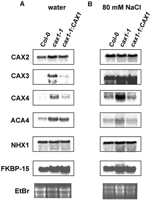
Because of the alterations in vacuolar Ca2+/H+ antiport, Ca2+-ATPase, and V-ATPase activity, we hypothesized that transporter genes that may be responsive to alterations in Ca2+ levels would be deregulated in the mutants. CAX3 RNA increases when exogenous Ca2+ and Na+ are in the medium but is expressed normally at relatively low levels (Shigaki and Hirschi, 2000). Under nonstressed conditions, CAX4 RNA can be detected only by reverse transcriptase–mediated PCR, and this gene is induced modestly by salt stress (Cheng et al., 2002). ACA4, which encodes a vacuolar Ca2+-ATPase, also is expressed at relatively low levels, and reverse transcriptase–mediated PCR shows a modest induction during salt stress (Geisler et al., 2000). Figure 5A shows that CAX3, CAX4, and ACA4 levels were significantly higher in cax1-1 than in both control plants and cax1-1 lines expressing deregulated CAX1. Specifically, CAX3 RNA levels increased fourfold, CAX4 RNA levels increased threefold, and ACA4 RNA levels increased twofold in cax1-1 compared with wild-type plants. Na+ treatment highly upregulated CAX3 in all plant backgrounds (12-, 14-, and 25-fold in wild-type, cax1-1, and CAX1-expressing mutant plants, respectively) (Figure 5B). Na+ treatment also produced an eightfold higher level of CAX4 RNA and a threefold higher level of ACA4 in cax1-1 compared with wild-type plants. Similar increases in the expression of ACA4 and CAX3 were seen upon Ca2+ treatment in cax1-1 compared with the wild type (data not shown). Comparable changes in CAX3, CAX4, and ACA4 transcripts also were seen in the cax1-2 allele (data not shown). In all cases, the increased expression of these transporters in cax1-1 was attenuated by the ectopic expression of deregulated CAX1. The RNA levels of the vacuolar metal/H+ antiporter CAX2 (Hirschi et al., 2000), the Na+/H+ antiporter AtNHX1 (Gaxiola et al., 1999), and FKBP15, a Ca2+ modulator protein (Luan et al., 1996), were not altered significantly during any stress treatment in cax1-1 and cax1-2 (Figures 5A and 5B and data not shown).
Figure 5.
RNA Gel Blot Analysis of Various Transporters.
mRNA expression of various genes in cax1 mutant plants was analyzed under normal (A) and Na+ stress (B) conditions. Total RNA was extracted from 3-week-old wild-type, cax1 mutant, and CAX1-expressing cax1 plants pretreated with water or 80 mM NaCl overnight. Ten micrograms of total RNA from each sample was loaded, blotted, and hybridized with 32P-labeled DNA probes corresponding to CAX2, CAX3, CAX4, ACA4, NHX1, and FKBP-15. Ethidium bromide (EtBr) staining of the agarose gel is shown as a loading control.
Perturbation of Ion Homeostasis in cax1 Mutant Plants
Ectopic expression of deregulated CAX1 increases Ca2+ content in transgenic tobacco plants (Hirschi, 1999). However, high-level CAX1 expression in tobacco causes numerous stress sensitivities associated with Ca2+ deficiencies. Although direct measurements of cytosolic Ca2+ levels have not been made in these CAX1-expressing plants, one hypothesis consistent with these phenotypes is that the sequestration of Ca2+ into the vacuole is highly upregulated and Ca2+ is “locked” inside the vacuole so that cytosolic Ca2+ levels are extremely low, thereby perturbing many Ca2+-dependent processes. To investigate the potential effects of increasing cytosolic Ca2+ levels by reducing vacuolar Ca2+ sequestration, we examined the ion content and stress sensitivities of CAX1–deficient lines.
When cax1-1 and cax1-2 were grown in either standard medium or medium containing high levels of Ca2+, there were no measurable changes in the levels of 29 different elements as measured by inductively coupled plasma atomic emission spectrometry (data not shown). This analysis suggested that the disruption of CAX1-mediated Ca2+/H+ exchange did not alter the total ion content of the plants significantly.
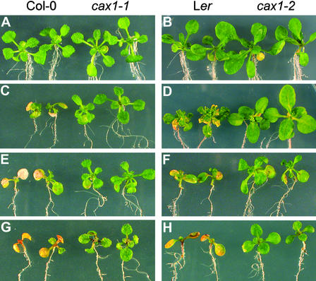
We examined the sensitivity of cax1 to a variety of different ion perturbations. Both alleles grew in a manner similar to wild-type plants on normal medium (Figures 6A and 6B) and medium containing Li+, Cd2+, Zn2+, Ni2+, and mannitol (data not shown); however, both cax1 alleles were more tolerant of Mg2+ and Mn2+ stresses than wild-type plants (Figures 6C to 6F). In addition, the cax1 alleles were more tolerant of medium lacking Ca2+ (Figures 6G and 6H) and medium containing increased levels of K+ and Na+ (data not shown). The ion sensitivities in the wild-type lines could be suppressed to levels similar to those in the cax1 alleles by the addition of exogenous Ca2+ to the growth medium (data not shown). The stress tolerances of the cax1-1 allele also could be suppressed by the expression of deregulated CAX1 (data not shown).
Figure 6.
Ion Sensitivity of cax1 Alleles.
Wild-type (Col-0) and cax1-1 plants are shown in (A), (C), (E), and (G). Wild-type (Ler) and cax1-2 plants are shown in (B), (D), (F), and (H). In each panel, the two wild-type plants are shown at left and the cax1 plants are shown at right. All photographs are representative of >100 plants grown in each condition.
(A) and (B) Five-day-old plants transferred to half-strength MS medium (Murashige and Skoog, 1962) and grown for 5 days.
(C) and (D) Five-day-old plants transferred to half-strength MS medium supplemented with 25 mM MgCl2 (C) or 10 mM MgCl2 (D) and grown for 5 days.
(E) and (F) Five-day-old plants transferred to half-strength MS medium supplemented with 1.5 mM MnCl2 and grown for 5 days.
(G) and (H) Five-day-old plants transferred to half-strength MS medium without Ca2+ and grown for 5 days.
Morphological Phenotypes of cax1 Alleles
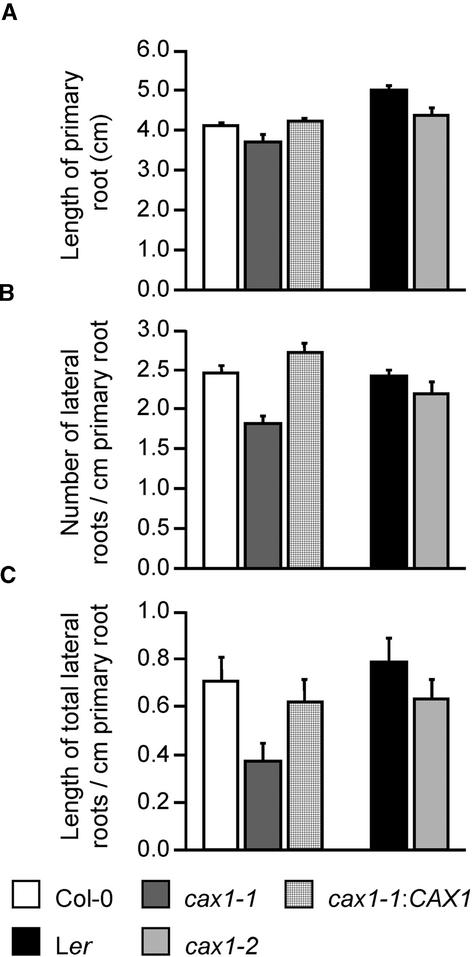
There were no differences in germination time and germination rate observable between the wild-type and the cax1-1 and cax1-2 lines (data not shown). The cax1-1 and cax1-2 seedlings displayed an ∼11% reduction in primary root length when grown on normal medium (Figure 7A). The number of lateral roots was reduced by 25% in cax1-1, and there was a 10% reduction in cax1-2 lateral root number (Figure 7B). Furthermore, the length of the lateral roots was reduced by almost 50% in cax1-1 and by ∼20% in cax1-2 (Figure 7C).
Figure 7.
Root Growth Measurements.
Primary roots (A) and lateral roots (B) and (C) of seedlings grown on the same plates at 22°C under continuous light were measured and counted after 10 days of growth on half-strength MS medium. All results are means ± se (n ≥ 12). These data are representative of four independent experiments.
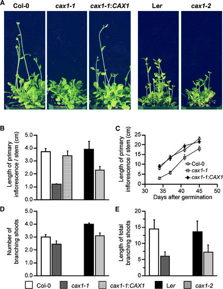
Under continuous light in soil, the transition of the cax1-1 and cax1-2 plants from the vegetative phase to the flowering phase was delayed by ∼5 to 7 days (Figure 8). At 27 days, the length of the primary inflorescence stem of cax1 plants was reduced by ∼70% in the cax1-1 line and by 40% in cax1-2 (Figure 8B). The number of branching shoots and the total length of the secondary shoots also were reduced significantly in both alleles. The cax1-1 and cax1-2 lines were reduced by 58 and 36%, respectively, in the total length of the secondary branches and by 20 and 23%, respectively, in the number of branches (Figures 8D and 8E). The expression of deregulated CAX1 in the cax1-1 line suppressed all of these phenotypes (Figure 8).
Figure 8.
Flowering and Apical Dominance Phenotypes.
(A) Twenty-seven-day-old wild-type and cax1 plants were grown in soil under continuous light. cax1 plants are delayed in flowering and shorter in the primary inflorescence stems, and cax1-1 plants expressing CAX1 driven by the 35S promoter are indistinguishable from wild-type plants. cax1-1 and CAX1-expressing cax1-1 are in the Col-0 background, and cax1-2 is in the Ler background.
(B) Quantitative analysis of the primary inflorescence stems of 27-day-old wild-type, cax1, and CAX1-expressing cax1-1 plants.
(C) Primary inflorescence length of cax1-1 and wild-type plants over the reproductive phase.
(D) Numbers of branching shoots.
(E) Lengths of branching shoots.
All results are means ± se (n ≥ 10). These photographs and data are representative of at least five independent experiments.
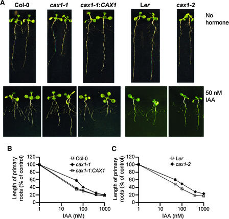
The alterations in lateral root growth and apical dominance in the cax1 alleles suggest that auxin levels might be altered in these plants. Given the complex interacting network of hormone response pathways, we analyzed cax1-1 and cax1-2 on numerous phytohormones. As shown in Figure 9, the cax1 roots were resistant to auxin but were tolerant to 1-aminocycloproprane-1-carboxylic acid (an ethylene precursor), abscisic acid, and benzyladenine (data not shown). This was apparent in the modest resistance to the inhibition of root elongation by exogenous phytohormones. In these conditions, cax1 roots were no longer shorter than wild-type roots. However, the cax1 alleles were not completely insensitive to these hormones; for example, auxin treatment did cause cax1 to form more lateral roots (data not shown). The cax1 alleles remained sensitive to gibberellic acid and epibrassinolide (data not shown). Furthermore, the expression of deregulated CAX1 in cax1-1 reduced root length significantly compared with that in cax1-1 in the presence of all phytohormones tested (Figures 9B and 9C and data not shown).
Figure 9.
Root Elongation Inhibition by Exogenous Auxin.
(A) cax1 and wild-type seedlings were germinated and grown on the same plates on standard medium (top row) or standard medium containing 50 nM IAA (bottom row) for 10 days at 22°C under yellow-filtered light.
(B) and (C) Quantitative analysis of root elongation inhibition by exogenous auxin. After 10 days of growth under yellow-filtered light on the medium supplemented with various concentrations of IAA, plants were removed from the medium and the lengths of the primary roots were measured. All data are standardized against growth on standard medium. All results are means ± se (n ≥ 12). These data are representative of four independent experiments.
IAA28 Expression in the cax1 Mutant
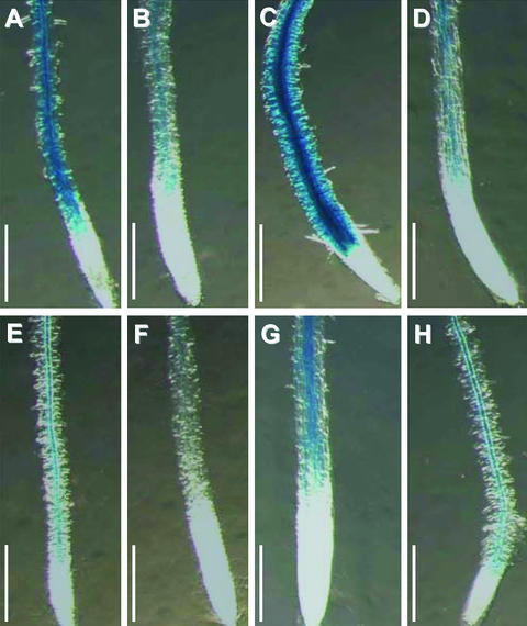
Reporter elements can be used to infer some of the alterations in the cax1-1 lines that cannot be detected immediately through whole-plant phenotype analysis. A number of well-characterized auxin reporter lines are available with which to dissect the impact of cax1 on auxin levels (Abel et al., 1995). These reporters may be especially useful, because several lines of evidence have suggested a relationship between Ca2+ homeostasis and auxin regulation (Hasenstein and Evans, 1988). The Aux/Indoleacetic Acid (IAA) gene family member IAA28 was used to further characterize the cax1-1 allele. This reporter is expressed preferentially in roots and inflorescence stems and demonstrates developmental and environmental regulation (Rogg et al., 2001). The IAA28 promoter–β-glucuronidase (GUS) fusion is expressed strongly from the distal root elongation zone to the root-hypocotyl junction, with the most intense staining in the root hair initiation zone. As shown in Figure 10, incubation of seedlings grown under yellow-filtered light with the GUS substrate 5-bromo-4-chloro-3-indolyl-β-d-glucuronic acid resulted in intense root staining. Unlike other characterized Aux/IAA genes, the IAA28 gene is downregulated by exogenous auxin treatment (Figure 10D) (Rogg et al., 2001). In addition, this reporter appeared to be responsive to Ca2+ conditions. As shown in Figures 10B and 10C, under yellow-filtered light, this reporter was downregulated by exogenous Ca2+ (Figure 10B) and upregulated in the absence of Ca2+ (Figure 10C). IAA28 promoter–GUS activity was reduced significantly in the cax1-1 mutant background when grown under various conditions (Figures 10E to 10H). When cax1-1 harboring the IAA28 promoter–GUS fusion was grown in normal medium under yellow-filtered light, GUS activity was lower than that in wild-type plants containing the reporter (cf. Figures 10A and 10E). Unlike in the wild-type background, the addition of IAA to cax1-1 had no obvious effect on IAA28 expression (Figure 10H). Although the expression of IAA28 in the cax1-1 background was increased by the removal of Ca2+ (Figure 10G) and decreased by the addition of Ca2+ (Figure 10F), the levels still were lower than those of similarly treated controls (Figures 10B and 10C).
Figure 10.
IAA28-GUS Expression in Wild-Type and cax1-1 Plants.
IAA28-GUS wild-type and cax1-1 seedlings grown under yellow-filtered light on half-strength MS medium for 5 days were transferred onto half-strength MS medium ([A] and [E]), 10 mM CaCl2 containing half-strength MS medium ([B] and [F]), or Ca2+-depleted medium ([C] and [G]) and then grown for another 3 days. Half of the seedlings from half-strength MS medium were treated with 20 μM IAA solution ([D] and [H]) for 4 h. All seedlings were stained with 1 mM 5-bromo-4-chloro-3-indolyl-β-d-glucuronic acid for 3 h at 37°C. Results from a representative experiment are shown. Approximately 100 F2 progeny were analyzed under each condition in the cax1-1 lines. The GUS staining shown here correlated with cax1-1 segregation in all 100 F2 progeny of the cax1-1 lines analyzed. Bars = 0.5 mm.
DISCUSSION
The data presented here offer insight into the biochemical and molecular mechanisms of Ca2+ homeostasis in plants. Cytosolic Ca2+ levels are controlled in part by a diverse collection of Ca2+ transporters and channels localized on both the plasma membrane and various internal organelles (Sanders et al., 1999, 2002). The vacuole is the predominant Ca2+ store within the cell (Marty, 1999), and both Ca2+-ATPase transporters and Ca2+/H+ antiporters exist at this organelle (Sze et al., 2000; Sanders et al., 2002) to help regulate intracellular Ca2+ levels. The work described here provides further evidence that CAX1, a Ca2+/H+ antiporter, localizes to the vacuolar membrane in Arabidopsis (Figure 1) and plays a central role in modulating myriad plant responses.
CAX1 Localization
An NRR on CAX1 has been shown to regulate Ca2+ transport by the mechanism of N-terminal autoinhibition (Pittman and Hirschi, 2001; Pittman et al., 2002). Here, we demonstrate that a HA-tagged version of CAX1 that lacks the NRR may localize to the tonoplast of transgenic Arabidopsis and tobacco (Figure 1). Using an antibody directed against the NRR, we also demonstrate that full-length CAX1 localizes to the Arabidopsis tonoplast. The Mg2+-shift experiment suggests that CAX1 does not localize to the endoplasmic reticulum; furthermore, chlorophyll levels (a chloroplast marker) peaked in 41 to 43% Suc fractions in the presence or absence of Mg2+ (data not shown), indicating that CAX1 is not found at this organelle. However, we still cannot exclude CAX1 localization to the Golgi; thus, such clarification will have to wait for CAX1–green fluorescent protein fusion studies. Future work using CAX1–green fluorescent protein fusions also will be needed to determine if CAX1 is localized differentially on small vacuoles or large central vacuoles (Paris et al., 1996). For example, CAX1 may be localized to a different vacuolar subtype from that proposed for ACA4 (Geisler et al., 2000).
These expression studies also demonstrate that full-length CAX1 is expressed in Arabidopsis, suggesting that CAX1 is not alternatively spliced to remove the NRR, as is seen with the regulatory domains of some mammalian plasma membrane–type Ca2+-ATPases (Penniston and Enyedi, 1998). Moreover, this finding suggests that the NRR is not a cleavable signal peptide (Darley et al., 2000). In total, these data allow us to conclude unambiguously that the NRR is present on the CAX1 transporter.
Although the vacuole is an important Ca2+ store, other organelles, such as the endoplasmic reticulum and the mitochondrion, may be important intracellular Ca2+ stores as well (Sanders et al., 2002). Various abiotic stresses induce Ca2+ release from different stores (Knight and Knight, 2001). For example, mitochondria are required for Ca2+ release in response to anoxia (Subbaiah et al., 1998). Although cax1 showed increased tolerance to a variety of ionic stresses (see below), there was no observed difference between cax1 and the wild type for other abiotic stresses, such as temperature and mannitol (data not shown). This result may indicate that the disruption in Ca2+ loading into the vacuole may affect only some signaling pathways.
Altered Vacuolar H+-ATPase Activity in the cax1 Lines
The transport system of the vacuolar membrane includes H+ pumps and several secondary active transporters. Two distinct H+ pumps, the V-ATPase and the V-PPase, are found on the tonoplast, and each has been well characterized (Barkla and Pantoja, 1996; Drozdowicz and Rea, 2001; Sze et al., 2002). These pumps create an electrochemical gradient of protons across the tonoplast, and this gradient is used as a source of energy for a variety of antiporters. For example, a stoichiometry of three H+ to one Ca2+ has been estimated for a vacuolar Ca2+/H+ antiporter (Blackford et al., 1990). As measured by direct ATP-dependent H+-pumping activity, the cax1 lines showed an ∼40% decrease in V-ATPase activity (Figure 3). Meanwhile, the deregulated CAX1-expressing lines showed a 26% increase in V-ATPase activity. Measurements of V-ATPase hydrolytic activity also supported these results (data not shown). These findings suggest that when CAX1 activity is high, V-ATPase activity is increased in response to the enhanced requirement for protons to drive CAX1-mediated Ca2+/H+ antiport. In the absence of CAX1, V-ATPase activity is reduced similarly. The changes observed in V-ATPase activity may result from the direct regulation by both transporters whereby CAX1 interacts physically with a V-ATPase subunit to modulate activity; alternatively, the V-ATPase activity may be reduced because of increased consumption of ATP used to drive Ca2+-ATPase activity (Figure 4) (see below).
Potentially, the numerous subtle phenotypes shown in the cax1-1 and cax1-2 alleles could be attributable to minor alterations in vacuolar biogenesis (Rojo et al., 2001); however, preliminary studies suggest that this is not the case. For example, cax1-1 root cells do not exhibit significant alterations in cell and vacuole morphology, as shown by visualization of wild-type and cax1-1 vacuoles upon staining with a vacuole-loaded fluorescent dye (Emans et al., 2002) and by comparisons of wild-type and cax1-1 protoplast morphology (data not shown). Furthermore, the alterations in vacuolar transporter expression and activity are specific and do not affect numerous vacuolar proteins. Although there are alterations in V-ATPase and Ca2+-ATPase activity and CAX3 and CAX4 expression, there is no significant alteration in V-PPase activity (data not shown) or AtNHX1 and CAX2 gene expression (Figure 5). Together, these findings suggest that only a subset of vacuolar transporters are perturbed, and these alterations are not caused by alterations in vacuolar morphology.
CAX1 and the Hierarchy of Vacuolar Ca2+ Transporters
In yeast, a null mutant in VCX1, the Ca2+/H+ antiporter, has no severe consequences on normal yeast growth despite these strains lacking all vacuolar Ca2+/H+ antiport activity (Cunningham and Fink, 1996; Miseta et al., 1999). Recently it was demonstrated that a hypertonic shock in yeast can release Ca2+ from the vacuole, and resetting this Ca2+ spike requires VCX1 but not the vacuolar Ca2+-ATPase PMC1 (Denis and Cyert, 2002). This finding suggests that vacuolar Ca2+/H+ transport is important in a subset of Ca2+ signal transduction events.
Although additional vacuolar Ca2+ sequestration pathways exist in plants, CAX1 contributes substantially to Ca2+ homeostasis. RNA analysis demonstrates that CAX1 is expressed in all tissues (Hirschi, 1999). In the two transposon-insertion mutants of cax1 reported here, CAX1 RNA and protein were not detected (Figure 2) and CAX1-mediated Ca2+/H+ antiport activity was abolished completely, as demonstrated by the lack of inhibition of antiport activity by a CAX1-specific synthetic peptide (Figure 4C). The two null alleles of CAX1 have various alterations in plant growth and development (detailed below) and vacuolar Ca2+ transport, even though vacuolar Ca2+-ATPase activity was enhanced and CAX3 and CAX4 RNA expression was increased (Figures 4 and 5). The enhanced expression of other transporters in the cax1 alleles is similar to the heightened ferric chelate reductase activity and IRT2 expression seen in the irt1-1 iron transport mutant (Vert et al., 2002). However, irt1-1 has a dramatic phenotype that cannot be compensated for despite upregulation of a homologous transporter. The induction of other putative Ca2+ transport systems, through some unknown regulatory network, must try to compensate for the loss of CAX1, suggesting the presence of a global mechanism of transcriptional regulation for vacuolar Ca2+ transporters. However, as shown by our Ca2+ transport data, this compensation is incomplete and resulted in an increase in high-affinity Ca2+-ATPase–mediated Ca2+ transport but a net decrease in low-affinity, high-capacity Ca2+/H+ antiporter–mediated Ca2+ transport (Figure 4).
The increased vacuolar Ca2+-ATPase activity (Figure 4B) may be mediated by the increased expression of ACA4 (Figure 5A). ACA4 is localized only on small vacuoles rather than on the large central vacuole (Geisler et al., 2000). Alternatively, other Ca2+-ATPases on the tonoplast, such as ACA11, which is the closest related transporter to ACA4 (Axelsen and Palmgren, 2001), may be responsible for the transport increases. It is the Ca2+/H+ antiporter rather than Ca2+-ATPase that efficiently sequesters Ca2+ when cytosolic Ca2+ levels are high (Miseta et al., 1999); therefore, an increase in Ca2+-ATPase activity may not be enough to efficiently reduce cytosolic Ca2+ in cax1.
Although the precise functions of CAX3 and CAX4 remain unknown (Shigaki and Hirschi, 2000; Cheng et al., 2002), the increased expression of these transporters in the cax1 alleles suggests that they play a role in Ca2+ homeostasis. This assumption is strengthened by the lack of induction of the characterized Na+ and metal antiporters in the cax1 alleles. These results also suggest that detailed expression profiling of the cax1 alleles may help identify other genes involved in Ca2+ homeostasis. Despite the increased expression of CAX3 and CAX4, the reduction in Ca2+/H+ transport in roots indicated that no antiport activity in the roots can compensate completely for the loss of CAX1. It is possible that a reduced H+ gradient attributable to decreased V-ATPase activity prevented Ca2+/H+ antiport by CAX3 and CAX4.
Phenotypes of cax1 Alleles
Many Arabidopsis knockout mutants fail to display any morphological phenotype, presumably because of functional redundancy (Krysan et al., 1999). CAX1 is a member of a large multigene family (Mäser et al., 2001) and just one of many Ca2+ efflux transporter genes present in Arabidopsis (Sze et al., 2000; Sanders et al., 2002). That we have identified a number of phenotypes, although subtle, for cax1 highlights its importance in plant growth. Although the knockout mutant of another endomembrane Ca2+ transporter, the Ca2+-ATPase ECA1, also showed subtle phenotypes, only a modest reduction in Ca2+ transport was observed (Wu et al., 2002), whereas the lack of CAX1 resulted in a significant disruption in Ca2+/H+ antiport activity.
The cax1 mutant displayed a significant alteration in flowering time (Figure 8). We have yet to determine the mechanism for this alteration. It is interesting that the Arabidopsis V-ATPase mutant det3 displays a variety of phenotypes that include reductions in hypocotyl, petiole, and inflorescence stem cell elongation (Schumacher et al., 1999). The det3 mutant has a 60% reduction in V-ATPase activity, which also is correlated with a reduction in plant height during the reproductive phase. However, the det3 phenotypes are much more pronounced than those in the cax1 lines, and at a later age the cax1 plants are indistinguishable from wild-type plants (data not shown). Currently, it is unclear which of the det3 phenotypes are attributable directly to a reduction in V-ATPase activity or to secondary effects caused by a reduction in the H+ gradient. For example, stomatal closure phenotypes of the det3 mutant attributable to various abiotic stresses have been suggested to be caused by a disruption of vacuolar Ca2+/H+ antiporter activity that may require energization by the V-ATPase (Allen et al., 2000). The modest 20% difference in V-ATPase activity between the cax1 lines and the det3 lines is difficult to reconcile with these dramatic phenotypic differences between the mutants. However, the mutants may differ in spatial and temporal reduction in V-ATPase activity. The cause of the det3 V-ATPase reduction is known: a reduction in the C-subunit. Additional work is required to determine the mechanism of diminished V-ATPase activity in the cax1 lines.
Phenotypes of Altered Vacuolar Ca2+/H+ Antiport: More versus Less
The phenotypes associated with the disruption of CAX1 can be compared and contrasted with the phenotypes obtained in CAX1 gain-of-function studies (Hirschi, 1999). A greater than twofold increase in root Ca2+ levels was measured in tobacco plants ectopically expressing deregulated CAX1 (Hirschi, 1999). This alteration in total ion content, presumably causing altered Ca2+ partitioning, was associated with the plants being more sensitive to environmental perturbations, such as high levels of Mg2+, K+, Na+, and cold. Although the cax1 alleles did not show altered ion content in any tissue analyzed (data not shown), they did exhibit the opposing stress phenotype: the plants were more tolerant of an array of ion imbalances, including K+, Na+ (data not shown), Mg2+, and Mn2+ (Figure 6). The growth of the cax1 alleles in medium containing Mg2+ and Mn2+ resembles the growth of wild-type plants in medium containing both Mg2+ and Ca2+ or medium containing Mn2+ and Ca2+ (data not shown). Exogenous Ca2+ can buffer the toxicity of numerous ions in the growth medium (Epstein, 1998). These ion tolerances exhibited by the cax1 alleles (which are reversible when sCAX1 is expressed) suggest that the cax1 lines have growth phenotypes that mimic the growth of wild-type plants in the presence of exogenous Ca2+.
However, the modest ion tolerances of the cax1 alleles are puzzling in the context of the reduced V-ATPase activity (Figure 3). Increased V-PPase activity increases Na+ tolerance significantly (Gaxiola et al., 2001), so it may follow that reduced V-ATPase activity should reduce ion tolerance dramatically (Gaxiola et al., 2002). As mentioned above, the altered V-ATPase activity found in the cax1 lines may be conditional, and the V-ATPase actually may be upregulated during these ion perturbations (Dietz et al., 2001). Alternatively, the altered expression of the other transporters (Figure 5) may compensate for the alterations in V-ATPase activity.
As with the ion tolerances, the cax1 lines were resistant to the inhibition of root elongation by a variety of hormones, particularly auxin (Figure 9). Many abiotic stresses, such as salinity (Knight et al., 1997), and various hormones, such as auxin (Felle, 1988), abscisic acid (Staxén et al., 1999), and gibberellin (Bush and Jones, 1988), stimulate increased cytosolic Ca2+. Ca2+ is a central component in many abiotic signaling pathways, and cytosolic Ca2+ increase is an early event response (Knight and Knight, 2001). One model for these phenotypes may be that if cytosolic Ca2+ increases above a certain threshold, which can be expected when CAX1 is disrupted, then a response that usually is induced by a stimulus via Ca2+ increase may not occur. Thus, in the cax1 lines, the duration of the cytosolic Ca2+ pulse is prolonged, so the response to a particular stimulus, such as auxin, does not occur. Conversely, in the CAX1-expressing plants, the duration of the cytosolic Ca2+ burst is attenuated and the stress response is heightened. Future studies, particularly Ca2+ imaging studies, should be directed at discerning the mechanistic cause of these different stress phenotypes.
Wild-type Arabidopsis displays a Ca2+ deficiency response when grown on Ca2+-deplete medium, but cax1 plants are tolerant of the lack of Ca2+ (Figures 6G and 6H). Various organelles, such as the endoplasmic reticulum and the Golgi, require Ca2+ to support biological functions (Sanders et al., 2002). The reduction in vacuolar Ca2+ sequestration in cax1 presumably allows some Ca2+ to be available for other endomembrane compartments. The importance of Ca2+ efflux into other endomembrane compartments has been highlighted in recent studies. For example, a T-DNA mutation in the endoplasmic reticulum Ca2+-ATPase ECA1 caused the growth of the eca1 plants to be impaired on low-Ca2+ medium (Wu et al., 2002).
IAA28 Levels Are Reduced in cax1
The cax1 phenotypes, particularly the alterations in apical dominance and lateral root development, led us to further investigate the relationship between auxin and CAX1. We obtained several promoter–GUS reporter constructs of various auxin-regulated genes and response elements, including the synthetic auxin response element DR5 (Ulmasov et al., 1997) and IAA28, a member of the Aux/IAA gene family (Rogg et al., 2001). Although we observed no difference in DR5 expression between cax1 and the wild type, IAA28 expression was more interesting. Whereas most Aux/IAA genes are upregulated by IAA, IAA28 is downregulated (Rogg et al., 2001) (Figure 10D). However, we observed that the IAA28 reporter is sensitive to Ca2+ perturbations in the medium (Figure 10), with exogenous Ca2+ repressing IAA28 expression. The use of the IAA28 promoter–GUS construct thus allows us to use another tool to infer the Ca2+ status within cax1 plants. The observation that IAA28 expression is decreased in cax1-1 plants on normal medium suggests conditions within the plants that phenocopy the application of high exogenous Ca2+. Furthermore, we found no differences in IAA28 expression in plants grown in medium containing increased levels of Mn2+ and Mg2+, suggesting that this reporter does not respond to general changes in ion homeostasis (data not shown). This finding agrees with the induction of CAX3 RNA levels in cax1-1 plants (Figure 5), because in a previous study we have shown CAX3 induction by exogenous Ca2+ (Shigaki and Hirschi, 2000). We believe that these alterations in IAA28 expression are not mediated by auxin, because cax1 lines did not display drastic alterations in other auxin-responsive reporters (data not shown) and the lateral root phenotypes displayed by the mutants are not consistent with increased levels of auxin.
Summary
The cax1 phenotypes reported here are especially noteworthy when one considers that 50% of the Ca2+/H+ antiport activity remains at the plant tonoplast (Figure 4B). Despite this residual Ca2+/H+ antiport, there is a 36% increase in vacuolar Ca2+-ATPase activity and a 40% reduction in V-ATPase activity. Given the compensatory responses in vacuolar transporters seen in the cax1 alleles, further analysis of Ca2+ sequestration into the plant vacuole will require the creation of multiple CAX mutations to fully attenuate vacuolar Ca2+/H+ antiporter activity.
METHODS
Isolation of CAX1 Knockout Mutants
For the isolation of a mutant carrying a dSpm transposon insertion in CAX1 (ecotype Columbia), pools of genomic DNA representing a population of SLAT lines (Sainsbury Laboratory Arabidopsis thaliana Transposants) (Tissier et al., 1999) were screened by PCR using CAX1- and dSpm-specific primers. Four pairs of primers were used, each pair including a dSpm-specific primer (5′-CTTATTTCAGTAAGA-GTGTGGGGTTTGG-3′ for the left border of dSpm and 5′-GGT-GCAGCAAAACCCACACTTTTACTTC-3′ for the right border of dSpm) and a CAX1 gene-specific primer (5′-AAAAAATCAGACCTCCGAGTG-ATTCAGAA-3′ for the 5′ end of CAX1 and 5′-CCTTTCTCCATT-GTCTCTGCTTTGGAAA-3′ for the 3′ end of CAX1). A pool from which a PCR product gave a positive signal after DNA gel blot hybridization with the CAX1 gene probe was rescreened by PCR using the four pairs of primers. After three rounds of PCR screening, individual plants were identified from the pool. One homozygous and one heterozygous plant carrying the dSpm insertion in CAX1 were isolated. The homozygous plant line was termed cax1-1. The location of the dSpm insertion was determined by sequencing of the PCR product. Using the same approach, another cax1 allele carrying a Ds transposon insertion in CAX1 (SGT [Singapore Gene Trap]; Ds insertion lines, ecotype Landsberg erecta) (Parinov et al., 1999) was isolated from seed pools that were obtained from the ABRC Seed Stock Center (Columbus, OH). The Ds-specific primers used in this screening were Ds5′-2 (5′-TCCGTTCGTTTTCGTTTTTTAC-3′ for the 5′ end of Ds) and Ds3′-2 (5′-CGATTACCGTATTTATCCCGTTC-3′ for the 3′ end of Ds). The homozygous plant line was termed cax1-2. Both cax1 alleles were backcrossed to their parental plants to remove any potential unlinked mutations (Vitart et al., 2001).
Plant Materials and Growth Conditions
Arabidopsis thaliana ecotypes Columbia (Col-0) and Landsberg erecta (Ler) were used in this study. Wild-type and cax1 mutant seeds were surface-sterilized, germinated, and grown on half-strength MS medium (Murashige and Skoog, 1962) containing 0.5% Suc solidified with 0.8% agar. All plates were sealed with paper surgical tape (Tenderskin; The Kendall Company, Mansfield, MA) and incubated at 22°C under continuous cool-fluorescent illumination for various intervals under various environmental conditions. For the ion sensitivity assays, 5-day-old wild-type and cax1 seedlings grown under normal conditions were transferred onto half-strength MS medium and the identical medium supplemented with various metal ions (as described in the legend to Figure 6) and grown for 5 additional days. To make the medium deficient in Ca2+, we deleted the CaCl2 from the nutrient solution (Hirschi, 1999).
To examine root growth, surface-sterilized seeds were plated on half-strength MS medium and grown for 10 days under standard conditions. The lengths of the primary roots were measured. The numbers and total lengths of lateral roots were examined with a dissecting microscope. A lateral root was scored if a visible primordium was formed, even if it had not yet emerged from the primary root (Rogg et al., 2001).
For hormone sensitivity assays, surface-sterilized wild-type and cax1 seeds were plated side by side on standard medium and medium with various concentrations of phytohormones (Rogg et al., 2001). All phytohormones were purchased from Sigma (St. Louis, MO). After 10 days of growth at 22°C under yellow-filtered light, the lengths of the primary roots were measured. Root elongation inhibition was calculated as the percentage of primary root growth of seedlings on supplemented medium versus those on standard medium.
To examine the morphological phenotypes of mature wild-type and cax1 plants, seeds were sown in artificial soil (Metromix 200; Scotts, Marysville, OH), kept at 4°C for 2 days to synchronize germination, and then grown at 24°C under continuous light. Plants were observed and measured once per week from 3 weeks after germination until 6 weeks of age. All photographs were taken using a Nikon digital camera (Coolpix 995; Nikon Corp., Tokyo, Japan).
Plasmid DNA Constructs and Plant Transformation
The deregulated form of CAX1 (sCAX1) was cloned previously in a yeast suppression screen (Hirschi et al., 1996). The triple hemagglutinin (HA) epitope–tagged sCAX1 (HA-sCAX1) was constructed as described previously (Shigaki et al., 2001). Both constructs were subcloned into the plant expression vector pBin19 (BD Bioscience Clontech, Palo Alto, CA), which contained the 35S promoter fragment of Cauliflower mosaic virus (Hull et al., 2000). The recombinant plasmids or vector controls were transformed into Agrobacterium tumefaciens GV3101 (Sambrook et al., 1989). These strains were used to transform various lines of Arabidopsis using the floral dip method (Clough and Bent, 1998) or tobacco (Nicotiana tabacum cv BY-2) suspension cells according to published methods (Matsuoka and Nakamura, 1991). For Arabidopsis transformation, T1 seeds were screened on the selection medium and resistant seedlings were transferred to soil, and T2 seeds were harvested. Homozygous lines were selected by examining the kanamycin resistance of T2 seedlings.
Preparation of Membrane Vesicles, Ca2+ Uptake, and V-ATPase Activity
For the measurement of Ca2+ uptake, vacuole-enriched membrane vesicles were prepared from root tissue obtained from 4-week-old Col-0 and cax1-1 mutant plants cultured in 1 × Gamborg's B5 medium (Invitrogen, Carlsbad, CA) and pretreated with 100 mM CaCl2 for 18 h before harvest. All membrane isolation steps were conducted at 4°C. Tissue was homogenized with a mortar and pestle in 4 mL/g fresh weight of homogenization solution containing 0.5 M sorbitol, 50 mM 3-(N-morpholino)-propanesulfonic acid–KOH, pH 7.6, 5 mM EDTA, 5 mM EGTA, 1.5% polyvinylpyrrolidone 40,000, 0.5% BSA, 1 mM phenylmethylsulfonyl fluoride (PMSF), and 1 mM DTT. The homogenate was filtered through four layers of cheesecloth and then centrifuged at 12,000g for 10 min. The supernatant was diluted in 0.3 M sorbitol, 10% glycerol, 2 mM Tris-Mes, pH 7.6, 1 mM PMSF, and 1 mM DTT and then centrifuged at 120,000g for 45 min. The microsomal pellet was resuspended in 15% (w/w) Suc solution (containing 10 mM Tris-Mes, pH 7.6, 1 mM EGTA, 25 mM KCl, 1.1 M glycerol, 0.2% BSA, 2 mM DTT, 1 mM PMSF, and 1 mg/L leupeptin), layered onto a 35% (w/w) Suc solution, and then centrifuged at 150,000g for 30 min. Endomembrane-enriched vesicles were collected at the interface and diluted in 5 mM Tris-Mes, pH 7.6, 0.3 M sorbitol, 1 mM EGTA, 0.1 M KCl, 1 mM DTT, 1 mM PMSF, and 1 mg/L leupeptin. The membranes were centrifuged at 150,000g for 30 min, resuspended in 0.3 M sorbitol, 5 mM Tris-Mes, pH 7.6, 1 mM PMSF, and 1 mM DTT, and stored in liquid N2 until use.
For the measurement of V-ATPase activity, microsomal membranes were isolated according to Barkla et al. (1999) from root tissue obtained from 4-week-old wild-type plants (Col-0 and Ler), cax1 mutant plants (cax1-1 and cax1-2), and the transgenic line cax1-1:CAX1. All plants were cultured in 1 × Gamborg's B5 medium (Invitrogen). Microsomal membranes then were subjected to Suc density gradient centrifugation, and purified tonoplast vesicles were collected according to Apse et al. (1999). Membranes were frozen directly in liquid N2 and stored at −80°C. Those used for quinacrine fluorescence measurements were subjected to only a single freeze/thaw cycle, because additional cycles increased the leakiness of the vesicles.
Time-dependent Ca2+ uptake into vacuolar membrane vesicles by Ca2+/H+ antiport and calmodulin-stimulated Ca2+-ATPase activity was measured using the filtration method (Pittman and Hirschi, 2001). Membrane vesicles were incubated in a reaction mixture containing 0.3 M sorbitol, 5 mM Tris-Mes, pH 7.6, 25 mM KCl, 0.1 mM NaN3, and 1 mM Mg2+-ATP. Uptake was initiated by the addition of 10 μM 45CaCl2 (6 mCi/mL; American Radiolabeled Chemicals, St. Louis, MO). At the times indicated in Figure 4, 70-μL aliquots of the reaction mixture were filtered through premoistened 0.45-μm (pore size) cellulose acetate GS-type filters (Millipore, Bedford, MA) and washed with 1 mL of ice-cold wash buffer containing 0.3 M sorbitol, 5 mM Tris-Mes, pH 7.6, 25 mM KCl, and 1 mM CaCl2. The filters were air-dried, and radioactivity was determined by liquid scintillation counting. For some experiments, Ca2+ uptake was measured in the presence of 0.2 mM sodium orthovanadate, 5 μM carbonyl cyanide p-(trifluoromethoxy)phenylhydrazone, or 0.5 μM bovine brain calmodulin (Sigma).
V-ATPase H+ transport activity was measured by the fluorescence quenching of quinacrine (6-chloro-9-{[4-(diethylamino)-1-methylbutyl]amino}-2-methoxyacridine dihydrochloride) by monitoring the formation and dissipation of inside-acid pH gradients across tonoplast vesicles, as described previously (Barkla et al., 1999). Fluorescence quenching was monitored in a thermostated cell at 25°C using a fluorescence spectrometer (model LS-50; Perkin-Elmer) at excitation and emission wavelengths of 427 and 495 nm, respectively, both with a slit width of 5 nm. As shown by Bennett and Spanswick (1983), the rate of fluorescence quenching is directly proportional to proton flux; thus, initial rates of fluorescence quenching represent initial rates of proton transport.
Preparation of Protoplasts and Visualization of Dye-Loaded Vacuoles
To generate protoplasts, 2-week-old wild-type and cax1 mutant plants cultured in 1 × Gamborg's B5 medium (Invitrogen) were harvested and incubated in culture medium containing 400 mM mannitol, 1% cellulose R10, and 0.25% Macerozyme R10 (Karlan, Inc., Santa Rosa, CA) for 4 h at 26°C with gentle shaking (Geisler et al., 2000). Protoplasts were filtered through two layers of cheesecloth and chilled on ice before use.
To examine the morphology of the vacuoles (central vacuoles and small vacuoles) in both protoplasts and intact root tissue cells of wild-type and cax1 mutant plants, a highly polar dye (Alexa 568 hydrazide, a fluid-phase vacuolar maker; Molecular Probes, Eugene, OR) was used to label the cells and protoplasts according to a published procedure (Emans et al., 2002) with minor modification. The root tissue (short cuttings of roots) and the protoplasts in the culture medium were chilled on ice for 30 min, supplemented with 100 μM Alexa 568 hydrazide (50 mM), and then incubated at 26°C for 4 h with gentle shaking. The cells or root tissues were washed three times with the ice-cold culture medium and imaged using an epifluorescence Axiophot microscope (Carl Zeiss, Inc., Thornwood, NY) equipped with a digital detection system (DEI-750D CE Digital Output, model S60675/S60674 PAL/NTSC; Optronics, Goleta, CA) using a 600-nm barrier filter.
RNA Gel Blot Analysis
RNA gel blot analysis was performed as described previously (Hirschi, 1999). Three-week-old Arabidopsis wild-type (Col-0 and Ler) and cax1 mutant plants were harvested and treated with water (as a control), 10 mM CaCl2, or 80 mM NaCl for 16 h. Total RNA was extracted, blotted, and hybridized with 32P-labeled gene-specific probes. Hybridization results were quantified with a PhosphorImager SI and ImageQuant software (Molecular Dynamics, Sunnyvale, CA).
Membrane Fractionation of HA-sCAX1–Expressing Arabidopsis and Tobacco BY-2 Cells
Microsomal membranes were prepared from HA-sCAX1–expressing Arabidopsis seedlings or tobacco BY-2 suspension cells as described previously (Cheng et al., 2002). Wild-type and HA-sCAX1 transgenic Arabidopsis grown in 1 × Gamborg's B5 medium were homogenized, and microsomes were prepared as described above. For some preparations, wild-type Arabidopsis also was homogenized with the addition of 5 mM MgCl2 in the homogenization buffer. Microsomes (0.5 mL) were layered onto a 15 to 50% (w/w) Suc gradient containing 10 mM Tris-Mes, pH 7.6, 1 mM EGTA, 25 mM KCl, 1.1 M glycerol, 0.2% BSA, 2 mM DTT, 1 mM PMSF, and 1 mg/L leupeptin and centrifuged at 150,000g for 16 h. For the Mg2+-shift Suc gradients, 5 mM MgCl2 was added to the centrifugation and Suc gradient buffers. Fractions (0.5 mL) were collected and stored at −80°C until use.
Preparation of the CAX1 Antibody and Protein Gel Blot Analysis
An anti-peptide antibody was raised against a synthetic peptide (CAX1-NRR) that corresponds to the first 36 amino acids of full-length CAX1 (Pittman et al., 2002). The antibody was raised (Alpha Diagnostic International, San Antonio, TX) in rabbits by injection of the peptide conjugated to keyhole limpet hemocyanin and then affinity purified against the CAX1-NRR peptide using Sepharose column chromatography.
Immunoblot analysis was performed as described previously (Pittman and Hirschi, 2001). The HA epitope and the membrane marker proteins were detected as described previously (Cheng et al., 2002). Monoclonal antibodies against HA (Berkely Antibody Co., Richmond, CA) and the plant endoplasmic reticulum luminal protein (BiP; StressGen Biotechnologies, Victoria, British Columbia, Canada) were used at dilutions of 1:1000 and 1:1500, respectively. Polyclonal antibodies against the mung bean (Vigna radiata) vacuolar pyrophosphatase (V-PPase) and radish plasma membrane aquaporin (PAQ1) were used at a dilution of 1:1000 (Nakanishi et al., 2001; Suga et al., 2001). The CAX1-NRR anti-peptide antibody was used at a dilution of 1:2000.
Reporter Gene Assay
To determine whether the knockout of the CAX1 gene perturbed the auxin signaling pathway, we performed backcrossing of cax1-1 plants with two reporting lines, DR5-GUS (Ulmasov et al., 1997) and IAA28-GUS (Rogg et al., 2001). F2 plants were used to examine GUS expression under different treatments. Histochemical analysis of GUS expression was performed as described previously (Oono et al., 1998) with minor modifications. Five-day-old seedlings grown under yellow-filtered light were transferred onto standard medium with or without 10 mM CaCl2 and Ca2+-depleted medium and then grown for an additional 3 days. The yellow-filtered light decreased the fluence of light (approximately twofold) and slowed the photochemical breakdown of indolic compounds (Rogg et al., 2001). Half of the seedlings from the standard medium were treated with 20 μM indoleacetic acid for 4 h before being examined for GUS expression. The seedlings were rinsed three times with staining buffer lacking 5-bromo-4-chloro-3-indolyl-β-d-glucuronide [50 mM sodium phosphate, pH 7.2, 0.5 mM K4Fe(CN)6, 0.5 mM K3Fe(CN)6] and then incubated for 3 h at 37°C in staining buffer containing 1 mM 5-bromo-4-chloro-3-indolyl-β-d-glucuronic acid. To clear chlorophyll from the green tissues, the stained seedlings were incubated in 70% ethanol overnight at 4°C and then kept in 95% ethanol. GUS staining patterns were recorded using a Zeiss Axiophot microscope, and images were processed using Adobe Photoshop software (version 6.0; Adobe Systems, San Jose, CA).
Upon request, all novel materials described in this article will be made available in a timely manner for noncommercial research purposes.
Acknowledgments
We thank Masayoshi Maeshima for the V-PPase peptide and PAQ1 peptide antibodies and Bonnie Bartel for IAA28-GUS seeds. We acknowledge Karen Hirschi for the use of the dissecting microscope. We also thank Roberto Gaxiola, Sherry LeClere, and Paul Nakata for their comments. This work was supported by the U.S. Department of Agriculture/Agricultural Research Service under Cooperative Agreement 58-6250-6001 and by National Institutes of Health Grants CHRC 5 P30 and 1R01 GM57427.
Article, publication date, and citation information can be found at www.plantcell.org/cgi/doi/10.1105/tpc.007385.
References
- Abel, S., Nguyen, M.D., and Theologis, A. (1995). The PS-IAA4/5-like family of early auxin-inducible mRNAs in Arabidopsis thaliana. J. Mol. Biol. 251, 533–549. [DOI] [PubMed] [Google Scholar]
- Allen, G.J., Chu, S.P., Schumacher, K., Shimazaki, C.T., Vafeados, D., Kemper, A., Hawke, S.K., Tallman, G., Tsien, R.Y., Harper, J.F., Chory, J., and Schroeder, J.I. (2000). Alteration of stimulus-specific guard cell calcium oscillations and stomatal closing in Arabidopsis det3 mutant. Science 289, 2338–2342. [DOI] [PubMed] [Google Scholar]
- Allen, G.J., Muir, S.R., and Sanders, D. (1995). Release of Ca2+ from individual plant vacuoles by both InsP3 and cyclic ADP-ribose. Science 268, 735–737. [DOI] [PubMed] [Google Scholar]
- Apse, M.P., Aharon, G.S., Snedden, W.A., and Blumwald, E. (1999). Salt tolerance conferred by overexpression of a vacuolar Na+/H+ antiport in Arabidopsis. Science 285, 1256–1258. [DOI] [PubMed] [Google Scholar]
- Axelsen, K.B., and Palmgren, M.G. (2001). Inventory of the superfamily of P-type ion pumps in Arabidopsis. Plant Physiol. 126, 696–706. [DOI] [PMC free article] [PubMed] [Google Scholar]
- Barbier-Brygoo, H., Gaymard, F., Rolland, N., and Joyard, J. (2001). Strategies to identify transport systems in plants. Trends Plant Sci. 6, 577–585. [DOI] [PubMed] [Google Scholar]
- Barkla, B.J., and Pantoja, O. (1996). Physiology of ion transport across the tonoplast of higher plants. Annu. Rev. Plant Physiol. Plant Mol. Biol. 47, 159–183. [DOI] [PubMed] [Google Scholar]
- Barkla, B.J., Vera-Estrella, R., Maldonado-Gamma, M., and Pantoja, O. (1999). Abscisic acid induction of vacuolar H+-ATPase activity in Mesembryanthemum crystallinum is developmentally regulated. Plant Physiol. 120, 811–819. [DOI] [PMC free article] [PubMed] [Google Scholar]
- Bennett, A.B., and Spanswick, R.M. (1983). Optical measurements of ΔpH and ΔΨ in corn root membrane vesicles: Kinetic analysis of Cl− effects of proton-translocating ATPases. J. Membr. Biol. 71, 95–107. [Google Scholar]
- Blackford, S., Rea, P.A., and Sanders, D. (1990). Voltage sensitivity of H+/Ca2+ antiport in higher plant tonoplast suggests a role in vacuolar calcium accumulation. J. Biol. Chem. 265, 9617–9620. [PubMed] [Google Scholar]
- Bush, D.S., and Jones, R.L. (1988). Cytoplasmic calcium and a-amy-lase secretion from barley aleurone protoplasts. Eur. J. Cell Biol. 46, 466–469. [Google Scholar]
- Cheng, N.-H., Pittman, J.K., Shigaki, T., and Hirschi, K.D. (2002). Characterization of CAX4: An Arabidopsis H+/cation antiporter. Plant Physiol. 128, 1245–1254. [DOI] [PMC free article] [PubMed] [Google Scholar]
- Clough, S.J., and Bent, A.F. (1998). Floral dip: A simplified method for Agrobacterium-mediated transformation of Arabidopsis thaliana. Plant J. 16, 735–743. [DOI] [PubMed] [Google Scholar]
- Cunningham, K.W., and Fink, G.R. (1996). Calcineurin inhibits VCX1-dependent H+/Ca2+ exchange and induces Ca2+-ATPases in Saccharomyces cerevisiae. Mol. Cell. Biol. 16, 2226–2237. [DOI] [PMC free article] [PubMed] [Google Scholar]
- Darley, C.P., van Wuytswinkel, O.C.M., van der Woude, K., Mager, W.H., and De Boer, A.H. (2000). Arabidopsis thaliana and Saccharomyces cerevisiae NHX1 genes encode amiloride sensitive electroneutral Na+/H+ exchangers. Biochem. J. 351, 241–249. [DOI] [PMC free article] [PubMed] [Google Scholar]
- Denis, V., and Cyert, M.S. (2002). Internal Ca2+ release in yeast is triggered by hypertonic shock and mediated by a TRP channel homologue. J. Cell Biol. 156, 29–34. [DOI] [PMC free article] [PubMed] [Google Scholar]
- Dietz, K.J., Tavakoli, N., Kluge, C., Mimura, T., Sharma, S.S., Harris, G.C., Chardonnens, A.N., and Golldack, D. (2001). Significance of the V-type ATPase for the adaptation to stressful growth conditions and its regulation on the molecular and biochemical level. J. Exp. Bot. 52, 1969–1980. [DOI] [PubMed] [Google Scholar]
- Drozdowicz, Y.M., and Rea, P.A. (2001). Vacuolar H+-pyrophosphatases: From the evolutionary backwaters into the mainstream. Trends Plant Sci. 6, 206–211. [DOI] [PubMed] [Google Scholar]
- Emans, N., Zimmermann, S., and Fischer, R. (2002). Uptake of a fluorescent marker in plant cells is sensitive to brefeldin A and wortmannin. Plant Cell 14, 71–86. [DOI] [PMC free article] [PubMed] [Google Scholar]
- Epstein, E. (1998). How calcium enhances plant salt tolerance. Science 280, 1906–1907. [DOI] [PubMed] [Google Scholar]
- Felle, H. (1988). Auxin causes oscillations of cytosolic free calcium in Zea mays coleoptiles. Planta 174, 495–499. [DOI] [PubMed] [Google Scholar]
- Gaxiola, R.A., Fink, G.R., and Hirschi, K.D. (2002). Genetic manipulation of vacuolar proton pumps and transporters. Plant Physiol. 129, 967–973. [DOI] [PMC free article] [PubMed] [Google Scholar]
- Gaxiola, R.A., Li, J., Undurraga, S., Dang, L.M., Allen, G.J., Alper, S.L., and Fink, G.R. (2001). Drought- and salt-tolerant plants result from overexpression of the AVP1 H+-pump. Proc. Natl. Acad. Sci. USA 98, 11444–11449. [DOI] [PMC free article] [PubMed] [Google Scholar]
- Gaxiola, R.A., Rao, R., Sherman, A., Grisafi, P., Alper, S.L., and Fink, G.R. (1999). The Arabidopsis thaliana proton transporters, AtNhx1 and Avp1, can function in cation detoxification in yeast. Proc. Natl. Acad. Sci. USA 96, 1480–1485. [DOI] [PMC free article] [PubMed] [Google Scholar]
- Geisler, M., Frangne, N., Gomes, E., Martinoia, E., and Palmgren, M.G. (2000). The ACA4 gene of Arabidopsis encodes a vacuolar membrane calcium pump that improves salt tolerance in yeast. Plant Physiol. 124, 1814–1827. [DOI] [PMC free article] [PubMed] [Google Scholar]
- Hasenstein, K.H., and Evans, M.L. (1988). Effects of cations on hormone transport in primary roots of Zea mays. Plant Physiol. 86, 890–894. [DOI] [PMC free article] [PubMed] [Google Scholar]
- Hirschi, K. (2001). Vacuolar H+/Ca2+ transport: Who's directing the traffic? Trends Plant Sci. 6, 100–104. [DOI] [PubMed] [Google Scholar]
- Hirschi, K.D. (1999). Expression of Arabidopsis CAX1 in tobacco: Altered calcium homeostasis and increased stress sensitivity. Plant Cell 11, 2113–2122. [DOI] [PMC free article] [PubMed] [Google Scholar]
- Hirschi, K.D., Korenkov, V., Wilganowski, N., and Wagner, G. (2000). Expression of Arabidopsis CAX2 in tobacco: Altered metal accumulation and increased manganese tolerance. Plant Physiol. 124, 125–134. [DOI] [PMC free article] [PubMed] [Google Scholar]
- Hirschi, K.D., Zhen, R., Cunningham, K.W., Rea, P.A., and Fink, G.R. (1996). CAX1, an H+/Ca2+ antiporter from Arabidopsis. Proc. Natl. Acad. Sci. USA 93, 8782–8786. [DOI] [PMC free article] [PubMed] [Google Scholar]
- Hull, A., Vij, R., and Celenza, J.L. (2000). Arabidopsis cytochrome P450s that catalyze the first step of tryptophan-dependent indole-3-acetic acid biosynthesis. Proc. Natl. Acad. Sci. USA 97, 2379–2384. [DOI] [PMC free article] [PubMed] [Google Scholar]
- Knight, H., and Knight, M.R. (2001). Abiotic stress signaling pathways: Specificity and cross-talk. Trends Plant Sci. 6, 262–267. [DOI] [PubMed] [Google Scholar]
- Knight, H., Trewavas, A.J., and Knight, M.R. (1997). Calcium signaling in Arabidopsis thaliana responding to drought and salinity. Plant J. 12, 1067–1078. [DOI] [PubMed] [Google Scholar]
- Krysan, P.J., Young, J.C., and Sussman, M.R. (1999). T-DNA as an insertional mutagen in Arabidopsis. Plant Cell 11, 2283–2290. [DOI] [PMC free article] [PubMed] [Google Scholar]
- Krysan, P.J., Young, J.C., Tax, F., and Sussman, M.R. (1996). Identification of transferred DNA insertions within Arabidopsis genes involved in signal transduction and ion transport. Proc. Natl. Acad. Sci. USA 93, 8145–8150. [DOI] [PMC free article] [PubMed] [Google Scholar]
- Luan, S., Kudla, J., Gruissem, W., and Schreiber, S.L. (1996). Molecular characterization of a FKBP-type immunophilin from higher plants. Proc. Natl. Acad. Sci. USA 93, 6964–6969. [DOI] [PMC free article] [PubMed] [Google Scholar]
- Marty, F. (1999). Plant vacuoles. Plant Cell 11, 587–599. [DOI] [PMC free article] [PubMed] [Google Scholar]
- Mäser, P., et al. (2001). Phylogenetic relationships within cation-transporter families of Arabidopsis thaliana. Plant Physiol. 126, 1646–1667. [DOI] [PMC free article] [PubMed] [Google Scholar]
- Matsuoka, K., and Nakamura, K. (1991). Properties of a precursor to a plant vacuolar protein required for vacuolar targeting. Proc. Natl. Acad. Sci. USA 88, 834–838. [DOI] [PMC free article] [PubMed] [Google Scholar]
- McKinney, E.C., Ali, N., Traut, A., Friedmann, K.A., Belostotsky, D.A., McDowell, J.M., and Meagher, R.B. (1995). Sequence-based identification of T-DNA insertion mutations in Arabidopsis actin mutants act2-1 and act4-1. Plant J. 8, 613–622. [DOI] [PubMed] [Google Scholar]
- Miseta, A., Kellermayer, R., Aiello, D.P., Fu, L., and Bedwell, D.M. (1999). The vacuolar Ca2+/H+ exchanger Vcx1p/Hum1p tightly controls cytosolic Ca2+ levels in S. cerevisiae. FEBS Lett. 451, 132–136. [DOI] [PubMed] [Google Scholar]
- Murashige, T., and Skoog, F. (1962). A revised medium for rapid growth and bioassays with tobacco tissue culture. Physiol. Plant. 15, 473–497. [Google Scholar]
- Nakanishi, Y., Saijo, T., Wada, Y., and Maeshima, M. (2001). Mutagenic analysis of functional residues in putative substrate-binding site and acidic domains of vacuolar H+-pyrophosphatase. J. Biol. Chem. 276, 7654–7660. [DOI] [PubMed] [Google Scholar]
- Oono, Y., Chen, Q.G., Overvoorde, P.J., Kohler, C., and Theologis, A. (1998). age mutants of Arabidopsis exhibit altered auxin-regulated gene expression. Plant Cell 10, 1649–1662. [DOI] [PMC free article] [PubMed] [Google Scholar]
- Palmgren, M.G. (2001). Plant plasma membrane H+-ATPases: Powerhouses for nutrient uptake. Annu. Rev. Plant Physiol. Plant Mol. Biol. 52, 817–845. [DOI] [PubMed] [Google Scholar]
- Parinov, S., Sevugan, M., Ye, D., Yang, W., Kumaran, M., and Sundaresan, V. (1999). Analysis of flanking sequences from Dissociation insertion lines: A database for reverse genetics in Arabidopsis. Plant Cell 11, 2263–2270. [DOI] [PMC free article] [PubMed] [Google Scholar]
- Paris, N., Stanley, M., Jones, R.L., and Rogers, J.C. (1996). Plant cells contain two functionally distinct vacuolar compartments. Cell 85, 563–572. [DOI] [PubMed] [Google Scholar]
- Penniston, J.T., and Enyedi, A. (1998). Modulation of the plasma membrane Ca2+/H+ pump. J. Membr. Biol. 165, 101–109. [DOI] [PubMed] [Google Scholar]
- Pittman, J.K., and Hirschi, K.D. (2001). Regulation of CAX1, an Arabidopsis Ca2+/H+ antiporter: Identification of an N-terminal autoinhibitory domain. Plant Physiol. 127, 1020–1029. [PMC free article] [PubMed] [Google Scholar]
- Pittman, J.K., Shigaki, T., Cheng, N.-H., and Hirschi, K.D. (2002). Mechanism of N-terminal autoinhibition in the Arabidopsis Ca2+/H+ antiporter CAX1. J. Biol. Chem. 277, 26452–26459. [DOI] [PubMed] [Google Scholar]
- Rogg, L.E., Lasswell, J., and Bartel, B. (2001). A gain-of-function mutation in IAA28 suppresses lateral root development. Plant Cell 13, 465–480. [DOI] [PMC free article] [PubMed] [Google Scholar]
- Rojo, E., Gillmor, C.S., Kovaleva, V., Somerville, C.R., and Raikhel, N.V. (2001). VACUOLELESS1 is an essential gene required for vacuole formation and morphogenesis in Arabidopsis. Dev. Cell 1, 303–310. [DOI] [PubMed] [Google Scholar]
- Sambrook, J., Fritsch, E.F., and Maniatis, T. (1989). Molecular Cloning: A Laboratory Manual. (Cold Spring Harbor, NY: Cold Spring Harbor Laboratory Press).
- Sanders, D., Brownlee, C., and Harper, J.F. (1999). Communicating with calcium. Plant Cell 11, 691–706. [DOI] [PMC free article] [PubMed] [Google Scholar]
- Sanders, D., Pelloux, J., Brownlee, C., and Harper, J.F. (2002). Calcium at the crossroads of signaling. Plant Cell 14 (suppl.), S401.–S417. [DOI] [PMC free article] [PubMed] [Google Scholar]
- Schumacher, K., Vafeados, D., McCarthy, M., Sze, H., Wilkins, T., and Chory, J. (1999). The Arabidopsis det3 mutant reveals a central role for the vacuolar H+-ATPase in plant growth and development. Genes Dev. 13, 3259–3270. [DOI] [PMC free article] [PubMed] [Google Scholar]
- Schumaker, K.S., and Sze, H. (1987). Inositol 1,4,5-trisphosphate releases Ca2+ from vacuolar membrane vesicles of oat roots. J. Biol. Chem. 262, 3944–3946. [PubMed] [Google Scholar]
- Shaul, O., Hilgemann, D.W., de-Almeida-Engler, J., Van Montagu, M., Inzé, D., and Galili, G. (1999). Cloning and characterization of a novel Mg2+/H+ exchanger. EMBO J. 18, 3973–3980. [DOI] [PMC free article] [PubMed] [Google Scholar]
- Shigaki, T., Cheng, N.-H., Pittman, J.K., and Hirschi, K. (2001). Structural determinants of Ca2+ transport in the Arabidopsis H+/Ca2+ antiporter CAX1. J. Biol. Chem. 276, 43152–43159. [DOI] [PubMed] [Google Scholar]
- Shigaki, T., and Hirschi, K.D. (2000). Cloning and characterization of CAX-like genes in plants: Implications for functional diversity. Gene 257, 291–298. [DOI] [PubMed] [Google Scholar]
- Staxén, I., Pical, C., Montgomery, L.T., Gray, J.E., Hetherington, A.M., and McAinsh, M.R. (1999). Abscisic acid induces oscillations in guard-cell cytosolic free calcium that involve phospho-inositide-specific phospholipase C. Proc. Natl. Acad. Sci. USA 96, 1779–1784. [DOI] [PMC free article] [PubMed] [Google Scholar]
- Subbaiah, C.C., Bush, D.S., and Sachs, M.M. (1998). Mitochondrial contribution to the anoxic Ca2+ signal in maize suspension cultured cells. Plant Physiol. 118, 759–771. [DOI] [PMC free article] [PubMed] [Google Scholar]
- Suga, S.S., Imagawa, S., and Maeshima, M. (2001). Specificity of the accumulation of mRNAs and proteins of the plasma membrane and tonoplast aquaporins in radish organs. Planta 212, 294–304. [DOI] [PubMed] [Google Scholar]
- Sze, H., Li, X., and Palmgren, M.G. (1999). Energization of plant cell membranes by H+-pumping ATPases: Regulation and biosynthesis. Plant Cell 11, 677–689. [DOI] [PMC free article] [PubMed] [Google Scholar]
- Sze, H., Liang, F., Hwang, I., Curran, A.C., and Harper, J. (2000). Diversity and regulation of Ca2+ pumps: Insights from expression in yeast. Annu. Rev. Plant Physiol. Plant Mol. Biol. 51, 433–462. [DOI] [PubMed] [Google Scholar]
- Sze, H., Schumacher, K., Müller, M.L., Padmanaban, S., and Taiz, L. (2002). A simple nomenclature for a complex proton pump: VHA genes encode the vacuolar H+-ATPase. Trends Plant Sci. 7, 157–161. [DOI] [PubMed] [Google Scholar]
- Tissier, A., Marillonnet, S., Klimyuk, V., Patel, K., Torres, M.A., Murphy, G.P., and Jones, J.D.G. (1999). Multiple independent defective Suppressor-mutator transposon insertions in Arabidopsis: A tool for functional genomics. Plant Cell 11, 1841–1852. [DOI] [PMC free article] [PubMed] [Google Scholar]
- Ueoka-Nakanishi, H., Tsuchiya, T., Sasaki, M., Nakanishi, Y., Cunningham, K.W., and Maeshima, M. (2000). Functional expression of mung bean Ca2+/H+ antiporter in yeast and its intracellular localization in the hypocotyl and tobacco cells. Eur. J. Biochem. 267, 3090–3098. [DOI] [PubMed] [Google Scholar]
- Ulmasov, T., Murfett, J., Hagen, G., and Guilfoyle, T.J. (1997). Aux/IAA proteins repress expression of reporter genes containing natural and highly active synthetic auxin response elements. Plant Cell 9, 1963–1971. [DOI] [PMC free article] [PubMed] [Google Scholar]
- Vert, G., Grotz, N., Dedaldechamp, F., Gaymard, F., Guerinot, M.L., Brait, J.-F., and Curie, C. (2002). IRT1, an Arabidopsis transporter essential for iron uptake from the soil and for plant growth. Plant Cell 14, 1223–1233. [DOI] [PMC free article] [PubMed] [Google Scholar]
- Vitart, V., Baxter, I., Doerner, P., and Harper, J.F. (2001). Evidence for a role in growth and salt resistance of a plasma membrane H+-ATPase in the root endodermis. Plant J. 27, 191–201. [DOI] [PubMed] [Google Scholar]
- Wu, Z., Liang, F., Hong, B., Young, J.C., Sussman, M.R., Harper, J.F., and Sze, H. (2002). An ER-bound Ca2+/Mn2+ pump, ECA1, supports plant growth and confers tolerance to Mn2+ stress. Plant Physiol. 130, 128–137. [DOI] [PMC free article] [PubMed] [Google Scholar]


