

Abstract
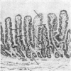
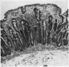
The histological appearances in 85 peroral small intestinal biopsies from children have been analysed quantitatively by the method of Dunnill and Whitehead (1972). As applied to such biopsy specimens, this technique provides indices of both the mucosal volume and of the mucosal surface-to-volume ratio.
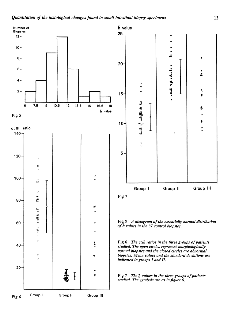
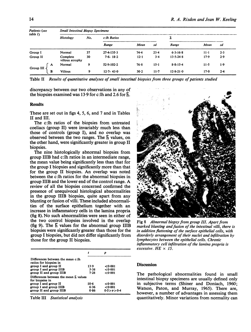
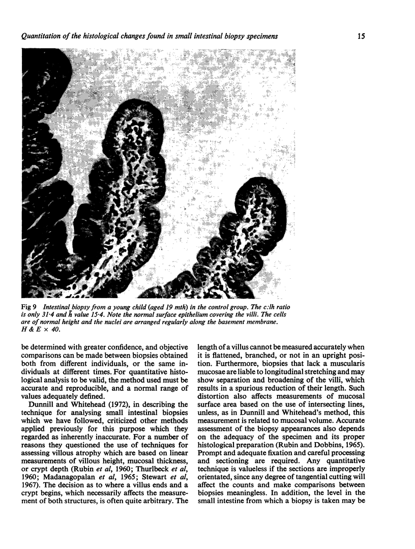
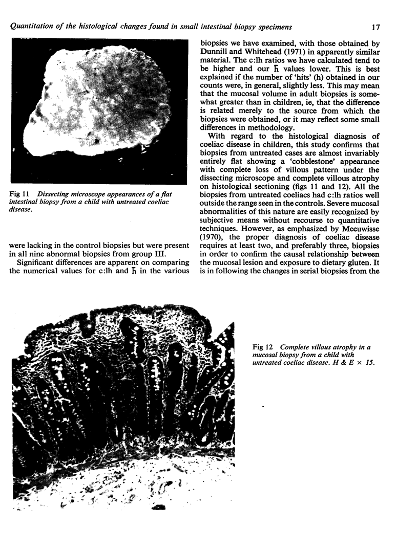
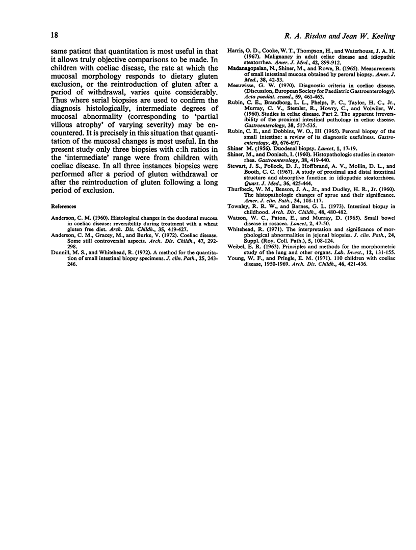
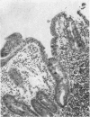
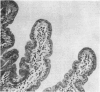
When compared with controls, `flat' biopsies from children with untreated coeliac disease invariably showed much smaller surface-to-volume (c:lh) ratios. The mucosal volumes (h) of these biopsies were, however, significantly increased. Specimens showing less marked abnormalities (socalled `partial villous atrophy') had c:lh ratios in an intermediate range, but, like the biopsies from untreated coeliacs, h values were significantly increased.
In morphologically normal biopsies correlation of surface-to-volume ratios with age showed that c:lh ratios were significantly smaller in younger children. Some of these lower c:lh ratios overlapped the range observed in abnormal biopsies showing partial villous atrophy. However, in the atrophic biopsies additional histological changes, such as abnormalities of the surface epithelial cells and an increased inflammatory cell infiltration of the lamina propria, were invariably present. Slight changes in biopsies from children under 2 years of age should, therefore, be interpreted cautiously, and should not necessarily be regarded as pathological on the evidence of the villous pattern alone.
The quantitative technique employed proved reproducible under normal working conditions, and its greatest practical value would appear to be in following accurately serial changes in biopsies from the same patient.
Full text
PDF
Images in this article

Selected References
These references are in PubMed. This may not be the complete list of references from this article.
- ANDERSON C. M. Histological changes in the duodenal mucosa in coeliac disease. Reversibility during treatment with a wheat gluten free diet. Arch Dis Child. 1960 Oct;35:419–427. doi: 10.1136/adc.35.183.419. [DOI] [PMC free article] [PubMed] [Google Scholar]
- Anderson C. M., Gracey M., Burke V. Coeliac disease. Some still controversial aspects. Arch Dis Child. 1972 Apr;47(252):292–298. doi: 10.1136/adc.47.252.292. [DOI] [PMC free article] [PubMed] [Google Scholar]
- Dunnill M. S., Whitehead R. A method for the quantitation of small intestinal biopsy specimens. J Clin Pathol. 1972 Mar;25(3):243–246. doi: 10.1136/jcp.25.3.243. [DOI] [PMC free article] [PubMed] [Google Scholar]
- Harris O. D., Cooke W. T., Thompson H., Waterhouse J. A. Malignancy in adult coeliac disease and idiopathic steatorrhoea. Am J Med. 1967 Jun;42(6):899–912. doi: 10.1016/0002-9343(67)90071-x. [DOI] [PubMed] [Google Scholar]
- MADANAGOPALAN N., SHINER M., ROWE B. MEASUREMENTS OF SMALL INTESTINAL MUCOSA OBTAINED BY PERORAL BIOPSY. Am J Med. 1965 Jan;38:42–53. doi: 10.1016/0002-9343(65)90158-0. [DOI] [PubMed] [Google Scholar]
- RUBIN C. E., BRANDBORG L. L., PHELPS P. C., TAYLOR H. C., Jr, MURRAY C. V., STEMLER R., HOWRY C., VOLWILER W. Studies of celiac disease. II. The apparent irreversibility of the proximal intestinal pathology in celiac disease. Gastroenterology. 1960 Apr;38:517–532. [PubMed] [Google Scholar]
- Rubin C. E., Dobbins W. O., 3rd Peroral biopsy of the small intestine. A review of its diagnostic usefulness. Gastroenterology. 1965 Dec;49(6):676–697. [PubMed] [Google Scholar]
- SHINER M., DONIACH I. Histopathologic studies in steatorrhea. Gastroenterology. 1960 Mar;38:419–440. [PubMed] [Google Scholar]
- SHINER M. Duodenal biopsy. Lancet. 1956 Jan 7;270(6906):17–19. doi: 10.1016/s0140-6736(56)91854-2. [DOI] [PubMed] [Google Scholar]
- Stewart J. S., Pollock D. J., Hoffbrand A. V., Mollin D. L., Booth C. C. A study of proximal and distal intestinal structure and absorptive function in idiopathic steatorrhoea. Q J Med. 1967 Jul;36(143):425–444. [PubMed] [Google Scholar]
- THURLBECK W. M., BENSON J. A., Jr, DUDLEY H. R., Jr The histopathologic changes of sprue and their significance. Am J Clin Pathol. 1960 Aug;34:108–117. doi: 10.1093/ajcp/34.2.108. [DOI] [PubMed] [Google Scholar]
- Townley R. R., Barnes G. L. Intestinal biopsy in childhood. Arch Dis Child. 1973 Jun;48(6):480–482. doi: 10.1136/adc.48.6.480. [DOI] [PMC free article] [PubMed] [Google Scholar]
- WATSON W. C., PATON E., MURRAY D. SMALL-BOWEL DISEASE IN ROSACEA. Lancet. 1965 Jul 10;1(7402):47–50. doi: 10.1016/s0140-6736(65)90128-5. [DOI] [PubMed] [Google Scholar]
- WEIBEL E. R. Principles and methods for the morphometric study of the lung and other organs. Lab Invest. 1963 Feb;12:131–155. [PubMed] [Google Scholar]
- Young W. F., Pringle E. M. 110 children with coeliac disease, 1950-1969. Arch Dis Child. 1971 Aug;46(248):421–436. doi: 10.1136/adc.46.248.421. [DOI] [PMC free article] [PubMed] [Google Scholar]