Abstract
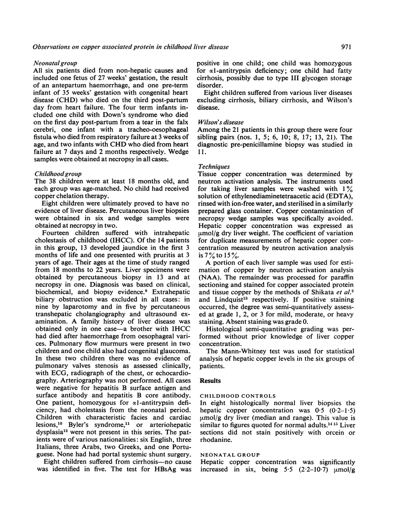
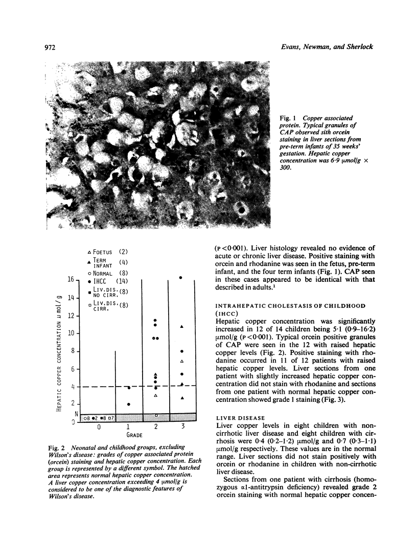
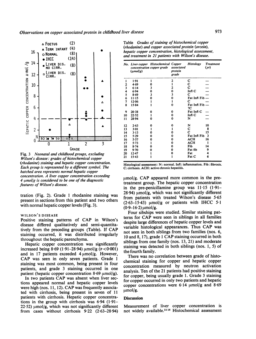
Hepatic copper concentrations were compared with staining grades of copper associated protein (CAP) and histochemical copper in liver sections from 44 patients (one fetus, one pre-term infant, four term infants, eight normal children, 16 children with various liver diseases, and 14 patients with intrahepatic cholestasis of childhood (IHCC)). A similar comparative study of hepatic copper concentration with CAP and histochemical copper was performed in 21 patients with Wilson's disease. CAP occurred in the fetus, pre-term infant, and term infants without liver disease. This suggests that CAP is a normal constituent of the hepatocyte and is not a consequence of liver disease or biliary obstruction. CAP was not seen when hepatic copper concentration was normal; it was absent in eight children with no evidence of liver disease, eight children with non-cirrhotic liver disease, and seven of eight children with cirrhosis. When hepatic copper concentration exceeded 4.0 mumol/g dry liver weight grade 2 or grade 3 staining for CAP and histochemical copper was found in the fetus, pre-term infant, infants, and IHCC. CAP was found in IHCC only in the presence of raised hepatic copper levels, supporting evidence of a relationship between copper and CAP. In 17 of 21 patients with Wilson's disease hepatic copper concentrations exceeded 4 mumol/g. Positive staining for CAP was seen in seven of these patients being usually grade 1. CAP is a normal associated protein, present when hepatic copper concentrations are increased in normal liver cells. It is usually absent in hepatocytes from Wilson's disease despite similar hepatic copper levels. CAP may represent material which protects the hepatocyte from the toxic effects of copper.
Full text
PDF
Images in this article
Selected References
These references are in PubMed. This may not be the complete list of references from this article.
- Alagille D., Odièvre M., Gautier M., Dommergues J. P. Hepatic ductular hypoplasia associated with characteristic facies, vertebral malformations, retarded physical, mental, and sexual development, and cardiac murmur. J Pediatr. 1975 Jan;86(1):63–71. doi: 10.1016/s0022-3476(75)80706-2. [DOI] [PubMed] [Google Scholar]
- Brückmann G., Zondek S. G. Iron, copper and manganese in human organs at various ages. Biochem J. 1939 Nov;33(11):1845–1857. doi: 10.1042/bj0331845. [DOI] [PMC free article] [PubMed] [Google Scholar]
- Dastur D. K., Manghani D. K. The liver in Wilson's disease. Some histochemical and histological aspects. Indian J Pathol Bacteriol. 1967 Apr;10(2):177–183. [PubMed] [Google Scholar]
- Evans J., Newman S., Sherlock S. Liver copper levels in intrahepatic cholestasis of childhood. Gastroenterology. 1978 Nov;75(5):875–878. [PubMed] [Google Scholar]
- Fleming C. R., Dickson E. R., Baggenstoss A. H., McCall J. T. Copper and primary biliary cirrhosis. Gastroenterology. 1974 Dec;67(6):1182–1187. [PubMed] [Google Scholar]
- Goldfischer S., Sternlieb I. Changes in the distribution of hepatic copper in relation to the progression of Wilson's disease (hepatolenticular degeneration). Am J Pathol. 1968 Dec;53(6):883–901. [PMC free article] [PubMed] [Google Scholar]
- Heathcote J., Deodhar K. P., Scheuer P. J., Sherlock S. Intrahepatic cholestasis in childhood. N Engl J Med. 1976 Oct 7;295(15):801–805. doi: 10.1056/NEJM197610072951503. [DOI] [PubMed] [Google Scholar]
- Jain S., Scheuer P. J., Archer B., Newman S. P., Sherlock S. Histological demonstration of copper and copper-associated protein in chronic liver diseases. J Clin Pathol. 1978 Aug;31(8):784–790. doi: 10.1136/jcp.31.8.784. [DOI] [PMC free article] [PubMed] [Google Scholar]
- Lindquist R. R. Studies on the pathogenesis of hepatolenticular degeneration. II. Cytochemical methods for the localization of copper. Arch Pathol. 1969 Apr;87(4):370–379. [PubMed] [Google Scholar]
- Porter H. The particulate half-cystine-rich copper protein of newborn liver. Relationship to metallothionein and subcellular localization in non-mitochondrial particles possibly representing heavy lysosomes. Biochem Biophys Res Commun. 1974 Feb 4;56(3):661–668. doi: 10.1016/0006-291x(74)90656-1. [DOI] [PubMed] [Google Scholar]
- Salaspuro M., Sipponen P. Demonstration of an intracellular copper-binding protein by orcein staining in long-standing cholestatic liver diseases. Gut. 1976 Oct;17(10):787–790. doi: 10.1136/gut.17.10.787. [DOI] [PMC free article] [PubMed] [Google Scholar]
- Sass-Kortsak A. Copper metabolism. Adv Clin Chem. 1965;8:1–67. doi: 10.1016/s0065-2423(08)60412-6. [DOI] [PubMed] [Google Scholar]
- Shaposhnikov A. M., Zubzitski Y. N., Shulman V. S. Identification of ceruloplasmin in human liver cells by fluorescent antibodies and absence of this protein in Wilson disease. Experientia. 1969 Apr 15;25(4):424–426. doi: 10.1007/BF01899962. [DOI] [PubMed] [Google Scholar]
- Shikata T., Uzawa T., Yoshiwara N., Akatsuka T., Yamazaki S. Staining methods of Australia antigen in paraffin section--detection of cytoplasmic inclusion bodies. Jpn J Exp Med. 1974 Feb;44(1):25–36. [PubMed] [Google Scholar]
- Sipponen P. Orcein positive hepatocellular material in long-standing biliary diseases. I. Histochemical characteristics. Scand J Gastroenterol. 1976;11(6):545–552. [PubMed] [Google Scholar]
- Sipponen P. Orcein positive hepatocellular material in long-standing biliary diseases. I. Histochemical characteristics. Scand J Gastroenterol. 1976;11(6):545–552. [PubMed] [Google Scholar]
- Sipponen P., Salaspuro M. P., Makkonen H. M. Orcein positive hepatocellular material in histological diagnosis of primary biliary cirrhosis. Ann Clin Res. 1975 Aug;7(4):273–277. [PubMed] [Google Scholar]
- Smallwood R. A., Williams H. A., Rosenoer V. M., Sherlock S. Liver-copper levels in liver disease: studies using neutron activation analysis. Lancet. 1968 Dec 21;2(7582):1310–1313. doi: 10.1016/s0140-6736(68)91814-x. [DOI] [PubMed] [Google Scholar]
- Sternlieb I., Harris R. C., Scheinberg I. H. Le cuivre dans la cirrhose biliaire de l'enfant. Rev Int Hepatol. 1966;16(5):1105–1110. [PubMed] [Google Scholar]
- Todd A. P., Thorpe M. E., Rosenoer V. M. Tissue copper determinations by neutron activation analysis. J Clin Pathol. 1967 May;20(3):276–279. doi: 10.1136/jcp.20.3.276. [DOI] [PMC free article] [PubMed] [Google Scholar]
- Watson G. H., Miller V. Arteriohepatic dysplasia: familial pulmonary arterial stenosis with neonatal liver disease. Arch Dis Child. 1973 Jun;48(6):459–466. doi: 10.1136/adc.48.6.459. [DOI] [PMC free article] [PubMed] [Google Scholar]


