

Abstract
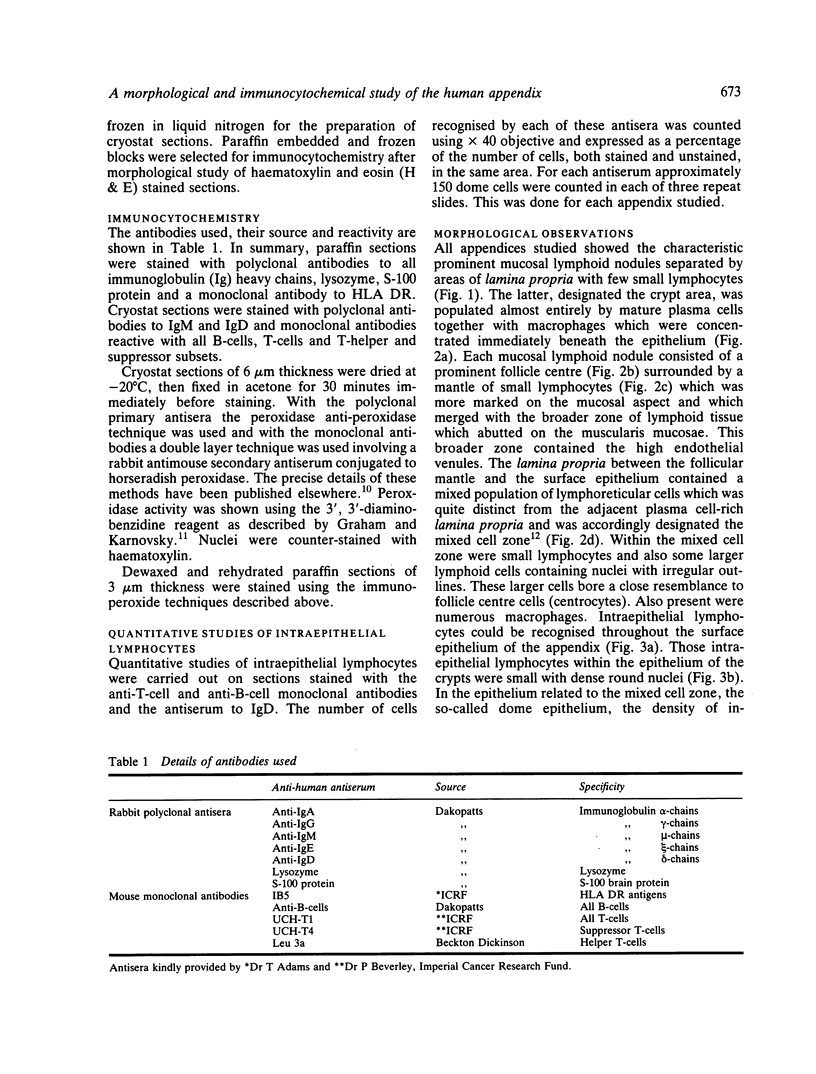
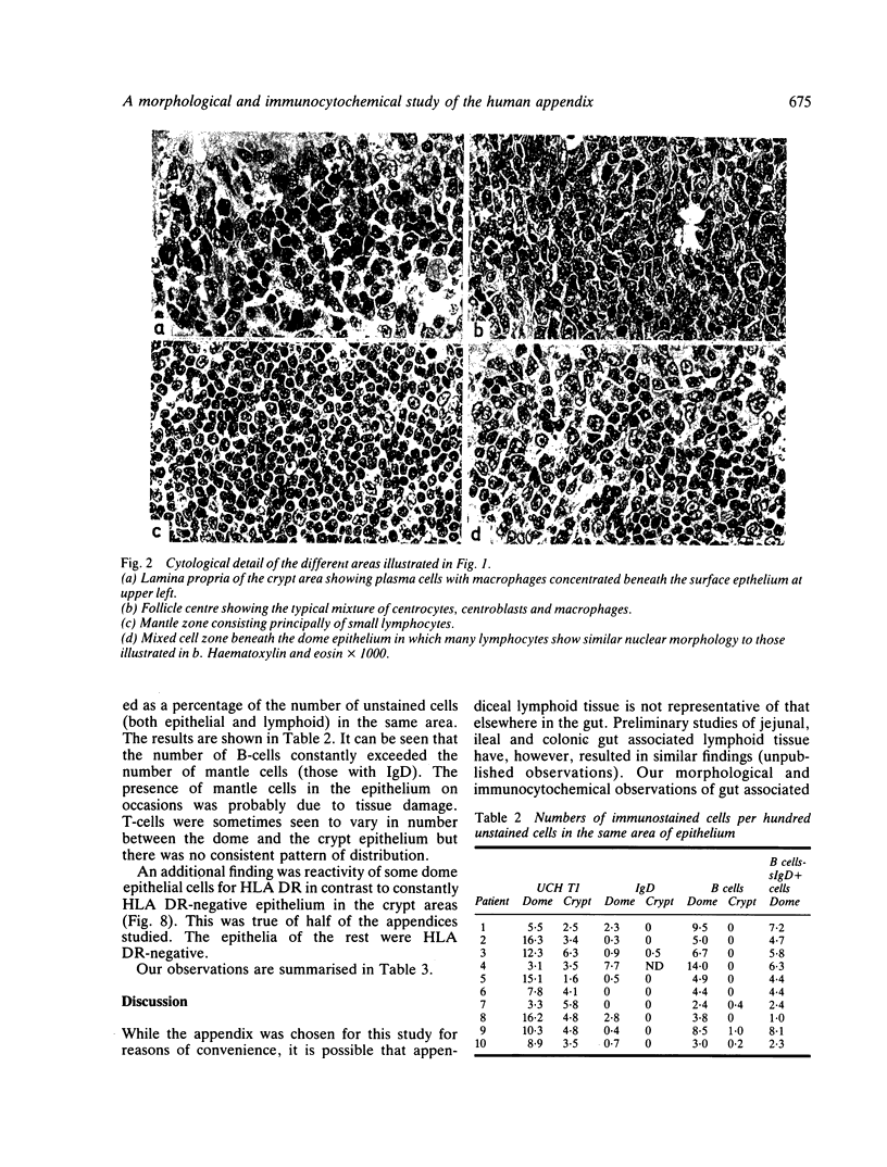
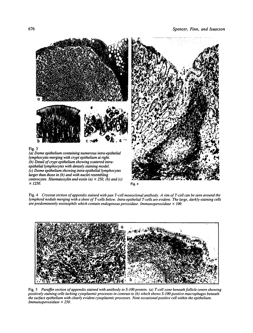
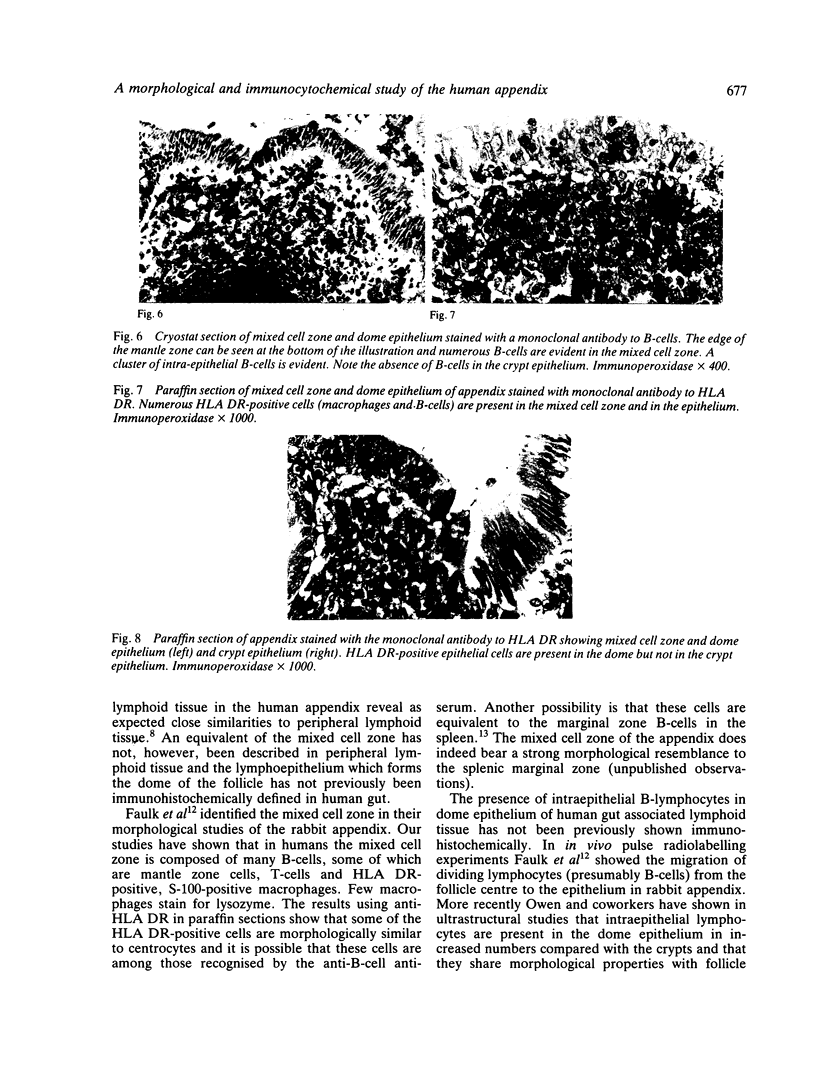
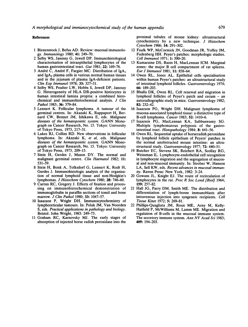
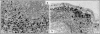
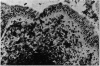
Gut associated lymphoid tissue in 15 normal appendices has been characterised in tissue sections using both morphological criteria and immunocytochemical techniques. A panel of monoclonal and polyclonal antibodies was used including antibodies to B-cells, T-cells, macrophages, HLA DR and immunoglobulins. The lymphoid tissue in the appendix was shown to bear a strong resemblance to that in lymph nodes with the exception of the region where the appendix follicles associate with the dome epithelium, which has no lymph node equivalent. This zone of cells between the lymphoid follicles and the dome epithelium termed the 'mixed cell zone' has been shown to contain an abundance of HLA DR-bearing cells, some of which have irregular nuclear morphology and resemble follicle centre cells. These cells were seen to extend into the epithelium of the dome but not the crypts. Using a monoclonal anti-B-cell antibody a population of B-cells was detected in the equivalent areas of mixed cell zone and epithelium and quantitative studies showed that these intraepithelial B-cells comprised approximately 4-5% of the cells in the epithelium. The mixed cell zone was also seen to contain T-cells, S-100 protein-containing macrophages and occasional lysozyme-containing macrophages. Plasma cells were rarely seen in this area.
Full text
PDF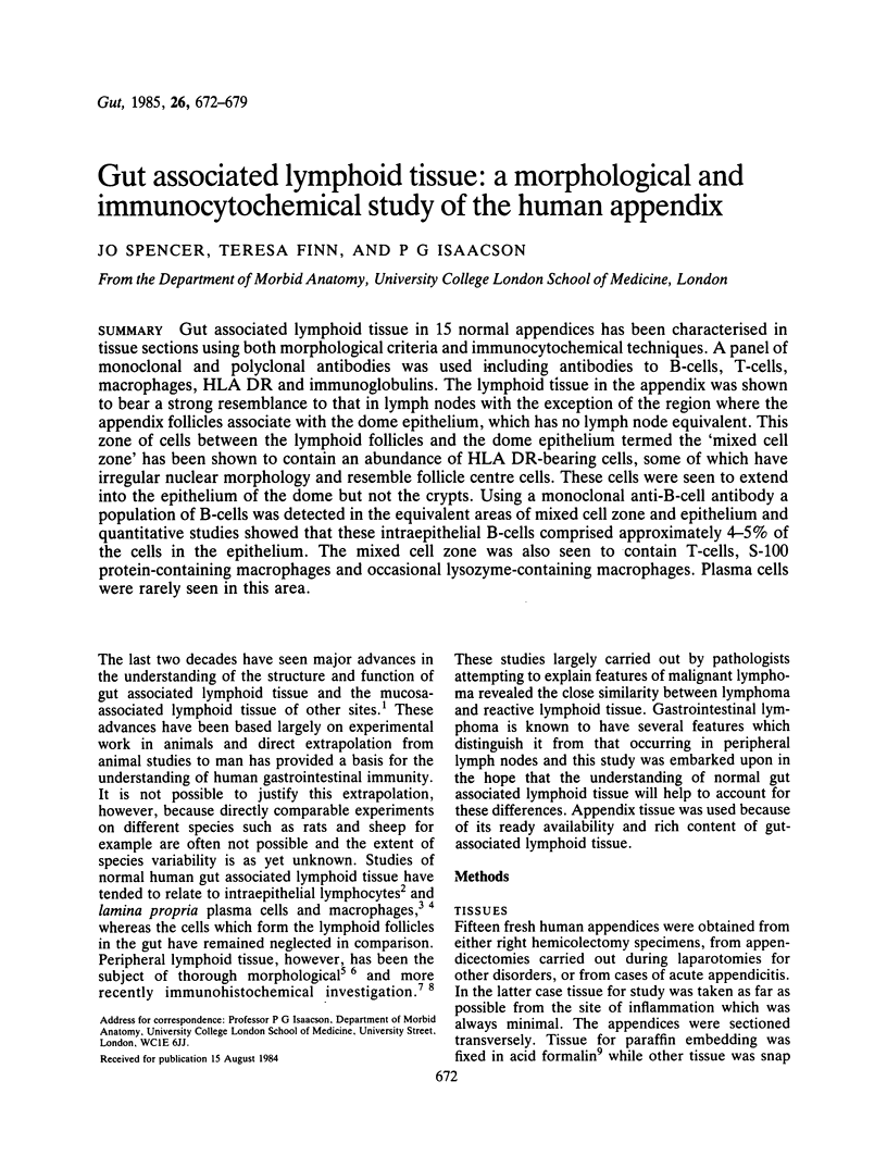


Images in this article


Selected References
These references are in PubMed. This may not be the complete list of references from this article.
- André C., André F., Fargier C. Distribution of IgA 1 and IgA 2 plasma cells in various normal human tissues and in the jejunum of plasma IgA-deficient patients. Clin Exp Immunol. 1978 Aug;33(2):327–331. [PMC free article] [PubMed] [Google Scholar]
- Bhalla D. K., Owen R. L. Cell renewal and migration in lymphoid follicles of Peyer's patches and cecum--an autoradiographic study in mice. Gastroenterology. 1982 Feb;82(2):232–242. [PubMed] [Google Scholar]
- Bienenstock J., Befus A. D. Mucosal immunology. Immunology. 1980 Oct;41(2):249–270. [PMC free article] [PubMed] [Google Scholar]
- Curran R. C., Gregory J. Effects of fixation and processing on immunohistochemical demonstration of immunoglobulin in paraffin sections of tonsil and bone marrow. J Clin Pathol. 1980 Nov;33(11):1047–1057. doi: 10.1136/jcp.33.11.1047. [DOI] [PMC free article] [PubMed] [Google Scholar]
- Faulk W. P., McCormick J. N., Goodman J. R., Yoffey J. M., Fudenberg H. H. Peyer's patches: morphologic studies. Cell Immunol. 1970 Nov;1(5):500–520. doi: 10.1016/0008-8749(70)90038-9. [DOI] [PubMed] [Google Scholar]
- GOWANS J. L., KNIGHT E. J. THE ROUTE OF RE-CIRCULATION OF LYMPHOCYTES IN THE RAT. Proc R Soc Lond B Biol Sci. 1964 Jan 14;159:257–282. doi: 10.1098/rspb.1964.0001. [DOI] [PubMed] [Google Scholar]
- Graham R. C., Jr, Karnovsky M. J. The early stages of absorption of injected horseradish peroxidase in the proximal tubules of mouse kidney: ultrastructural cytochemistry by a new technique. J Histochem Cytochem. 1966 Apr;14(4):291–302. doi: 10.1177/14.4.291. [DOI] [PubMed] [Google Scholar]
- Hall J. G., Parry D. M., Smith M. E. The distribution and differentiation of lymph-borne immunoblasts after intravenous injection into syngeneic recipients. Cell Tissue Kinet. 1972 May;5(3):269–281. doi: 10.1111/j.1365-2184.1972.tb00365.x. [DOI] [PubMed] [Google Scholar]
- Isaacson P. G., MacLennan K. A., Subbuswamy S. G. Multiple lymphomatous polyposis of the gastrointestinal tract. Histopathology. 1984 Jul;8(4):641–656. doi: 10.1111/j.1365-2559.1984.tb02377.x. [DOI] [PubMed] [Google Scholar]
- Isaacson P., Wright D. H. Malignant lymphoma of mucosa-associated lymphoid tissue. A distinctive type of B-cell lymphoma. Cancer. 1983 Oct 15;52(8):1410–1416. doi: 10.1002/1097-0142(19831015)52:8<1410::aid-cncr2820520813>3.0.co;2-3. [DOI] [PubMed] [Google Scholar]
- Kumararatne D. S., Bazin H., MacLennan I. C. Marginal zones: the major B cell compartment of rat spleens. Eur J Immunol. 1981 Nov;11(11):858–864. doi: 10.1002/eji.1830111103. [DOI] [PubMed] [Google Scholar]
- Owen R. L., Jones A. L. Epithelial cell specialization within human Peyer's patches: an ultrastructural study of intestinal lymphoid follicles. Gastroenterology. 1974 Feb;66(2):189–203. [PubMed] [Google Scholar]
- Owen R. L. Sequential uptake of horseradish peroxidase by lymphoid follicle epithelium of Peyer's patches in the normal unobstructed mouse intestine: an ultrastructural study. Gastroenterology. 1977 Mar;72(3):440–451. [PubMed] [Google Scholar]
- Phillips-Quagliata J. M., Roux M. E., Arny M., Kelly-Hatfield P., McWilliams M., Lamm M. E. Migration and regulation of B-cells in the mucosal immune system. Ann N Y Acad Sci. 1983 Jun 30;409:194–203. doi: 10.1111/j.1749-6632.1983.tb26869.x. [DOI] [PubMed] [Google Scholar]
- Selby W. S., Janossy G., Jewell D. P. Immunohistological characterisation of intraepithelial lymphocytes of the human gastrointestinal tract. Gut. 1981 Mar;22(3):169–176. doi: 10.1136/gut.22.3.169. [DOI] [PMC free article] [PubMed] [Google Scholar]
- Selby W. S., Poulter L. W., Hobbs S., Jewell D. P., Janossy G. Heterogeneity of HLA-DR-positive histiocytes in human intestinal lamina propria: a combined histochemical and immunohistological analysis. J Clin Pathol. 1983 Apr;36(4):379–384. doi: 10.1136/jcp.36.4.379. [DOI] [PMC free article] [PubMed] [Google Scholar]
- Stein H., Bonk A., Tolksdorf G., Lennert K., Rodt H., Gerdes J. Immunohistologic analysis of the organization of normal lymphoid tissue and non-Hodgkin's lymphomas. J Histochem Cytochem. 1980 Aug;28(8):746–760. doi: 10.1177/28.8.7003001. [DOI] [PubMed] [Google Scholar]
- Stein H., Gerdes J., Mason D. Y. The normal and malignant germinal centre. Clin Haematol. 1982 Oct;11(3):531–559. [PubMed] [Google Scholar]