Abstract
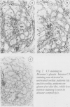
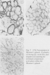
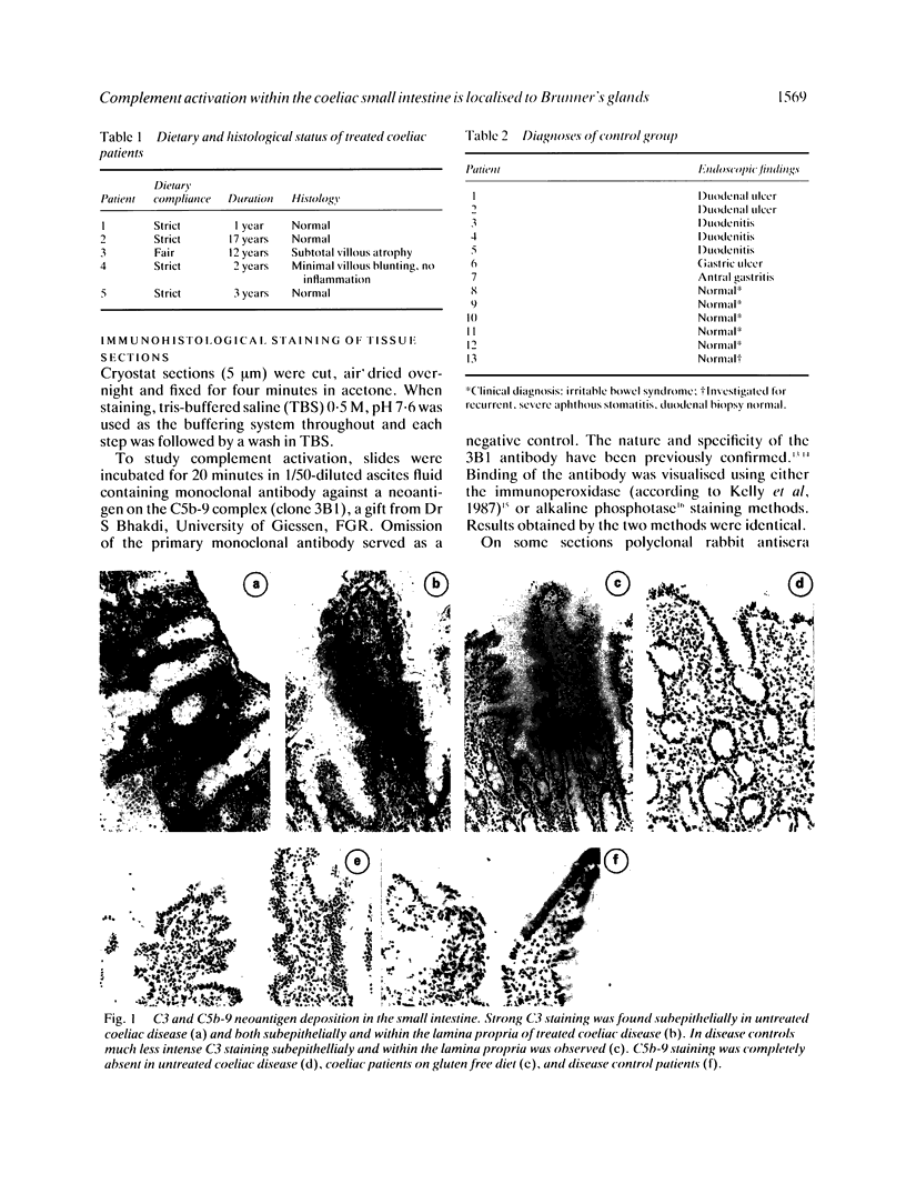
Complement activation may play an important role in the pathogenesis of coeliac disease. In the present study immunohistochemical localisation of C3 and of a neoantigen exposed only on the terminal C5b-9 complement complex has been performed on small intestinal biopsy sections from newly diagnosed untreated coeliac patients, from coeliac patients on long-term gluten-free diet and from disease controls. Levels of C3 were markedly increased in treated coeliac patients compared with controls. Staining of C3 was concentrated subepithelially and within the centre of the lamina propria. No staining was detected at these sites using antibody to the neoantigen, however, strongly suggesting that the increased levels of C3 seen in the coeliac patients was the result of increased extravasation of serum proteins rather than complement activation. Surprisingly, complement activation was detected within the glands of Brunner. Positive staining using anti-C5b-9 neoantigen was found in all coeliac patients, both treated and untreated. Three of the 13 disease controls also showed reactivity with this antibody. This novel finding suggests that Brunner's glands, hitherto largely neglected structures, may play an important role in the development of coeliac disease.
Full text
PDF
Images in this article
Selected References
These references are in PubMed. This may not be the complete list of references from this article.
- Baklien K., Brandtzaeg P. Letter: Immunohistochemical localization of complement in intestinal mucosa. Lancet. 1974 Nov 2;2(7888):1087–1088. doi: 10.1016/s0140-6736(74)92198-9. [DOI] [PubMed] [Google Scholar]
- Bhakdi S., Muhly M., Roth M. Preparation and isolation of specific antibodies to complement components. Methods Enzymol. 1983;93:409–420. doi: 10.1016/s0076-6879(83)93054-9. [DOI] [PubMed] [Google Scholar]
- Boackle R. J., Pruitt K. M., Mestecky J. The interactions of human complement with interfacially aggregated preparations of human secretory IgA. Immunochemistry. 1974 Sep;11(9):543–548. doi: 10.1016/0019-2791(74)90245-6. [DOI] [PubMed] [Google Scholar]
- Carney D. F., Koski C. L., Shin M. L. Elimination of terminal complement intermediates from the plasma membrane of nucleated cells: the rate of disappearance differs for cells carrying C5b-7 or C5b-8 or a mixture of C5b-8 with a limited number of C5b-9. J Immunol. 1985 Mar;134(3):1804–1809. [PubMed] [Google Scholar]
- Cordell J. L., Falini B., Erber W. N., Ghosh A. K., Abdulaziz Z., MacDonald S., Pulford K. A., Stein H., Mason D. Y. Immunoenzymatic labeling of monoclonal antibodies using immune complexes of alkaline phosphatase and monoclonal anti-alkaline phosphatase (APAAP complexes). J Histochem Cytochem. 1984 Feb;32(2):219–229. doi: 10.1177/32.2.6198355. [DOI] [PubMed] [Google Scholar]
- Ferguson A., Murray D. Quantitation of intraepithelial lymphocytes in human jejunum. Gut. 1971 Dec;12(12):988–994. doi: 10.1136/gut.12.12.988. [DOI] [PMC free article] [PubMed] [Google Scholar]
- Hugo F., Jenne D., Bhakdi S. Monoclonal antibodies against neoantigens of the terminal C5b-9 complex of human complement. Biosci Rep. 1985 Aug;5(8):649–658. doi: 10.1007/BF01116996. [DOI] [PubMed] [Google Scholar]
- Imai H., Chen A., Wyatt R. J., Rifai A. Lack of complement activation by human IgA immune complexes. Clin Exp Immunol. 1988 Sep;73(3):479–483. [PMC free article] [PubMed] [Google Scholar]
- Kelly J., O'Farrelly C., O'Mahony C., Weir D. G., Feighery C. Immunoperoxidase demonstration of the cellular composition of the normal and coeliac small bowel. Clin Exp Immunol. 1987 Apr;68(1):177–188. [PMC free article] [PubMed] [Google Scholar]
- Kelly J., Whelan C. A., Weir D. G., Feighery C. Removal of endogenous peroxidase activity from cryostat sections for immunoperoxidase visualisation of monoclonal antibodies. J Immunol Methods. 1987 Jan 26;96(1):127–132. doi: 10.1016/0022-1759(87)90376-0. [DOI] [PubMed] [Google Scholar]
- Marsh M. N., Hinde J. Inflammatory component of celiac sprue mucosa. I. Mast cells, basophils, and eosinophils. Gastroenterology. 1985 Jul;89(1):92–101. doi: 10.1016/0016-5085(85)90749-8. [DOI] [PubMed] [Google Scholar]
- Mohammed I., Holborow E. J., Fry L., Thompson B. R., Hoffbrand A. V., Stewart J. S. Multiple immune complexes and hypocomplementaemia in dermatitis herpetiformis and coeliac disease. Lancet. 1976 Sep 4;1(7984):487–490. doi: 10.1016/s0140-6736(76)90787-x. [DOI] [PubMed] [Google Scholar]
- Schäfer H., Mathey D., Hugo F., Bhakdi S. Deposition of the terminal C5b-9 complement complex in infarcted areas of human myocardium. J Immunol. 1986 Sep 15;137(6):1945–1949. [PubMed] [Google Scholar]
- Scott B. B., Goodall A., Stephenson P., Jenkins D. Small intestinal plasma cells in coeliac disease. Gut. 1984 Jan;25(1):41–46. doi: 10.1136/gut.25.1.41. [DOI] [PMC free article] [PubMed] [Google Scholar]
- Scott B. B., Scott D. G., Losowsky M. S. Jejunal mucosal immunoglobulins and complement in untreated coeliac disease. J Pathol. 1977 Apr;121(4):219–223. doi: 10.1002/path.1711210405. [DOI] [PubMed] [Google Scholar]
- Shiner M., Ballard J. Antigen-antibody reactions in jejunal mucosa in childhood coeliac disease after gluten challenge. Lancet. 1972 Jun 3;1(7762):1202–1205. doi: 10.1016/s0140-6736(72)90924-5. [DOI] [PubMed] [Google Scholar]
- Shorrock C. J., Rees W. D. Overview of gastroduodenal mucosal protection. Am J Med. 1988 Feb 22;84(2A):25–34. doi: 10.1016/0002-9343(88)90251-3. [DOI] [PubMed] [Google Scholar]
- Wingren U., Hallert C., Norrby K., Enerbäck L. Histamine and mucosal mast cells in gluten enteropathy. Agents Actions. 1986 Apr;18(1-2):266–268. doi: 10.1007/BF01988038. [DOI] [PubMed] [Google Scholar]


