

Abstract
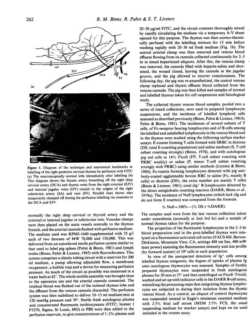
The emigration of labelled thymus cells in the pig was studied directly in blood draining the large right distal cervical lobe of the thymus after controlled labelling with FITC delivered through cannulated branches of a main thymic artery and vein by temporary ex vivo perfusion at body temperature. Roughly 1% of thymic cells emigrated per day. Unlike most thymocytes, which are small, the size spectrum of thymic emigrants is slightly larger than that of typical blood lymphocytes. Surface-marker studies show that the surface phenotypes of the emigrants differ from both typical thymus and peripheral blood lymphocytes. Although the emigrants resemble thymocytes in the high proportion of strong rosettes formed with sheep red blood cells (RBC), they rosette poorly with pig red cells, particularly in the unenhanced saline test, in this respect behaving like blood lymphocytes. The peripheral T-cell subset bearing a Fc receptor is almost absent in thymus, but is well represented among the emigrants which thus resemble corticosteroid-resistant thymocytes in the pig. The large population of thymus-dependent Null lymphocytes in young pig blood apparently arise in thymus since they constitute 1/3 of emigrants, although only forming less than 10% of thymus cells. This emigration of thymic cells is discussed in relation to its implications for the turnover of known functional peripheral T-cell populations.
Full text
PDF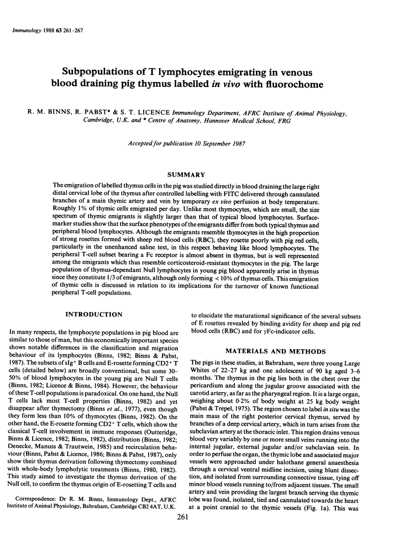

Images in this article
Selected References
These references are in PubMed. This may not be the complete list of references from this article.
- Alexander E. L., Titus J. A., Segal D. M. Quantitation of Fc receptors and surface immunoglobulin is affected by cell isolation procedures using plasmagel and ficoll-hypaque. J Immunol Methods. 1978;22(3-4):263–272. doi: 10.1016/0022-1759(78)90034-0. [DOI] [PubMed] [Google Scholar]
- Binns R. M., Licence S. T. A major subpopulation of Fc receptor-bearing lymphocytes revealed by rosette formation in dextran media: studies with pig, sheep and rat lymphocytes. J Immunol Methods. 1981;43(2):153–162. doi: 10.1016/0022-1759(81)90018-1. [DOI] [PubMed] [Google Scholar]
- Binns R. M., Licence S. T. Patterns of migration of labelled blood lymphocyte subpopulations: evidence for two types of Peyer's patch in the young pig. Adv Exp Med Biol. 1985;186:661–668. doi: 10.1007/978-1-4613-2463-8_81. [DOI] [PubMed] [Google Scholar]
- Binns R. M., Licence S. T., Symons D. B., Gurner B. W., Coombs R. R., Walters D. E. Comparison of the direct antiglobulin rosetting reaction (DARR) and direct immunofluorescence (DIF) for demonstration of sIg-bearing lymphocytes in pigs, sheep and cattle. Immunology. 1979 Mar;36(3):549–555. [PMC free article] [PubMed] [Google Scholar]
- Binns R. M. Organisation of the lymphoreticular system and lymphocyte markers in the pig. Vet Immunol Immunopathol. 1982 Jan;3(1-2):95–146. doi: 10.1016/0165-2427(82)90033-2. [DOI] [PubMed] [Google Scholar]
- Binns R. M., Pabst R., Licence S. T. Classification of lymphocytes recirculating in the spleen. Immunology. 1981 Oct;44(2):273–279. [PMC free article] [PubMed] [Google Scholar]
- Binns R. M., Pabst R., Licence S. T. Lymphocyte emigration from lymph nodes by blood in the pig and efferent lymph in the sheep. Immunology. 1985 Jan;54(1):105–111. [PMC free article] [PubMed] [Google Scholar]
- Binns R. M., Pallares V., Symons D. B., Sibbons P. Effect of thymectomy on lymphocyte subpopulations in the pig. Demonstration of a thymus-dependent 'null' cell. Int Arch Allergy Appl Immunol. 1977;55(1-6):96–101. doi: 10.1159/000231915. [DOI] [PubMed] [Google Scholar]
- Binns R. M. Sheep erythrocyte rosettes in pigs, sheep, cattle and goats demonstrated in the presence of dextran. J Immunol Methods. 1978;21(3-4):197–210. doi: 10.1016/0022-1759(78)90146-1. [DOI] [PubMed] [Google Scholar]
- Binns R. M., Vaiman M., Davies H., Symons D. B. Characterization of pig lymphocyte subpopulations by adherence to nylon wool. Int Arch Allergy Appl Immunol. 1979;58(2):128–134. doi: 10.1159/000232184. [DOI] [PubMed] [Google Scholar]
- Denecke R., Mannuss B., Trautwein G. Immunological and enzyme histochemical identification of lymphocyte subpopulations in pig lymphoreticular tissues. Zentralbl Veterinarmed B. 1985 Oct;32(9):660–678. doi: 10.1111/j.1439-0450.1985.tb02008.x. [DOI] [PubMed] [Google Scholar]
- Georgieva R. Dynamics of T-suppressor and T-helper lymphocytes and haemolytic plaque-forming cells during normal pregnancy in the sow. J Reprod Immunol. 1984 May;6(3):151–156. doi: 10.1016/0165-0378(84)90020-2. [DOI] [PubMed] [Google Scholar]
- Joel D. D., Chanana A. D., Cronkite E. P. Thymus cell migration. Ser Haematol. 1974;7(4):464–481. [PubMed] [Google Scholar]
- Licence S. T., Binns R. M. Analysis of a sheep anti-pig T lymphoblast serum with specificity for E rosette-forming lymphocytes. Vet Immunol Immunopathol. 1984 Oct;7(3-4):255–273. doi: 10.1016/0165-2427(84)90084-9. [DOI] [PubMed] [Google Scholar]
- Outteridge P. M., Binns R. M., Licence S. T. Subpopulations of pig blood E-rosette-forming lymphocytes and thymus-dependent null cells: separation by nylon wool columns, rosette formation and macrophage-dependent mitogen and antigen responsiveness. Int Arch Allergy Appl Immunol. 1982;67(1):18–24. doi: 10.1159/000232982. [DOI] [PubMed] [Google Scholar]
- Pabst R., Binns R. M. In vivo labelling of the spleen and mesenteric lymph nodes with fluorescein isothiocyanate for lymphocyte migration studies. Immunology. 1981 Oct;44(2):321–329. [PMC free article] [PubMed] [Google Scholar]
- Pabst R., Binns R. M., Licence S. T. Surface markers on lymphocytes leaving pig lymph nodes. Immunology. 1985 Oct;56(2):301–306. [PMC free article] [PubMed] [Google Scholar]
- Pabst R., Licence S. T., Binns R. M. Classification of newly formed and migrating splenic lymphocytes. Clin Exp Immunol. 1983 Feb;51(2):333–337. [PMC free article] [PubMed] [Google Scholar]
- Pescovitz M. D., Lunney J. K., Sachs D. H. Murine anti-swine T4 and T8 monoclonal antibodies: distribution and effects on proliferative and cytotoxic T cells. J Immunol. 1985 Jan;134(1):37–44. [PubMed] [Google Scholar]
- Salmon H. Rosette-formation of pig thymic lymphocytes with sheep and pig erythrocytes. II. Markers for cortical and medullary thymocytes. Thymus. 1983 Mar;5(2):105–113. [PubMed] [Google Scholar]
- Scollay R. G., Butcher E. C., Weissman I. L. Thymus cell migration. Quantitative aspects of cellular traffic from the thymus to the periphery in mice. Eur J Immunol. 1980 Mar;10(3):210–218. doi: 10.1002/eji.1830100310. [DOI] [PubMed] [Google Scholar]
- Scollay R., Wilson A., Shortman K. Thymus cell migration: analysis of thymus emigrants with markers that distinguish medullary thymocytes from peripheral T cells. J Immunol. 1984 Mar;132(3):1089–1094. [PubMed] [Google Scholar]