Abstract
Helicobacter pylori babA encodes an outer membrane protein that binds to fucosylated Lewis b blood group antigen. We analyzed a panel of 35 H. pylori strains and identified three possible chromosomal loci for babA. There was a significant association between the presence of babA and the presence of cagA (P = 0.0001). Phylogenetic analysis of babA alleles revealed two divergent families of signal sequences. Among 17 strains in which an intact in-frame babA allele was identified, 10 expressed a detectable BabA protein. Expression of a BabA protein and the Lewis b-binding phenotype were not dependent on the chromosomal locus of babA. These data indicate that there is marked heterogeneity among H. pylori strains in babA genetic content and BabA expression.
Helicobacter pylori is a gram-negative bacterium that colonizes the human stomach. H. pylori genomes contain about 30 related hop genes, which are predicted to encode outer membrane proteins (1). Several Hop proteins, including BabA, SabA, AlpA, AlpB, and HopZ, can mediate adherence of H. pylori to gastric epithelial cells. The BabA adhesin binds to the fucosylated Lewis b blood group antigen on the surfaces of these cells (3, 8).
Some H. pylori strains bind Lewis b antigen, whereas other strains do not (3, 5, 8). Differences among H. pylori strains in the Lewis b binding phenotype can potentially be explained by the observation that only some of them express a BabA protein (7, 15). Peptic ulcer disease and gastric adenocarcinoma occur more commonly in persons infected with BabA-expressing H. pylori strains than in persons infected with strains that do not express BabA (5, 15). The molecular basis for variation among strains in expression of the BabA protein has not yet been investigated in any detail. In a previous study, we analyzed BabA expression by immunoblotting with two anti-BabA ScFv antibodies and detected expression of BabA in only about half of the H. pylori strains tested (7). In the current study, we studied the same panel of strains in order to analyze babA genetic content and investigate the molecular basis for variation among strains in BabA expression.
Genome sequence analyses of H. pylori strains J99 and 26695 indicated that babA can be present in one of two possible chromosomal loci (2, 14), which we designate locus A and locus B, respectively. Strain J99 expresses BabA and binds to Lewis b antigen, whereas strain 26695 does not express a detectable BabA protein and does not bind to Lewis b antigen (7, 8, 11). The mechanistic basis for the absence of BabA expression in strain 26695 is not known. As a first step in analyzing babA, we designed PCR primers that could amplify locus A and locus B (Table 1), and we analyzed a panel of 35 previously described H. pylori strains (7). PCR products of the expected sizes were successfully amplified from locus A (>3.5 kb) and from locus B (>3 kb) in 32 and 33 of the H. pylori strains tested, respectively (Table 2). These amplicons were analyzed initially by nucleotide sequencing using primers A1-S, A3-AS, or A3b-AS (Table 1). The sequence data (about 600 to 800 nucleotides from each strain) were then compared with nucleotide sequences deposited in GenBank in order to identify the most closely related sequences.
TABLE 1.
Oligonucleotide primers used for analysis of H. pylori babA
| Primer name | Primer sequence | Descriptiona |
|---|---|---|
| A99-S | 5′GTATTTTGTGTAGTCTTTGTTGGTGG | jhp0832 (HP0893); locus A |
| A99-AS | 5′GCAGTTGCATGGTCAGCTCTGAGG | jhp0835 (HP0898); locus A |
| AHp-S | 5′CCACAGCATGATCATAGAGTATAGAG | jhp1163 (HP1242); locus B |
| AHp-AS | 5′GGCTTTAATCCCCTACATTGTGGA | jhp1165 (HP1244); locus B |
| Hp1-S | 5′GAAGCTTTGGTGTGGGGTTA | jhp0298 (HP0313); locus C |
| Hp1-AS | 5′ACCCTAATGGGCATGTGGTA | jhp0301 (HP0318); locus C |
| OPE1243 | 5′ACCATCTTCAACAACGAGCCAGGG | jhp0833 (babA), bp 418-441 (forward) |
| OP9647 | 5′GGTATCGATCCACTTCCATCAC | jhp0833 (babA), bp 440-461 (forward) |
| OPE1244 | 5′AGCTCTTGCGCGTCCGTGATC | jhp0833 (babA), bp 1002-1022 (reverse) |
| OPE1245 | 5′GTGAAAGGGTTGAAAGGCTTGC | jhp0833 (babA), bp 1085-1106 (reverse) |
| A1-S | 5′CCCGGGAGACGACGGCTTTTACACAb | jhp0833 (babA), bp 63-81 (forward) |
| A3-AS | 5′CTCGAGGATGCTGTTTTTAAGATTTb | jhp0833 (babA), bp 1380-1362 (reverse) |
| A3b-AS | 5′GCTGTATCTGCTGCTCTTGAGTGCC | jhp0833 (babA), bp 1414-1390 (reverse) |
Primers were derived from the indicated H. pylori chromosomal loci in H. pylori strain 26695 or J99. Locus A corresponds to the site of babA (jhp0833) in strain J99 and the site of babB (HP0896) in strain 26695. Locus B corresponds to the site of babA (HP1243) in strain 26695 and the site of babB (jhp1164) in strain J99. Locus C corresponds to the site of omp9 (HP0317) in strain 26695.
Underlined sequences represent restriction sites for SmaI and XhoI.
TABLE 2.
Analysis of three potential chromosomal loci for babA in a panel of 35 H. pylori strains
| Locusa | No. of strainsb | No. of strains in which the following gene was present in locus A, B, or C
|
||
|---|---|---|---|---|
| babA | babB | omp9 | ||
| A | 32 | 19 | 8 | 5 |
| B | 33 | 4 | 24 | 5 |
| C | 7 | 3 | 2 | 2 |
Chromosomal loci A, B, and C are defined as described in the text.
Total number of strains containing a detectable hop gene in locus A, B, or C.
As shown in Tables 2 and 3, 19 of the 32 sequences from locus A and 4 of the 33 sequences from locus B were most closely related to babA. Two strains (92-25 and J195) contained babA in both locus A and locus B. The other sequences were most closely related either to babB (exemplified by the genes designated HP0896 and jhp1164 in strains 26695 and J99, respectively) or to omp9 (hopU) (exemplified by HP0317 in strain 26695) (Table 2). These experiments demonstrated that babA is found more commonly in locus A than in locus B and that babA chromosomal loci frequently contain one of two alternate hop genes (babB or omp9).
TABLE 3.
Analysis of BabA expression and babA genetic content in 35 H. pylori strains
| Strain | cagAa | BabA proteinb | Lewis b bindingc | babA localizationd | BabA lengthe |
|---|---|---|---|---|---|
| J99 | + | + | 2.3 | A | 744 |
| 87-29 | + | + | 1.7 | A | 741 |
| 87-33 | + | + | 2.5 | A | 740 |
| J258 | + | + | 1.4 | A | 744 |
| J133 | + | + | 1.7 | A | ND |
| 87-199 | + | + | 1.8 | B | 744 |
| C | 744 | ||||
| 92-25 | + | + | 2.3 | A | ND |
| B | ND | ||||
| 87-81 | + | + | 1.8 | C | 735 |
| J262 | − | + | 1.1 | A | ND |
| J190 | − | + | 1.0 | — | NA |
| 87-91 | + | + | 0.9 | ID | ND |
| 87-203 | − | + | 1.0 | ID | ND |
| 92-18 | + | + | 0.9 | A | 742 |
| 92-26 | + | + | 1.0 | A | 737 |
| J116 | + | + | 1.0 | A | 742 |
| J123 | + | + | 0.9 | A | 744 |
| 92-19 | + | − | 1.1 | A | 738 |
| 92-21 | + | − | 1.1 | A | 736 |
| J166 | + | − | 0.9 | A | ND |
| J195 | − | − | 1.0 | A | 55 |
| B | 55 | ||||
| J223 | + | − | 1.0 | A | 745 |
| 26695 | + | − | 1.0 | B | 733 |
| 86-338 | − | − | 1.1 | A | 736 |
| J178 | + | − | 0.9 | A | 737 |
| 60190 | + | − | 1.2 | A | 721 |
| C | 721 | ||||
| 92-28 | − | − | 1.2 | — | NA |
| J154 | − | − | 1.1 | — | NA |
| 86-313 | − | − | 1.0 | — | NA |
| J128 | + | − | 0.9 | — | NA |
| 92-20 | − | − | 1.1 | — | NA |
| 92-23 | − | − | 1.0 | — | NA |
| 87-230 | − | − | 0.9 | — | NA |
| 87-75 | − | − | 0.9 | — | NA |
| J63 | − | − | 1.1 | — | NA |
| Tx30a | − | − | 0.9 | — | NA |
Presence or absence of cagA nucleotide sequences (7).
Immunoreactivity with anti-BabA ScFv antibodies (7).
Values represent a ratio of optical density values, comparing binding of H. pylori strains to Lewis b versus binding to an albumin control. In this assay, values of >1.5 have been considered indicative of binding to Lewis b (5, 9, 13).
Chromosomal loci A, B, and C are defined as described in the text. ID (indeterminate), babA sequences were detected in these strains but the babA chromosomal locus was not identified. —, babA sequences were not detected.
Deduced lengths (in amino acids) of the encoded BabA proteins. Five previously determined babA sequences correspond to GenBank accession numbers AY549174 to AY549178 (7), and new babA sequences have been assigned GenBank accession numbers DQ225153 to DQ225165. ND, not determined; NA, not applicable.
Based on the detection of omp9 in H. pylori chromosomal loci known to commonly contain babA or babB, we reasoned that intragenomic recombination events might involve babA and omp9 sequences, similar to what has been described for babA and babB sequences (10, 13). To analyze the locus corresponding to the site of omp9 in H. pylori strain 26695 (designated locus C), appropriate primers (derived from genes flanking the omp9 locus in H. pylori strain 26695) were designed (Table 1) and used for PCR assays. Amplicons of >4 kb were obtained from seven strains, and the remaining strains yielded amplicons of ≤2.8 kb. Sequence analysis revealed that three of the larger amplicons contained babA, two contained omp9, and two contained babB (Table 2). These data indicate that babA may be found in one of at least three possible chromosomal loci (Table 3).
In the analyses described above, 13 of the 35 strains did not contain detectable babA sequences in locus A, B, or C (Table 3). To investigate by another approach whether babA sequences were present, we attempted to PCR amplify fragments of babA from these strains, using primers OPE1243, OP9647, OPE1244, and OPE1245 in all four possible combinations (Table 1). These primers were designed, based on aligned sequences from many different H. pylori strains, to amplify babA but not related H. pylori hop gene sequences. PCR products of the expected size were amplified from 2 strains (87-91 and 87-203) but not from the remaining 11 strains. Sequence analysis confirmed that these two amplicons were fragments of babA. We hypothesize that there is an unidentified chromosomal locus for babA in these two strains. Thus, among the 35 strains analyzed in the current study, 24 contained babA sequences and 11 did not. Our conclusion that some strains lack babA is consistent with results obtained in other studies using microarray analysis (12), Southern hybridization (13), or PCR using different primers (5, 15). Twenty of the 24 strains that contained babA contained cagA (a marker for the cag pathogenicity island); cagA was present in only 1 of the 11 strains that lacked babA (P = 0.0001) (Table 3).
To gain further insight into babA genetic diversity, we analyzed the complete babA nucleotide sequences from 18 different H. pylori strains, including 3 strains (87-199, 60190, and J195) that contained two copies of babA (Table 3). This was accomplished by sequencing the PCR products described above on both strands with appropriate primers. In strain 87-199 (one of the strains that contained multiple copies of babA), the babA sequences found in loci B and C were identical and encoded full-length BabA proteins. In strain 60190, the babA sequences from loci A and C were almost identical except for three substitutions near the 5′ ends of the genes; both copies of babA in this strain encoded full-length BabA proteins. In strain J195, the babA sequences found in loci A and B were identical, and each contained a frameshift mutation that prevented expression of a full-length BabA protein. The multiple copies of babA in these strains presumably resulted from gene conversion (intragenomic nonreciprocal recombination) events (4, 10, 13). With the exception of the two copies of babA in strain J195, all of the babA alleles analyzed were predicted to encode full-length BabA proteins, ranging from 721 to 745 amino acids (Table 3).
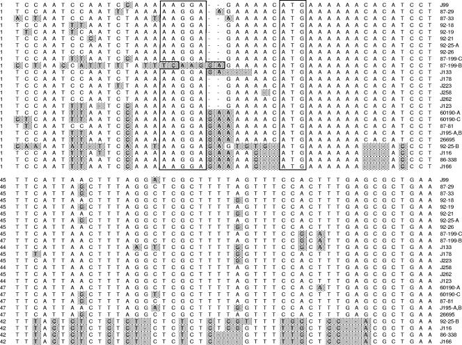
Analysis of a consensus alignment of the 5′ portion of babA alleles revealed two divergent families of sequences (Fig. 1). Four sequences (from strains J116, 86-338, J166, and 92-25 locus B) were markedly different in this region from babA sequences found in the other strains. However, further downstream, all of the sequences were closely related (data not shown). Multiple sequential CT repeats were present in one family of sequences (Fig. 1). Such regions are predicted to be subject to frameshift mutations, which could result in phase-variable expression of the encoded proteins.
FIG. 1.
Nucleotide diversity in the 5′ region of babA. Nucleotide sequences in the 5′ region of babA from 22 H. pylori strains (including 4 strains with multiple copies of babA) were aligned. For strains with multiple copies of babA, the locus of the sequence (A, B, or C) is given next to the strain designation. ATG start codons and putative Shine-Dalgarno sequences are boxed. The babA sequences in 4 strains (J166, 86-338, J116, and one copy of babA from strain 92-25) were markedly different from those in the other 18 strains. Shading indicates nucleotides different from the consensus.
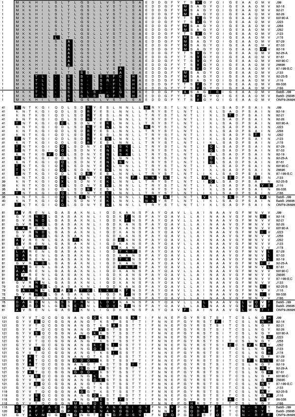
To analyze the conservation and diversity of BabA amino acid sequences, we examined a consensus alignment of deduced BabA sequences. Consistent with the nucleotide sequence data shown in Fig. 1, two divergent families of amino acid sequences were detected near the amino terminus of BabA (Fig. 2). The region of divergence (amino acids 4 to 19) is predicted to constitute part of an amino-terminal signal peptide. The signal peptides encoded by four strains (J116, 86-338, J166, and 92-25-B) were closely related to signal peptides of paralogous BabB proteins (exemplified by HP0896 in strain 26695 and jhp1164 in strain J99) (Fig. 2). Notably, other parts of these four proteins were most closely related to BabA sequences (Fig. 2). It is striking that all four proteins had BabB-type signal peptides but sequences further downstream were typical BabA sequences.
FIG. 2.
Divergence among BabA signal peptides. Deduced N-terminal amino acid sequences of BabA from 21 H. pylori strains (including 3 strains with multiple copies of babA) were aligned. Deduced amino acid sequences of BabB (from strains J99 and 26695) and Omp9 (from strain 26695) are included in the alignment. Putative signal peptides are shaded and boxed. Two distinct families of BabA signal peptides can be distinguished. The signal peptides encoded by four strains are identical to BabB signal peptides. Black highlighting indicates amino acids different from the consensus.
We next sought to investigate potential links between babA genetic features, BabA protein expression, and binding to Lewis b antigen. BabA protein expression in this panel of strains was characterized previously by immunoblot analysis (7). Sixteen of the 35 strains expressed a detectable BabA protein (Table 3). Nearly all of these 16 strains contained babA in locus A, but 1 contained babA in locus C and 1 contained two copies of babA in loci B and C. Among 17 strains in which an intact in-frame babA allele (predicted to encode a BabA protein of >700 amino acids) was identified, only 10 expressed a detectable BabA protein (Table 3). Potentially, BabA expression was not detected in some strains due to BabA amino sequence diversity and failure of anti-BabA antibodies to recognize certain epitopes. However, in a phylogenetic analysis, the babA alleles from BabA-expressing strains did not cluster separately from strains that failed to express BabA (data not shown).
We then tested all 35 strains for the ability to bind Lewis b antigen, using an in vitro adhesion assay (9). Among strains that expressed BabA, there was considerable variation in the level of binding to Lewis b antigen (Table 3). In previous studies, Lewis b/control binding ratios of >1.5 have been considered indicative of binding to Lewis b antigen (5, 9, 13). Based on this criterion, 7 strains exhibited detectable binding to Lewis b antigen and the other 28 strains did not (Table 3). As expected, all seven strains that bound to Lewis b antigen expressed a detectable BabA protein. Five of these seven strains contained babA in locus A. Among the four strains with divergent sequences near the 5′ end of babA (J166, 86-338, J116, and 92-25) (Fig. 1 and 2), two strains produced a detectable BabA protein and two did not. Strain J116 expressed a detectable BabA protein but did not exhibit detectable binding to Lewis b antigen. Strain 92-25 expressed a BabA protein and bound to Lewis b antigen, but this strain contained two nonidentical copies of babA. Therefore, it is not possible to reach any conclusions about the functional role of the divergent copy of babA in this strain.
It has been reported previously that the babA1 gene in strain CCUG17875 lacks a translation initiation codon (8). To determine whether the lack of BabA expression in some strains was due to this phenomenon, we analyzed the 5′ regions of babA alleles. Each of the babA alleles contained an ATG start codon (Fig. 1); therefore, the absence of a translational initiation codon, as described for babA1 from CCUG17875, is probably uncommon. Each of the genes analyzed contained an AAGGA sequence, corresponding to a putative ribosomal binding site, upstream of the ATG start codon (Fig. 1). There was variation among strains in the spacing between the ribosomal binding site and the BabA translational start codon (Fig. 1), but there was no significant correlation between the spacing of the Shine-Dalgarno sequence and expression of a BabA protein (compare Fig. 1 and Table 3). A TGNTATAAT consensus sequence, corresponding to an extended −10 motif, was present upstream of the babA translation initiation codon in all of the strains analyzed, with the exception of the locus B copy of babA in strain 87-199 (data not shown). Further upstream of the putative −10 sequence, there was considerable nucleotide sequence variation among strains, but we did not identify any motifs that correlated with the presence or absence of BabA protein expression.
In summary, these data indicate that there is extensive variation among H. pylori strains in babA genetic content. There are multiple possible chromosomal loci for babA. Individual strains can contain anywhere from 0 to 2 copies of babA. Frameshift mutations can result in the absence of BabA expression. Chimeric babB/babA alleles are commonly detected, and polymorphisms are present throughout babA alleles. In addition to this extensive variation among strains in babA content, a recent study showed that there can be metastability in babA sequences within the same strain (4). Based on the observation that many strains do not contain babA and do not bind to Lewis b antigen, it may be presumed that BabA expression is not required for persistent H. pylori colonization of the human stomach. However, adherence of H. pylori to gastric epithelial cells via the BabA adhesin may be a factor that contributes to the development of gastroduodenal disease (5, 6).
Nucleotide sequence accession numbers.
The babA sequences newly determined in this study have been assigned GenBank accession numbers DQ225153 to DQ225165.
Acknowledgments
This work was supported by NIH R01 DK53623, the Medical Research Service of the Department of Veterans Affairs, and CMKP grant 501-1-1-09-16/05, Poland.
Editor: J. B. Bliska
REFERENCES
- 1.Alm, R. A., J. Bina, B. M. Andrews, P. Doig, R. E. Hancock, and T. J. Trust. 2000. Comparative genomics of Helicobacter pylori: analysis of the outer membrane protein families. Infect. Immun. 68:4155-4168. [DOI] [PMC free article] [PubMed] [Google Scholar]
- 2.Alm, R. A., L. S. Ling, D. T. Moir, B. L. King, E. D. Brown, P. C. Doig, D. R. Smith, B. Noonan, B. C. Guild, B. L. deJonge, G. Carmel, P. J. Tummino, A. Caruso, M. Uria-Nickelsen, D. M. Mills, C. Ives, R. Gibson, D. Merberg, S. D. Mills, Q. Jiang, D. E. Taylor, G. F. Vovis, and T. J. Trust. 1999. Genomic-sequence comparison of two unrelated isolates of the human gastric pathogen Helicobacter pylori. Nature 397:176-180. [DOI] [PubMed] [Google Scholar]
- 3.Aspholm-Hurtig, M., G. Dailide, M. Lahmann, A. Kalia, D. Ilver, N. Roche, S. Vikstrom, R. Sjostrom, S. Linden, A. Backstrom, C. Lundberg, A. Arnqvist, J. Mahdavi, U. J. Nilsson, B. Velapatino, R. H. Gilman, M. Gerhard, T. Alarcon, M. Lopez-Brea, T. Nakazawa, J. G. Fox, P. Correa, M. G. Dominguez-Bello, G. I. Perez-Perez, M. J. Blaser, S. Normark, I. Carlstedt, S. Oscarson, S. Teneberg, D. E. Berg, and T. Boren. 2004. Functional adaptation of BabA, the Helicobacter pylori ABO blood group antigen binding adhesin. Science 305:519-522. [DOI] [PubMed] [Google Scholar]
- 4.Backstrom, A., C. Lundberg, D. Kersulyte, D. E. Berg, T. Boren, and A. Arnqvist. 2004. Metastability of Helicobacter pylori bab adhesin genes and dynamics in Lewis b antigen binding. Proc. Natl. Acad. Sci. USA 101:16923-16928. [DOI] [PMC free article] [PubMed] [Google Scholar]
- 5.Gerhard, M., N. Lehn, N. Neumayer, T. Boren, R. Rad, W. Schepp, S. Miehlke, M. Classen, and C. Prinz. 1999. Clinical relevance of the Helicobacter pylori gene for blood-group antigen-binding adhesin. Proc. Natl. Acad. Sci. USA 96:12778-12783. [DOI] [PMC free article] [PubMed] [Google Scholar]
- 6.Guruge, J. L., P. G. Falk, R. G. Lorenz, M. Dans, H. P. Wirth, M. J. Blaser, D. E. Berg, and J. I. Gordon. 1998. Epithelial attachment alters the outcome of Helicobacter pylori infection. Proc. Natl. Acad. Sci. USA 95:3925-3930. [DOI] [PMC free article] [PubMed] [Google Scholar]
- 7.Hennig, E. E., R. Mernaugh, J. Edl, P. Cao, and T. L. Cover. 2004. Heterogeneity among Helicobacter pylori strains in expression of the outer membrane protein BabA. Infect. Immun. 72:3429-3435. [DOI] [PMC free article] [PubMed] [Google Scholar]
- 8.Ilver, D., A. Arnqvist, J. Ogren, I. M. Frick, D. Kersulyte, E. T. Incecik, D. E. Berg, A. Covacci, L. Engstrand, and T. Boren. 1998. Helicobacter pylori adhesin binding fucosylated histo-blood group antigens revealed by retagging. Science 279:373-377. [DOI] [PubMed] [Google Scholar]
- 9.Olfat, F. O., Q. Zheng, M. Oleastro, P. Voland, T. Boren, R. Karttunen, L. Engstrand, R. Rad, C. Prinz, and M. Gerhard. 2005. Correlation of the Helicobacter pylori adherence factor BabA with duodenal ulcer disease in four European countries. FEMS Immunol. Med. Microbiol. 44:151-156. [DOI] [PubMed] [Google Scholar]
- 10.Pride, D. T., and M. J. Blaser. 2002. Concerted evolution between duplicated genetic elements in Helicobacter pylori. J. Mol. Biol. 316:629-642. [DOI] [PubMed] [Google Scholar]
- 11.Pride, D. T., R. J. Meinersmann, and M. J. Blaser. 2001. Allelic variation within Helicobacter pylori babA and babB. Infect. Immun. 69:1160-1171. [DOI] [PMC free article] [PubMed] [Google Scholar]
- 12.Salama, N., K. Guillemin, T. K. McDaniel, G. Sherlock, L. Tompkins, and S. Falkow. 2000. A whole-genome microarray reveals genetic diversity among Helicobacter pylori strains. Proc. Natl. Acad. Sci. USA 97:14668-14673. [DOI] [PMC free article] [PubMed] [Google Scholar]
- 13.Solnick, J. V., L. M. Hansen, N. R. Salama, J. K. Boonjakuakul, and M. Syvanen. 2004. Modification of Helicobacter pylori outer membrane protein expression during experimental infection of rhesus macaques. Proc. Natl. Acad. Sci. USA 101:2106-2111. [DOI] [PMC free article] [PubMed] [Google Scholar]
- 14.Tomb, J.-F., O. White, A. R. Kerlavage, R. A. Clayton, G. G. Sutton, R. D. Fleischmann, K. A. Ketchum, H. P. Klenk, S. Gill, B. A. Dougherty, K. Nelson, J. Quackenbush, L. Zhou, E. F. Kirkness, S. Peterson, B. Loftus, D. Richardson, R. Dodson, H. G. Khalak, A. Glodek, K. McKenney, L. M. Fitzegerald, N. Lee, M. D. Adams, E. K. Hickey, D. E. Berg, J. D. Gocayne, T. R. Utterback, J. D. Peterson, J. M. Kelley, M. D. Cotton, J. M. Weidman, C. Fujii, C. Bowman, L. Watthey, E. Wallin, W. S. Hayes, M. Borodovsky, P. D. Karp, H. O. Smith, C. M. Fraser, and J. C. Venter. 1997. The complete genome sequence of the gastric pathogen Helicobacter pylori. Nature 388:539-547. [DOI] [PubMed] [Google Scholar]
- 15.Yamaoka, Y., J. Souchek, S. Odenbreit, R. Haas, A. Arnqvist, T. Boren, T. Kodama, M. S. Osato, O. Gutierrez, J. G. Kim, and D. Y. Graham. 2002. Discrimination between cases of duodenal ulcer and gastritis on the basis of putative virulence factors of Helicobacter pylori. J. Clin. Microbiol. 40:2244-2246. [DOI] [PMC free article] [PubMed] [Google Scholar]


