Abstract
It is an open question how ion channel subunits that lack protein–protein binding motifs become targeted and covalently modified by cellular signaling enzymes. Here, we show that Src-family protein tyrosine kinases (PTKs) bind to heteromultimeric Shaker-family voltage-gated potassium (Kv) channels by interactions between the Src homology 3 (SH3) domain and the proline-rich SH3 domain ligand sequence in the Shaker-family subunit Kv1.5. Once bound to Kv1.5, Src-family PTKs phosphorylate adjacent subunits in the Kv channel heteromultimer that lack proline-rich SH3 domain ligand sequences. This SH3-dependent tyrosine phosphorylation contributes to significant suppression of voltage-evoked currents flowing through the heteromultimeric channel. These results demonstrate that Kv1.5 subunits function as SH3-dependent adaptor proteins that marshal Src-family kinases to heteromultimeric potassium channel signaling complexes, and thereby confer functional sensitivity upon coassembled channel subunits that are themselves not bound directly to Src-family kinases by allowing their phosphorylation. This is a mechanism for information transfer between subunits in heteromultimeric ion channels that is likely to underlie the generation of combinatorial signaling diversity in the control of cellular electrical excitability.
Present understanding of the interaction between regulatory signaling enzymes and ion channels is limited in several respects. First, although almost all ion channel subunits that have been tested are biophysically modulated in response to covalent modification by a variety of protein kinases (1–12), most ion channel subunits do not exhibit known protein–protein binding module ligand sequences. It is thus an open question how such ion channel subunits that lack protein–protein binding motifs become targeted by particular signaling enzymes. Second, although the majority of ion channels found in native tissues are heteromultimers (13–16), mechanistic analyses of ion channel interactions with signaling enzymes have been limited to homomultimers consisting of multiple copies of a single ion channel subunit. Thus, the functional regulation of heteromultimeric channels by signaling enzymes and the nature of the interaction between distinct subunits within a heteromultimer and stably associated signaling enzymes remains unexplored.
It has been demonstrated that voltage-gated potassium (Kv)1.5-containing channels are associated with Src-family protein tyrosine kinases (PTKs) by means of Src homology 3 (SH3) domain interactions with the Kv1.5 proline-rich SH3 domain ligand sequence (ref. 1; T.C.H., J. Marquez, and I. B. Levitan, personal communication). When coexpressed in mammalian cells in culture, Kv1.5 is tyrosine phosphorylated by Src, and voltage-evoked currents are dramatically suppressed as a consequence (1). It is known that members of the mammalian Shaker family form heteromultimers in the mammalian brain (13, 14, 16). Whether heteromultimer formation provides a mechanism for the functionally relevant transfer of sensitivity to Src-family PTKs from mammalian Shaker subunits containing SH3 domain ligand sequences to those that lack such sequences remains unknown.
Here, we show that the Src-family PTK Fyn is associated in mammalian hippocampus with Kv1.5-containing heteromultimeric channels that also contain Kv1.2 or Kv1.4 subunits, which lack SH3 domain ligand sequences. We further demonstrate that Kv1.5 subunits act as SH3-dependent adaptors that marshal Src-family PTKs to heteromultimeric Shaker family channels and promote functionally relevant tyrosine phosphorylation of neighboring subunits lacking SH3 domain ligand sequences.
Materials and Methods
Preparation and Immunoprecipitation of Synaptic Membranes.
Synaptic membranes were prepared from rabbit hippocampus as described (16). Synaptic membranes derived from one rabbit hippocampus were solubilized in 4 ml modified RIPA lysis buffer (25 mM Tris, pH 7.5/150 mM NaCl/100 mM NaF/5 mM EDTA/1 mM Na3VO4/1% Triton X-100/1 mM PMSF/1 μg/ml leupeptin/2 μg/ml aprotinin) (2). Solubilized membranes were precleared with 50 μl of protein A/G beads (Pierce) per 1 ml solubilized membranes. Proteins were immunoprecipitated overnight at 4°C from 1 ml precleared solubilized membranes using either 2.5 μl of serum, or 2.5 μg of purified mAbs or polyclonal Abs (pAbs), and 50 μl of protein A/G beads. Immunoprecipitating antibodies were as follows: α-Kv1.2 antiserum (17), α-Kv1.4 antiserum raised against a glutathione S-transferase (GST) fusion protein comprising amino acids 588–655 of Kv1.4 (GenBank accession no. P15385), α-Kv1.5 antiserum raised against a GST fusion protein comprising amino acids 561–613 of Kv1.5 (GenBank accession no. P22460), preimmune serum from the rabbit in which the α-Kv1.5 antiserum was raised, affinity-purified α-Fyn pAbs (Santa Cruz Biotechnology), affinity-purified α-Fyn pAbs preabsorbed against a 5-fold mass excess of the cognate Fyn peptide antigen, α-Kv1.2 mAb (18) (Upstate Biotechnology, Lake Placid, NY), and α-prepro-insulin C-peptide (α-CP) mAb (BiosPacific, Emeryville, CA). α-Kv1.2, α-Kv1.4, and α-Kv1.5 antisera only immunoprecipitated Kv1.2, Kv1.4, or Kv1.5 homomultimers, respectively, that were expressed in HEK 293 cells, thus establishing lack of crossreactivity between these antibodies and their respective antigens (data not shown). Immunoprecipitated proteins bound to pelleted protein A/G beads were washed thoroughly in modified RIPA buffer containing 0.1% Triton X-100, separated by using SDS/PAGE, and electroblotted onto nitrocellulose membranes. Immunoblots were stained with α-Kv1.2 mAb, α-Kv1.4 mAb (18) (Upstate Biotechnology), α-Fyn mAb (Transduction Laboratories, Lexington, KY), or α-Kv1.5 antiserum. Specific protein signals on the Western blots were visualized by enhanced chemiluminescence (Supersignal, Pierce).
cDNA Constructs, Transfection of HEK 293 Cells, and Immunoprecipitation.
All cDNA constructs were under the control of the mammalian cytomegalovirus CMV promoter. Kv1.5 fused at its C terminal to the CP epitope tag was provided by L. Philipson (University of Chicago, Chicago), and Kv1.4 was provided by Morgan Sheng (Massachusetts General Hospital, Boston). Catalytically impaired Src (SrcCI) was generated by introducing the R385G point mutation into v-Src cDNA. A point mutation (D99N) that decreases 40- to 50-fold SH3 domain binding to proline-rich ligand sequences (19) was introduced into SrcCI, resulting in SrcCISH3KO. SrcCI-Tag was generated by inserting two repeats of the c-Myc epitope tag between amino acids 17 and 18 of SrcCI. The proline-rich region of Kv1.5 containing two RPLPPLP repeats, amino acids 64–82, was deleted, resulting in Kv1.5-ΔPro. The entire coding region of Kv1.5 was inserted between amino acids 169 and 170 of Kv1.4, resulting in a Kv1.4–Kv1.5 tandem dimer subunit. A synthetic gene fragment encoding the proline-rich region of Kv1.5 was inserted between amino acids 649 and 650 of Kv1.4, resulting in Kv1.4-Pro. HEK 293 cells were maintained and transfected as described (2), with the modification that the Fugene transfection reagent (Roche Molecular Biochemicals) was used. Cell lysis and immunoprecipitations (IPs) were performed as described (2). IP samples were separated on SDS/PAGE gels and electroblotted onto nitrocellulose membranes. Immunoblots were stained with α-Kv1.4 mAb, α-Src mAb (Upstate Biotechnology), α-CP mAb, α-Myc mAb (clone 9E10), or α-phosphotyrosine mAb (clone 4G10; Upstate Biotechnology). Specific protein signals on the Western blots were visualized by enhanced chemiluminescence (Super Signal, Pierce).
Electrophysiological Analysis of mRNA-Injected Xenopus Oocytes.
Xenopus laevis oocytes were prepared, injected, and recorded by using standard methods (20). Oocytes were injected with 50 nl in vitro transcribed cRNAs encoding various Kv subunits. Two days after Kv subunit cRNA injections, current evoked by a 1-s step from the holding potential of −80mV to +80mV was recorded by using a two-electrode voltage clamp. Each oocyte then was injected a second time, either with 50 nl water, SrcCI cRNA, or SrcCISH3KO cRNA, and then voltage-evoked current was again recorded 9–12 h after the reinjection. SrcCI and SrcCISH3KO cRNAs were of identical concentration, as assessed on an ethidium bromide-stained formaldehyde-agarose gel. The ratio for each oocyte of the peak current recorded after reinjection to that recorded before reinjection was analyzed among all of the experimental conditions using one-way ANOVA. Differences were further analyzed with the Scheffé paired-comparison test, with a threshold for significance of P < 0.01. Representative current traces depicted in the figures were chosen on the basis of exhibiting the closest peak current magnitude to that of the mean for the represented experimental condition.
Results
Stable Association of Src-Family PTK Fyn with Kv1.5-Containing Heteromultimers in Mammalian Hippocampus.
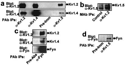
We examined the subunit composition of heteromultimeric Kv channels in mammalian hippocampus and determined whether the Src-family PTK Fyn associates with such heteromultimeric channels in vivo. Kv1.2 and Kv1.4 subunits coprecipitated with Kv1.5 subunits immunoprecipitated from solubilized rabbit hippocampus synaptic membranes using α−Kv1.5 antiserum (Fig. 1a). This indicates the existence of both Kv1.2/Kv1.5- and Kv1.4/Kv1.5-containing native heteromultimers. Kv1.2/Kv1.5 heteromultimers were demonstrated reciprocally by coprecipitation of Kv1.5 subunits with Kv1.2 subunits immunoprecipitated using α−Kv1.2 antiserum (Fig. 1b). Robust expression of Kv1.4/Kv1.5 heteromultimers has also been observed by others in mammalian pituitary cells (21). Fyn PTK associates with the identified native hippocampal Kv1.2/Kv1.5- and Kv1.4/Kv1.5-containing heteromultimers, as shown by the coprecipitation of both Kv1.2 and Kv1.4 subunits with Fyn immunoprecipitated using α−Fyn antibodies (Fig. 1c), as well as by the coprecipitation of Fyn with Kv1.5 subunits immunoprecipitated using α−Kv1.5 antiserum (Fig. 1d). These results suggest that Fyn PTK is a stable component of Kv1.2/Kv1.5- and Kv1.4/Kv1.5-containing heteromultimeric Kv channel signaling complexes in mammalian hippocampus.
Figure 1.
Stable association of Src-family PTK Fyn with Kv1.2/Kv1.5- and Kv1.4/Kv1.5-containing heteromultimeric potassium channels in mammalian hippocampus. (a) Kv1.5 subunits coassemble with Kv1.2 and Kv1.4 subunits in hippocampus. Heteromultimeric Kv channels were isolated from detergent solubilized rabbit hippocampus synaptic membranes by immunoprecipitation (IP) with α−Kv1.2, α−Kv1.4, or α−Kv1.5 antisera, using preimmune serum as a negative control. IP samples were probed on Western blots with α−Kv1.2 mAb (Upper, n = 6) or Kv1.4 mAb (Lower, n = 6). (b) Kv1.5 subunits that coimmunoprecipitate with Kv1.2 subunits are detected by probing α−Kv1.2 mAb IPs with α−Kv1.5 antiserum (n = 4), using α−CP mAb as a negative control. (c) Fyn associates with Kv1.2- and Kv1.4-containing channels in hippocampus. α−Fyn pAb IPs were probed with α−Kv1.2 mAb (Upper, n = 6), α−Kv1.4 mAb (Middle, n = 4), or α−Fyn mAb (Lower, n = 6). The negative control IP antibody used was prepared by complete preabsorption of α−Fyn pAb with an excess of the peptide antigen (PreAbs). (d) Fyn associates with Kv1.5-containing channels in hippocampus. Fyn that coimmunoprecipitates with Kv1.5 is detected by probing α−Kv1.5 IPs with α−Fyn mAb (n = 3), using preimmune serum as a negative control.
Marshaling of Src-Family PTK Src to Heteromultimeric Channel by SH3 Domain Interaction with Kv1.5 Subunit.
We undertook a mechanistic analysis of Src-family PTK interactions with Kv1.4/Kv1.5 heteromultimers in transfected HEK 293 cells. SrcCI, a catalytically impaired point mutant (R385G) of v-Src, was developed and used for these studies to avoid the robust promiscuous binding-independent tyrosine phosphorylation of Kv subunits exhibited by v-Src (data not shown). The R385G mutation of SrcCI disrupts an intramolecular activating interaction, resulting in a form of Src that exhibits greatly decreased binding-independent phosphorylation of cellular targets in comparison to v-Src or c-Src (data not shown). To examine the dependence of Src phosphorylation of Kv subunits on SH3-mediated interactions, we generated SrcCISH3KO by introducing a point mutation (D99N) into SrcCI that decreases SH3 domain binding to proline-rich ligand sequences by 40- to 50-fold (19). The Src-family PTKs Src and Fyn exhibit essentially identical SH3 domain ligand specificity and catalytic domain substrate specificity (22, 23).
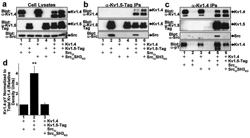
Kv1.4 and Kv1.5 subunits are readily expressed in transfected HEK 293 cells. The fully posttranslationally processed form of Kv1.4 exhibits an apparent molecular mass of ≈95 kDa on Western blots (Fig. 2a, Top) (24), and the fully posttranslationally processed form of Kv1.5 exhibits an apparent molecular mass of ≈80–85 kDa (Fig. 2a, Middle). Kv1.4/Kv1.5 heteromultimers form robustly in transfected cells, as demonstrated by the coprecipitation of Kv1.4 subunits with Kv1.5 subunits immunoprecipitated by using α−Kv1.5-tag antibodies (Fig. 2b, Top), and the reciprocal coprecipitation of Kv1.5 subunits with Kv1.4 subunits immunoprecipitated by using α−Kv1.4 antibodies (Fig. 2c, Upper Middle).
Figure 2.
Stable SH3-dependent association of Src with Kv1.4/Kv1.5 heteromultimeric potassium channels and consequent increased Kv1.4 phosphorylation. (a) Expression of Kv1.4/Kv1.5 heteromultimeric channels with Src PTK in HEK 293 cells. Combinations of Kv1.4, C-peptide epitope-tagged Kv1.5 (Kv1.5-Tag), catalytically impaired Src (SrcCI), and SrcCI with a point mutation in the SH3 domain that abolishes SH3 domain binding to its ligand (SrcCISH3KO) were transfected into HEK 293 cells, followed by Western blot of detergent solubilized cell lysates probed with α−Kv1.4 (Top, n = 6), α−Kv1.5-Tag (Middle, n = 6), or α−Src (Bottom, n = 6) mAbs. The fully processed plasma membrane-targeted forms of Kv1.4 (≈95 kDa) and Kv1.5 (≈80–85 kDa) are indicated by the arrows labeled “Kv1.4” and “Kv1.5,” respectively, and migrate slower than incompletely processed and targeted forms. (b) SrcCI coimmunoprecipitates with Kv1.5 homomultimers and Kv1.4/Kv1.5 heteromultimers, whereas SrcCISH3KO fails to coimmunoprecipitate with either Kv1.5 homomultimers or Kv1.4/Kv1.5 heteromultimers. Cell lysates analyzed in a were immunoprecipitated with α−Kv1.5-Tag mAb and analyzed by Western blot. (c) Stable Src association and increased phosphorylation of Kv1.4 subunits occurs only in Kv1.4/Kv1.5 heteromultimers and depends on Src SH3 domain interaction with the Kv1.5 proline-rich region. Cell lysates analyzed in a were immunoprecipitated with α−Kv1.4 mAb and analyzed by Western blot. SrcCI only coimmunoprecipitates with Kv1.4 when Kv1.4 is coassembled with Kv1.5 in Kv1.4/Kv1.5 heteromultimers. SrcCISH3KO fails to coimmunoprecipitate with Kv1.4, even when Kv1.4 is coassembled in Kv1.4/Kv1.5 heteromultimers. Phosphorylation of fully processed Kv1.4 subunits is increased when coassembled in Kv1.4/Kv1.5 heteromultimers, and this increase is abolished when SrcCI is replaced with SrcCISH3KO. (d) Quantitative comparison of Kv1.4 subunit phosphorylation in cells transfected with combinations of Kv1.4, Kv1.5-Tag, SrcCI, and SrcCISH3KO. Signal density of Kv1.4 phosphorylation detected with α−pY mAb is normalized to the corresponding signal density of Kv1.4 protein detected with α−Kv1.4 mAb. Bars represent relative Kv1.4 normalized phosphotyrosine density for each transfection condition determined within each experiment by dividing the normalized phosphotyrosine density for the indicated transfection condition by the normalized phosphotyrosine density for the Kv1.4/SrcCI transfection condition (mean ± SEM, n = 6; **, P < 0.001 by one-way ANOVA).
SrcCI coprecipitates by α−Kv1.5 immunoprecipitation of either Kv1.5 homomultimers or Kv1.4/Kv1.5 heteromultimers (Fig. 2b, Bottom). SrcCI coprecipitates by α−Kv1.4 immunoprecipitation only when Kv1.4 is coassembled with Kv1.5 in a Kv1.4/Kv1.5 heteromultimer, failing to coprecipitate with Kv1.4 when Kv1.4 is expressed as a homomultimer (Fig. 2c, Lower Middle). SH3 domain-disabled SrcCI (SrcCISH3KO) coprecipitates neither with Kv1.4/Kv1.5 heteromultimers (Fig. 2b, Bottom and 2c, Lower Middle), nor with Kv1.5 homomultimers (Fig. 2b, Bottom). This mutation-induced abolishment of Src/Kv channel binding is not because of inefficient expression of SrcCISH3KO, as comparable amounts of SrcCI and SrcCISH3KO are detected in cell lysates for all transfection conditions (Fig. 2a, Bottom). SrcCI fails to coprecipitate with heteromultimers assembled from Kv1.4 subunits and Kv1.5 subunits that have been modified to lack the proline-rich SH3 domain binding sequence (Kv1.5-ΔPro) (Fig. 3a, Bottom and 3b, Lower Middle). These results indicate that Src does not bind directly to Kv1.4 and is marshaled to Kv1.4/Kv1.5 heteromultimers exclusively by means of a direct interaction between the Src SH3 domain and the proline-rich region of Kv1.5.
Figure 3.
Increased phosphorylation of Kv1.4 subunits in Kv1.4/Kv1.5 heteromultimers mediated by the interaction of Src with the Kv1.5 proline-rich SH3 domain ligand sequence. (a) Stable association of Src PTK with Kv1.4/Kv1.5 heteromultimeric channels depends on the interaction of Src with the proline-rich 2xRPLPPLP sequence of Kv1.5. Combinations of Kv1.4, Kv1.5-Tag, Kv1.5-ΔPro-Tag, and c-Myc epitope-tagged catalytically impaired Src (SrcCI-Tag) were transfected into HEK 293 cells, followed by Western blot of α−Kv1.4 immunoprecipitates of cell lysates. SrcCI coimmunoprecipitates with Kv1.4/Kv1.5 heteromultimers, but fails to coimmunoprecipitate with either Kv1.4 homomultimers or Kv1.4/Kv1.5-ΔPro heteromultimers. (b) Increased phosphorylation of Kv1.4 subunits by Src in Kv1.4/Kv1.5 heteromultimers depends on the Kv1.5 2xRPLPPLP Src SH3 domain ligand. Cell lysates from a were immunoprecipitated with α−Kv1.5-Tag mAb and analyzed by Western blot. The Kv1.4 subunits coassembled with Kv1.5 exhibit greater tyrosine phosphorylation than the Kv1.4 subunits coassembled with Kv1.5-ΔPro. (c) Quantitative comparison of Kv1.4 subunit phosphorylation in cells transfected with combinations of Kv1.4, Kv1.5-Tag, Kv1.5-ΔPro-Tag, and SrcCI-Tag. Kv1.4 phosphorylation was quantitatively analyzed as in Fig. 2d, but with relative density defined in relation to the normalized density observed for the Kv1.4/Kv1.5-Tag/SrcCI-Tag transfection condition (mean ± SEM, n = 4; **, P < 0.001 by paired t test).
Kv1.5-Mediated SH3-Dependent Phosphorylation by Src of Non-Kv1.5 Subunits in Heteromultimers.
To determine whether Src-family PTKs, once marshaled to Kv1.5-containing heteromultimers, are able to phosphorylate other subunits within such heteromultimers, we examined the phosphorylation of Kv1.4 subunits by SrcCI in Kv1.4/Kv1.5 heteromultimers by probing immunoprecipitates from transfected HEK 293 cells with a highly specific α−phosphotyrosine (α−pY) mAb (Figs. 2c, Bottom and 3b, Bottom). Phosphorylated fully processed Kv1.4 can be readily distinguished from phosphorylated Kv1.5 on these α−pY Western blots, as fully processed Kv1.4 runs at an apparent molecular mass of ≈95 kDa, with fully processed Kv1.5 running at ≈80–85 kDa. Also, phosphorylation of Kv1.4 is so much greater than phosphorylation of Kv1.5 that Kv1.5 subunits could only be detected on α−pY Western blots with exposure times many-fold greater than used here (data not shown). Phosphorylation of fully processed Kv1.4 by SrcCI was 4-fold greater in Kv1.4/Kv1.5 heteromultimers than in Kv1.4 homomultimers (Fig. 2c, Bottom and 2d, P < 0.001). This Kv1.5-mediated increase in Kv1.4 subunit phosphorylation by SrcCI was abolished when SrcCI was replaced with SrcCISH3KO (Fig. 2c, Bottom and 2d, P < 0.001). In addition, Kv1.4 phosphorylation by SrcCI was 5-fold greater in heteromultimers containing wild-type Kv1.5 than in heteromultimers containing Kv1.5-ΔPro (Fig. 3b, Bottom and 3c, P < 0.001). The small SH3-independent component of Kv1.4 phosphorylation may be because of the fact that SrcCI, while catalytically impaired, derives from v-Src and is thus constitutively active. These results show that once marshaled to Kv1.5-containing heteromultimeric Kv channel signaling complexes by direct SH3 domain binding to the Kv1.5 proline-rich region, Src PTK distributes its signal throughout the heteromultimer by phosphorylating adjacent channel subunits that lack SH3 domain binding sites.
Kv1.5-Mediated SH3-Dependent Suppression by Src of Heteromultimeric Channel Currents.
The physiological consequence of the Src-marshaling adaptor function of Kv1.5 subunits in Kv1.4/Kv1.5 heteromultimeric channels was examined by using a two-electrode voltage clamp of X. laevis oocytes. Baseline unmodulated depolarization-evoked macroscopic channel currents were recorded 2 days after channel subunit cRNA injection. Each channel-expressing oocyte was reinjected with Src cRNA or H2O immediately after baseline current measurements. Depolarization-evoked channel currents were again recorded from each oocyte 9–12 h after the reinjection. This protocol allowed analysis in each oocyte of the ratio of peak channel current measured after expression of the Src PTK to that measured before (“peak current ratio”), thus controlling for the variability in channel expression between oocytes.
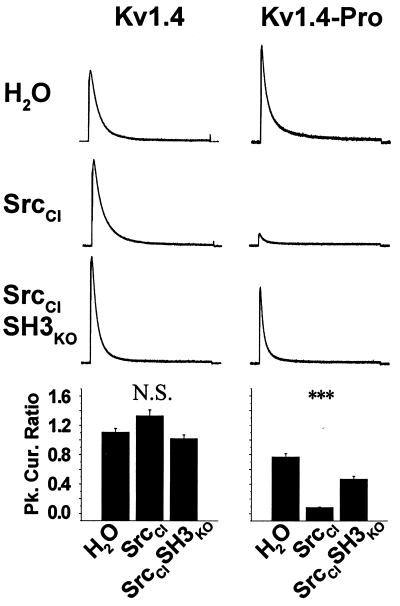
There is no significant difference in the peak current ratio for Kv1.4 homomultimeric channels in oocytes that were reinjected with either SrcCI or SrcCISH3KO as compared with oocytes reinjected with water (Fig. 4 Left). In contrast, both Kv1.5- and Kv1.4-Kv1.5-tandem-expressing oocytes exhibit significantly reduced peak current ratios when reinjected with SrcCI, as compared with oocytes reinjected with water (Fig. 4, Left Center and Right Center). The expression of Kv1.4-Kv1.5-tandem subunits generates Kv1.4/Kv1.5 heteromultimeric channels of defined 1:1 stoichiometry, with kinetic properties intermediate between the rapidly inactivating Kv1.4 homomultimer and the very slowly inactivating Kv1.5 homomultimer. Although Kv1.5- and Kv1.4-Kv1.5-tandem-expressing oocytes reinjected with SrcCISH3KO also exhibit significantly reduced peak current ratios, these decreases are at least 2-fold smaller than for SrcCI, thus indicating an SH3-dependent component to the Src-mediated suppression of Kv1.5-containing channels. The SH3-independent component may be because of the fact that SrcCI, while catalytically impaired, is constitutively active. Currents carried by Kv1.4-Kv1.5-ΔPro tandem are not suppressed by SrcCI (Fig. 4 Right), and currents carried by Kv1.5-ΔPro are suppressed significantly less than was unmodified Kv1.5 (Fig. 4 Center and Left Center). These results show that Src-induced suppression of Kv1.4/Kv1.5 heteromultimeric channel currents depends on the Kv1.5-mediated Src SH3 domain interaction.
Figure 4.
Physiological suppression of Kv1.4/Kv1.5 heteromultimeric channels mediated by the SH3-dependent interaction of Src with the Kv1.5 proline-rich SH3 domain ligand sequence. Currents evoked in Kv subunit cRNA-injected X. laevis oocytes by a 1-s voltage step from a holding potential of −80 mV to a test potential of +80 mV were measured by using a two-electrode voltage clamp both immediately before and 9–12 h after a second injection with either H20, SrcCI, or SrcCISH3KO cRNA. Current traces depicted were measured after the second injection and were chosen on the basis of exhibiting the closest peak current magnitude to the mean for the represented experimental condition (ND, not determined). Current traces within each column are drawn to the same scale, with the following peak current magnitudes after H20 treatment: Kv1.4, 13.6 μA; Kv1.5, 9.9 μA; Kv1.5-ΔPro, 5.0 μA; Kv1.4-Kv1.5-Tandem, 11.1 μA; Kv1.4-Kv1.5-tandem-ΔPro, 2.1 μA. Bar graphs depict the mean ± SEM peak current ratios for each oocyte recorded after reinjection to that recorded before reinjection, and were analyzed using one-way ANOVA and the Scheffé paired-comparison test. Group sizes were between 12 and 29 oocytes. NS, no significant differences; ***, P < 0.01.
We conferred direct sensitivity to Src SH3-dependent binding and phosphorylation upon Kv1.4 homomultimeric channels by inserting the Kv1.5 proline-rich Src SH3 domain ligand sequence into the Kv1.4 subunit (yielding the Kv1.4-Pro subunit). Although wild-type Kv1.4 homomultimeric channels are not physiologically suppressed by Src (Fig. 5 Left), currents recorded from homomultimeric Kv1.4-Pro channels are dramatically suppressed by Src-mediated tyrosine phosphorylation (Fig. 5 Right). This functional suppression is SH3-dependent, as Kv1.4-Pro-expressing oocytes reinjected with SrcCISH3KO exhibit peak current ratios that significantly differ from those of the SrcCI reinjected oocytes (Fig. 5 Right). These results show that SH3-dependent Src-mediated tyrosine phosphorylation of Kv1.4 subunits functionally suppresses Kv1.4-containing channels.
Figure 5.
SH3 domain-dependent physiological suppression by Src phosphorylation conferred upon otherwise insensitive Kv1.4 homomultimers modified to contain the Kv1.5 Src ligand sequence. Currents evoked in Kv subunit cRNA-injected X. laevis oocytes were measured both immediately before and 9–12 h after a second injection with H2O, SrcCI, or SrcCISH3KO cRNA. Details are as in Fig. 4. The unmodified Kv1.4 groups are reproduced from Fig. 4 for comparison. Current traces within each column are drawn to the same scale, with the following peak current magnitudes after H2O treatment: Kv1.4, 13.6 μA; Kv1.4-Pro, 14.0 μA. Group sizes were between 12 and 20 oocytes. NS, no significant differences; ***, P < 0.01.
Discussion
We have demonstrated that Src-family PTKs bind to heteromultimeric mammalian Shaker channels by SH3 domain interactions with the proline-rich region of Kv1.5. The functional consequence of Src/Kv1.5 binding within the heteromultimer is the increased phosphorylation of adjacent subunits that lack proline-rich SH3 domain ligand regions and are unable to bind directly to Src. We have also demonstrated that this SH3-dependent phosphorylation contributes to suppression of the depolarization-evoked currents carried by the heteromultimeric channel. These results show that pore-forming channel subunits can function as adaptor proteins that (i) specifically marshal signaling enzymes to heteromultimeric ion channels, (ii) induce covalent modification of neighboring subunits, and (iii) distribute physiologically important regulatory signals to subunits that would otherwise remain unmodified. This suggests a functional significance for heteromultimer formation that supplements the conventional view of combinatorial generation of a variety of different biophysical channel properties. We propose that heteromultimer formation is important for the generation of ion channels that exhibit sensitivities to a combinatorial variety of enzymatic signaling pathways by virtue of the signaling protein interaction domains possessed by the constituent subunits.
Rapidly inactivating transient potassium currents mediated by Kv1.4-containing heteromultimeric channels have been found in the presynaptic terminals of hippocampal neurons (13, 25). We have shown that Kv1.4/Kv1.5-containing heteromultimeric potassium channels form stable complexes with the Src-family PTK Fyn in hippocampus. We have also demonstrated that coassembly of Kv1.5 subunits confers sensitivity to Src-family PTK signaling upon Kv1.4-containing rapidly inactivating channels that carry transient potassium currents. Transient presynaptic potassium currents play an important role in controlling hippocampal action potential kinetics and consequent neurotransmitter release (26–28). Thus, the coassembly of Kv1.5 subunits into presynaptic Kv1.4-containing rapidly inactivating potassium channels in hippocampal neurons could couple modulation of action potential duration and neurotransmitter release to Src-family PTK activation.
Although our findings pertain directly to Shaker-family Kv channels, the biological principle that we have uncovered—that ion channel subunits can function as SH3-dependent adaptor proteins—is likely to have wide application. Subunits from a number of diverse ion channel families possess canonical proline-rich SH3 ligand sequences, many of which have been experimentally determined to interact directly with SH3 domains (1, 29–32) (T.C.H., J. Marquez, and I. B. Levitan, personal communication). It will be of interest to determine whether other ion channel subunits that possess potential modular binding sites can marshal signaling enzymes to heteromultimers, and thereby confer functionally relevant signaling upon coassembled adjacent subunits that lack modular binding sites. Indeed, a variety of protein families, in addition to ion channels, contain multiple members capable of multimerization and share great similarity in their core functional effector domains, while exhibiting considerable divergence in their regulatory domains (33). Thus, heteromultimer assembly could underlie the generation of combinatorial signaling diversity in a wide range of protein families, by mediating the integration of multiple enzyme-based regulatory pathways.
Acknowledgments
We thank J. Trimmer, L. Philipson, M. Sheng, X.-Y. Huang, and M. Resh for providing antibodies and cDNA constructs; P. Furmanski, G. Coruzzi, A. Wang, and T. Otis for their comments on the manuscript; and A. Bill and G. Gupta for technical assistance. This research was supported by funds provided to T.C.H. by New York University, including New York University Research Challenge Fund and Whitehead grants. M.N.N. was supported in part by a National Institutes of Health National Research Service Award fellowship.
Abbreviations
- Kv
voltage-gated potassium
- PTK
protein tyrosine kinase
- SH3
Src homology 3
- α-CP
α-prepro-insulin C-peptide
- SrcCI
catalytically impaired Src
- IP
immunoprecipitation
- α-pY
α-phosphotyrosine
Footnotes
This paper was submitted directly (Track II) to the PNAS office.
Article published online before print: Proc. Natl. Acad. Sci. USA, 10.1073/pnas.031446198.
Article and publication date are at www.pnas.org/cgi/doi/10.1073/pnas.031446198
References
- 1.Holmes T C, Fadool D A, Ren R, Levitan I B. Science. 1996;274:2089–2091. doi: 10.1126/science.274.5295.2089. [DOI] [PubMed] [Google Scholar]
- 2.Holmes T C, Fadool D A, Levitan I B. J Neurosci. 1996;16:1581–1590. doi: 10.1523/JNEUROSCI.16-05-01581.1996. [DOI] [PMC free article] [PubMed] [Google Scholar]
- 3.Fadool D A, Holmes T C, Berman K, Dagan D, Levitan I B. J Neurophysiol. 1997;78:1563–1573. doi: 10.1152/jn.1997.78.3.1563. [DOI] [PubMed] [Google Scholar]
- 4.Huang X Y, Morielli A D, Peralta E G. Cell. 1993;75:1145–1156. doi: 10.1016/0092-8674(93)90324-j. [DOI] [PubMed] [Google Scholar]
- 5.Szabo I, Gulbins E, Apfel H, Zhang X, Barth P, Busch A E, Schlottmann K, Pongs O, Lang F. J Biol Chem. 1996;271:20465–20469. doi: 10.1074/jbc.271.34.20465. [DOI] [PubMed] [Google Scholar]
- 6.Fadool D A, Levitan I B. J Neurosci. 1998;18:6126–6137. doi: 10.1523/JNEUROSCI.18-16-06126.1998. [DOI] [PMC free article] [PubMed] [Google Scholar]
- 7.Levitan I B. Annu Rev Physiol. 1994;56:193–212. doi: 10.1146/annurev.ph.56.030194.001205. [DOI] [PubMed] [Google Scholar]
- 8.Jonas E A, Kaczmarek L K. Curr Opin Neurobiol. 1996;6:318–323. doi: 10.1016/s0959-4388(96)80114-0. [DOI] [PubMed] [Google Scholar]
- 9.Smart T G. Curr Opin Neurobiol. 1997;7:358–367. doi: 10.1016/s0959-4388(97)80063-3. [DOI] [PubMed] [Google Scholar]
- 10.Wang Y T, Salter M W. Nature (London) 1994;369:233–235. doi: 10.1038/369233a0. [DOI] [PubMed] [Google Scholar]
- 11.Kohr G, Seeburg P H. J Physiol. 1996;492:445–452. doi: 10.1113/jphysiol.1996.sp021320. [DOI] [PMC free article] [PubMed] [Google Scholar]
- 12.Yu X M, Askalan R, Keil G J, Salter M W. Science. 1997;275:674–678. doi: 10.1126/science.275.5300.674. [DOI] [PubMed] [Google Scholar]
- 13.Sheng M, Liao Y J, Jan Y N, Jan L Y. Nature (London) 1993;365:72–75. doi: 10.1038/365072a0. [DOI] [PubMed] [Google Scholar]
- 14.Wang H, Kunkel D D, Martin T M, Schwartzkroin P A, Tempel B L. Nature (London) 1993;365:75–79. doi: 10.1038/365075a0. [DOI] [PubMed] [Google Scholar]
- 15.McBain C J, Mayer M L. Physiol Rev. 1994;74:723–760. doi: 10.1152/physrev.1994.74.3.723. [DOI] [PubMed] [Google Scholar]
- 16.Shamotienko O G, Parcej D N, Dolly J O. Biochemistry. 1997;36:8195–8201. doi: 10.1021/bi970237g. [DOI] [PubMed] [Google Scholar]
- 17.Zhou B Y, Ma W, Huang X Y. J Gen Physiol. 1998;111:555–563. doi: 10.1085/jgp.111.4.555. [DOI] [PMC free article] [PubMed] [Google Scholar]
- 18.Bekele-Arcuri Z, Matos M F, Manganas L, Strassle B W, Monaghan M M, Rhodes K J, Trimmer J S. Neuropharmacology. 1996;35:851–865. doi: 10.1016/0028-3908(96)00128-1. [DOI] [PubMed] [Google Scholar]
- 19.Feng S, Kasahara C, Rickles R J, Schreiber S L. Proc Natl Acad Sci USA. 1995;92:12408–12415. doi: 10.1073/pnas.92.26.12408. [DOI] [PMC free article] [PubMed] [Google Scholar]
- 20.Goldin A L. Methods Enzymol. 1992;207:266–279. doi: 10.1016/0076-6879(92)07017-i. [DOI] [PubMed] [Google Scholar]
- 21.Takimoto K, Levitan E S. Biochemistry. 1996;35:14149–14156. doi: 10.1021/bi961290s. [DOI] [PubMed] [Google Scholar]
- 22.Rickles R J, Botfield M C, Zhou X M, Henry P A, Brugge J S, Zoller M J. Proc Natl Acad Sci USA. 1995;92:10909–10913. doi: 10.1073/pnas.92.24.10909. [DOI] [PMC free article] [PubMed] [Google Scholar]
- 23.Cheng H C, Nishio H, Hatase O, Ralph S, Wang J H. J Biol Chem. 1992;267:9248–9256. [PubMed] [Google Scholar]
- 24.Manganas L N, Trimmer J S. J Biol Chem. 2000;275:29685–29693. doi: 10.1074/jbc.M005010200. [DOI] [PubMed] [Google Scholar]
- 25.Cooper E C, Milroy A, Jan Y N, Jan L Y, Lowenstein D H. J Neurosci. 1998;18:965–974. doi: 10.1523/JNEUROSCI.18-03-00965.1998. [DOI] [PMC free article] [PubMed] [Google Scholar]
- 26.Brown R E, Haas H L. J Physiol (London) 1999;515:777–786. doi: 10.1111/j.1469-7793.1999.777ab.x. [DOI] [PMC free article] [PubMed] [Google Scholar]
- 27.Simmons M L, Chavkin C. Mol Pharmacol. 1996;50:80–85. [PubMed] [Google Scholar]
- 28.Barish M E, Ichikawa M, Tominaga T, Mastumoto G, Iijima T. J Neurosci. 1996;16:5672–5687. doi: 10.1523/JNEUROSCI.16-18-05672.1996. [DOI] [PMC free article] [PubMed] [Google Scholar]
- 29.Rotin D, Bar-Sagi D, O'Brodovich H, Merilainen J, Lehto V P, Canessa C M, Rossier B C, Downey G P. EMBO J. 1994;13:4440–4450. doi: 10.1002/j.1460-2075.1994.tb06766.x. [DOI] [PMC free article] [PubMed] [Google Scholar]
- 30.Kanemitsu M Y, Loo L W, Simon S, Lau A F, Eckhart W. J Biol Chem. 1997;272:22824–22831. doi: 10.1074/jbc.272.36.22824. [DOI] [PubMed] [Google Scholar]
- 31.Santoro B, Grant S G N, Bartsch D, Kandel E R. Proc Natl Acad Sci USA. 1997;94:14815–14820. doi: 10.1073/pnas.94.26.14815. [DOI] [PMC free article] [PubMed] [Google Scholar]
- 32.Maximov A, Sudhof T C, Bezprozvanny I. J Biol Chem. 1999;274:24453–24456. doi: 10.1074/jbc.274.35.24453. [DOI] [PubMed] [Google Scholar]
- 33.Roach P J. J Biol Chem. 1991;266:14139–14142. [PubMed] [Google Scholar]


