

Abstract
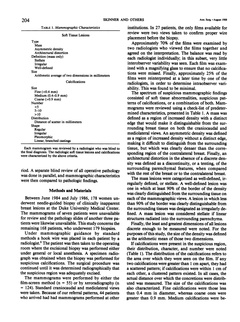
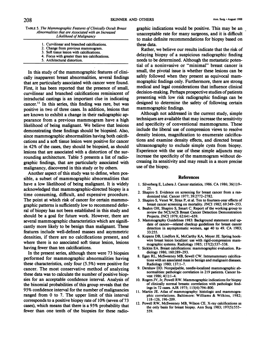
Several studies have demonstrated that mammographic screening of asymptomatic women results in a lower mortality rate where breast cancer is concerned. Often, screening mammograms reveal a nonpalpable radiographic abnormality and the diagnosis must be determined by an excisional biopsy after radiographic needle localization. The mammographic features associated with 179 nonpalpable breast abnormalities biopsied after radiographic needle localization were carefully characterized. There were 41 carcinomas (23%) in the series. The aim of this study was to determine which radiographic findings, if any, strongly portend the presence of either a malignant or benign lesion. Mammographic features that were commonly associated with malignancy include a change from a previous mammogram, a distortion of the surrounding architecture, the association of a soft tissue density and calcifications, and the presence of more than ten calcifications in the lesion. The radiographic abnormalities which were more commonly associated with benign disease include well-defined densities without calcifications, asymmetric densities without calcifications, and abnormalities consisting solely of a focus of mammographic calcifications that have fewer than ten concretions. The incidence of malignancy in lesions having these mammographic characteristics was only 5.5%. On the basis of these results alone, no firm threshold for biopsy can be recommended. The risks of deferring biopsy until there is worsening of the mammographic image remains to be determined.
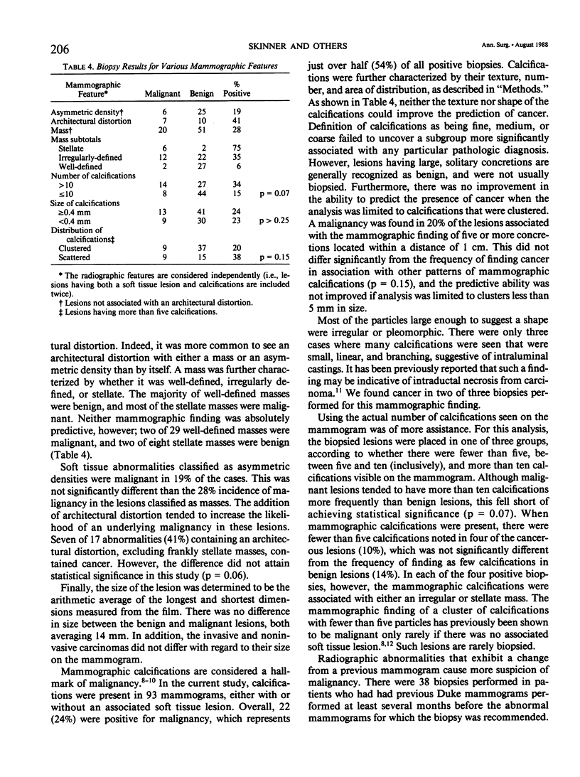
Full text
PDF
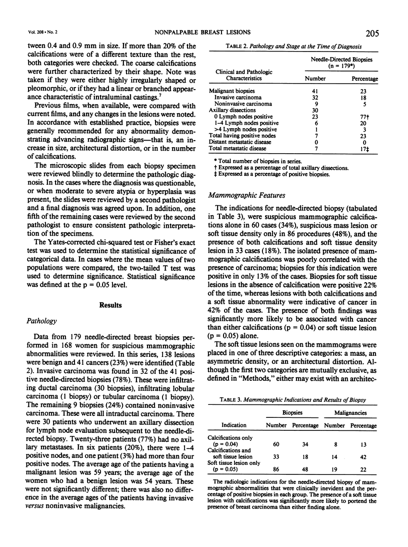
Selected References
These references are in PubMed. This may not be the complete list of references from this article.
- Dershaw D. D. Nonpalpable, needle-localized mammographic abnormalities: pathologic correlation in 219 patients:. Cancer Invest. 1986;4(1):1–4. doi: 10.3109/07357908609039821. [DOI] [PubMed] [Google Scholar]
- Egan R. L., McSweeney M. B., Sewell C. W. Intramammary calcifications without an associated mass in benign and malignant diseases. Radiology. 1980 Oct;137(1 Pt 1):1–7. doi: 10.1148/radiology.137.1.7422830. [DOI] [PubMed] [Google Scholar]
- Kopans D. B., Lindfors K., McCarthy K. A., Meyer J. E. Spring hookwire breast lesion localizer: use with rigid-compression mammographic systems. Radiology. 1985 Nov;157(2):537–538. doi: 10.1148/radiology.157.2.4048466. [DOI] [PubMed] [Google Scholar]
- Powell R. W., McSweeney M. B., Wilson C. E. X-ray calcifications as the only basis for breast biopsy. Ann Surg. 1983 May;197(5):555–559. doi: 10.1097/00000658-198305000-00009. [DOI] [PMC free article] [PubMed] [Google Scholar]
- Shapiro S. Evidence on screening for breast cancer from a randomized trial. Cancer. 1977 Jun;39(6 Suppl):2772–2782. doi: 10.1002/1097-0142(197706)39:6<2772::aid-cncr2820390665>3.0.co;2-k. [DOI] [PubMed] [Google Scholar]
- Shapiro S., Venet W., Strax P., Venet L., Roeser R. Ten- to fourteen-year effect of screening on breast cancer mortality. J Natl Cancer Inst. 1982 Aug;69(2):349–355. [PubMed] [Google Scholar]
- Sickles E. A. Breast calcifications: mammographic evaluation. Radiology. 1986 Aug;160(2):289–293. doi: 10.1148/radiology.160.2.3726103. [DOI] [PubMed] [Google Scholar]
- Silverberg E. Cancer statistics. 1986. CA Cancer J Clin. 1986 Jan-Feb;36(1):9–25. doi: 10.3322/canjclin.36.1.9. [DOI] [PubMed] [Google Scholar]