Abstract
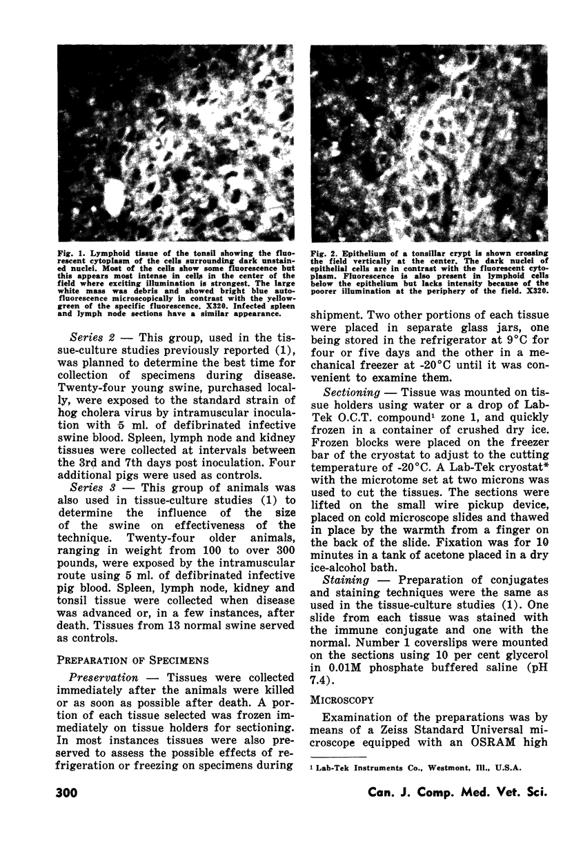
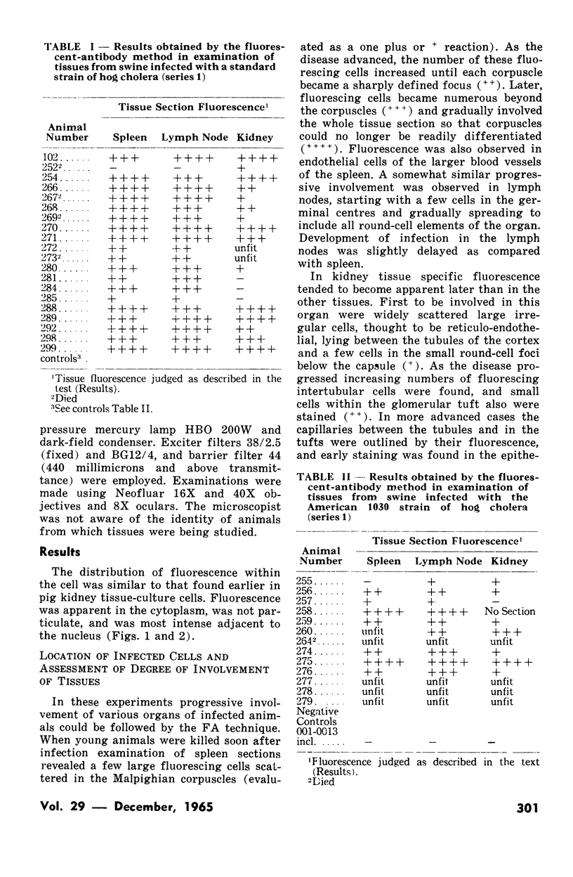
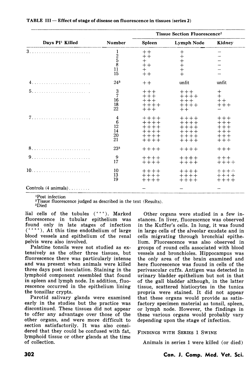
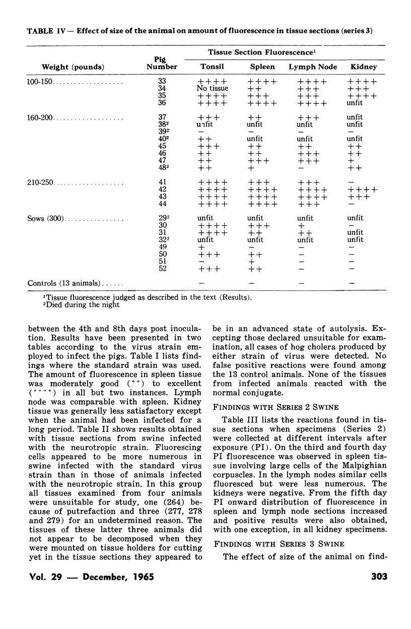
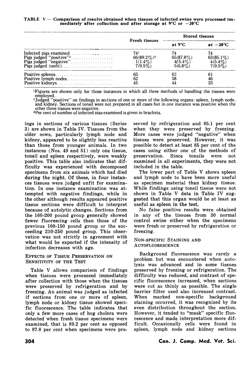
The fluorescent antibody technique was employed to detect hog cholera virus in tissue sections of various organs from experimentally infected swine. The method proved to be highly sensitive and infection could be detected in these animals as early as three days after inoculation with the virus. Best results were obtained when tissues were collected from young animals in advanced disease rather than from sows or from pigs in the early febrile phase. Tonsil, spleen and lymph node were the tissues of choice and were most satisfactory when removed from freshly killed animals rather than from those that had died.
Full text
PDF
Images in this article
Selected References
These references are in PubMed. This may not be the complete list of references from this article.
- Ruckerbauer G. M., Appel M., Bannister G. L., Mori K., Cochrane D., Boulanger P. Hog Cholera: I. Investigation of the Agar Double-Diffusion Precipitation Test for The Detection of the Virus in Swine Tissue. Can J Comp Med Vet Sci. 1964 Dec;28(12):297–303. [PMC free article] [PubMed] [Google Scholar]
- STAIR E. L., RHODES M. B., AIKEN J. M., UNDERDAHL N. R., YOUNG G. A. A hog cholera virus-fluorescent antibody system. Its potential use in study of embryonic infection. Proc Soc Exp Biol Med. 1963 Jul;113:656–660. doi: 10.3181/00379727-113-28455. [DOI] [PubMed] [Google Scholar]


