Abstract
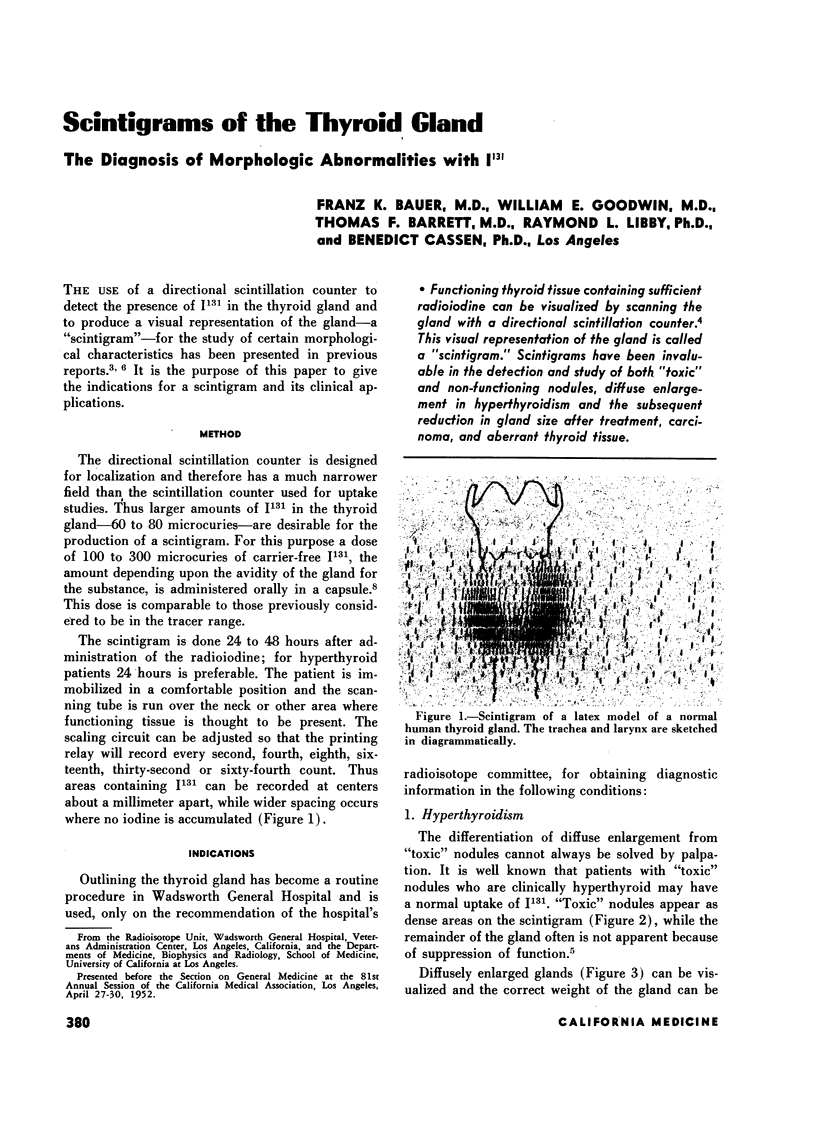
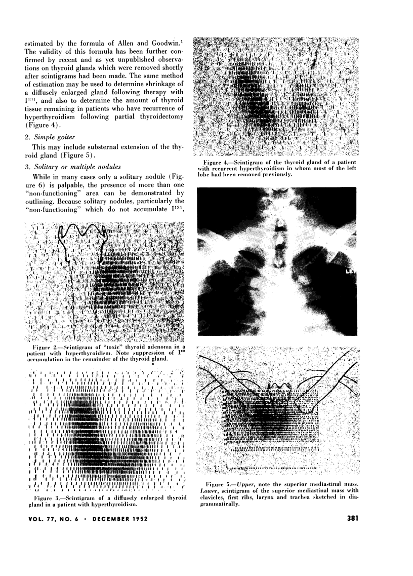
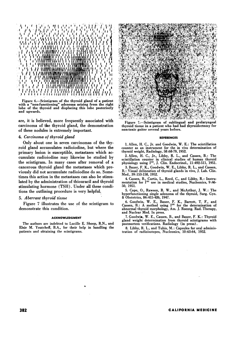
Functioning thyroid tissue containing sufficient radioiodine can be visualized by scanning the gland with a directional scintillation counter.4 This visual representation of the gland is called a “scintigram.” Scintigrams have been invaluable in the detection and study of both “toxic” and non-functioning nodules, diffuse enlargement in hyperthyroidism and the subsequent reduction in gland size after treatment, carcinoma, and aberrant thyroid tissue.
Full text
PDF


Images in this article
Selected References
These references are in PubMed. This may not be the complete list of references from this article.
- ALLEN H. C., Jr, GOODWIN W. E. The scintillation counter as an instrument for in vivo determination of thyroid weight. Radiology. 1952 Jan;58(1):68–79. doi: 10.1148/58.1.68. [DOI] [PubMed] [Google Scholar]
- ALLEN H. C., Jr, LIBBY R. L., CASSEN B. The scintillation counter in clinical studies of human thyroid physiology using I131. J Clin Endocrinol Metab. 1951 May;11(5):492–511. doi: 10.1210/jcem-11-5-492. [DOI] [PubMed] [Google Scholar]
- BAUER F. K., GOODWIN W. E., LIBBY R. L., CASSEN B. Visual delineation of thyroid glands in vivo. J Lab Clin Med. 1952 Jan;39(1):153–158. [PubMed] [Google Scholar]


