Abstract
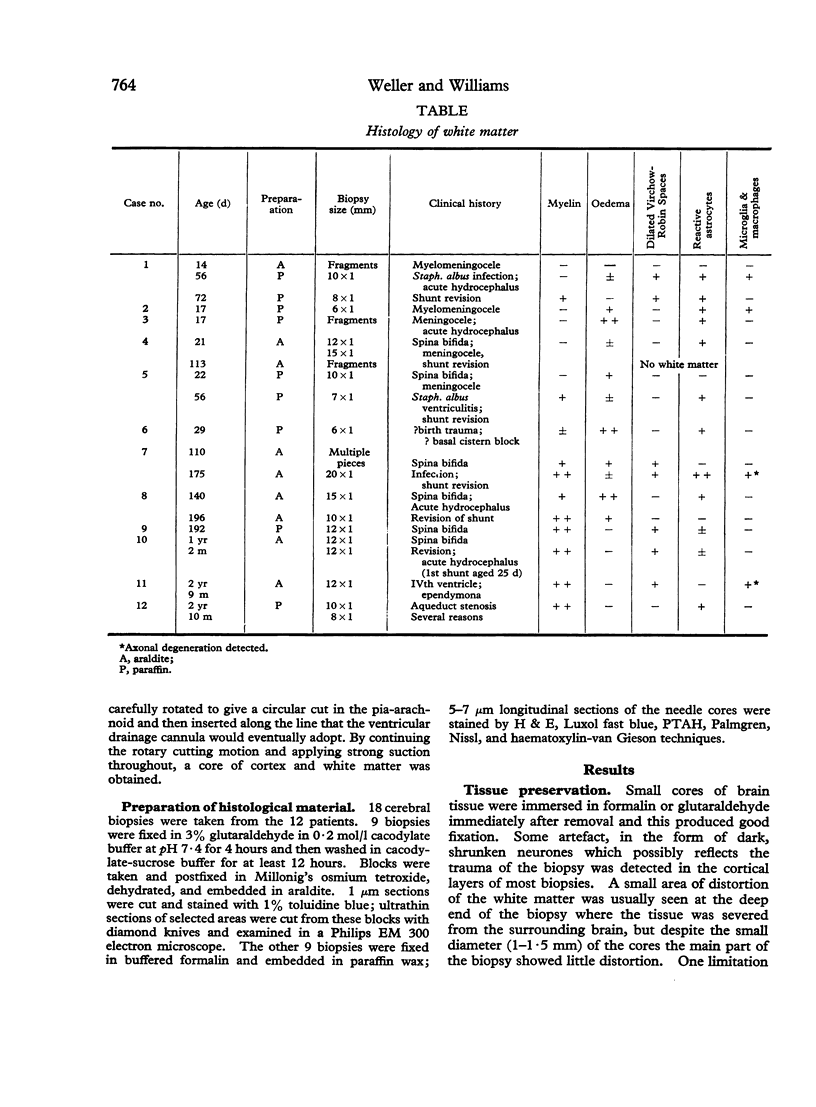
Needle biopsies of the cerebral mantle were taken from 12 hydrocephalic children aged between 14 days and 3 years. 5 children were biopsied twice or more often during subsequent shunting operations. Histological studies of biopsies embedded in epoxy resin for light and electron microscope examination revealed more useful information than those embedded in paraffin wax. There was evidence of axonal degeneration in the white matter of patients with acute hydrocephalus. Progressive gliosis was seen in more chronic hydrocephalus together with signs of cerebral atrophy. No measurable effect of hydrocephalus on myelination was detected. This histological study of needle biopsies taken at shunt operations could be useful in assessing brain damage and thus in predicting future intellectual development in hydrocephalic children.
Full text
PDF
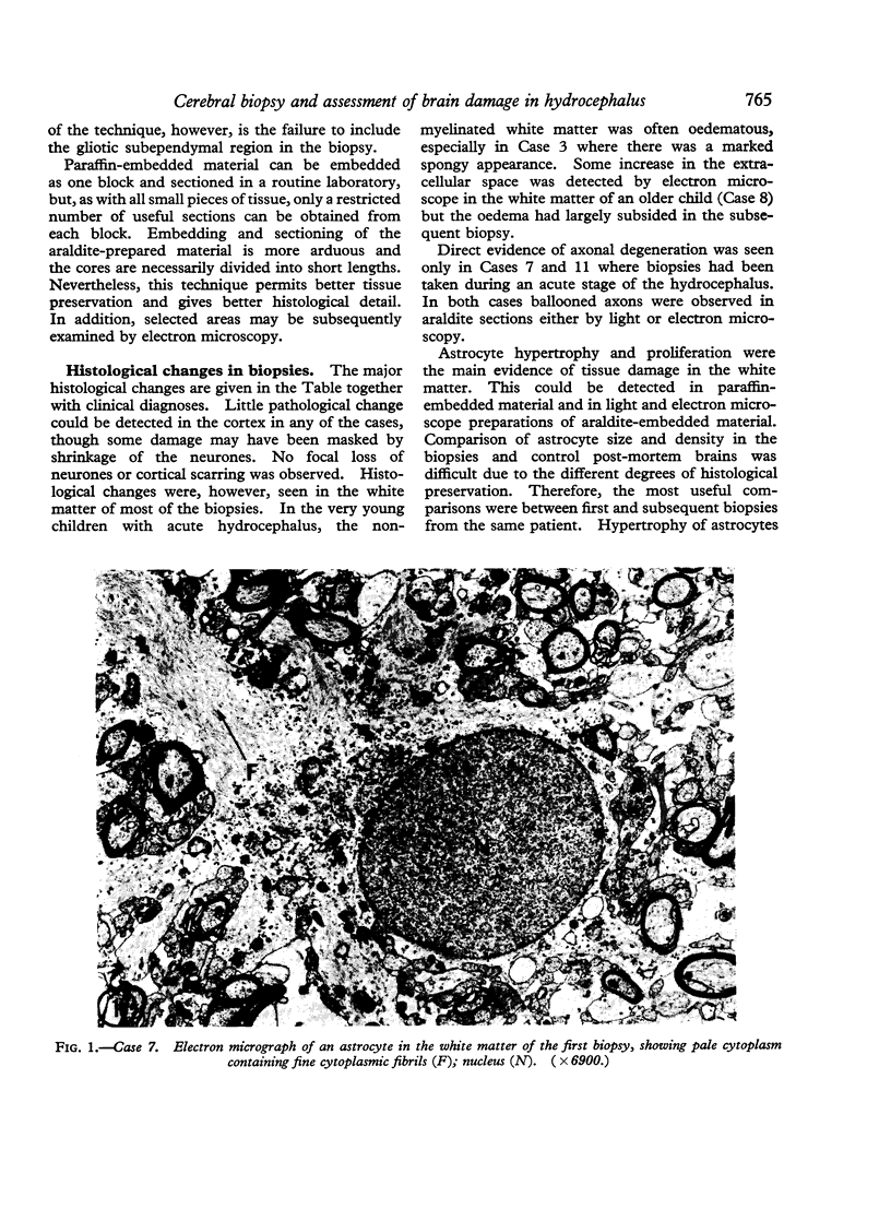
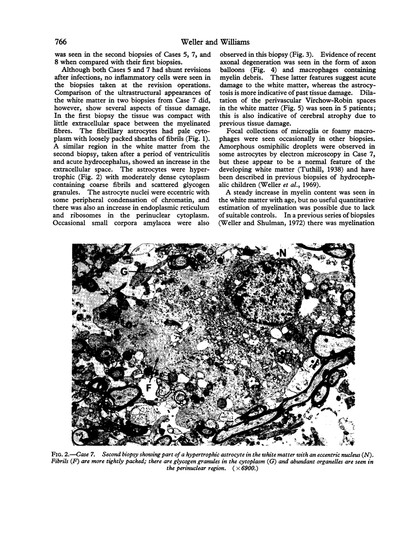
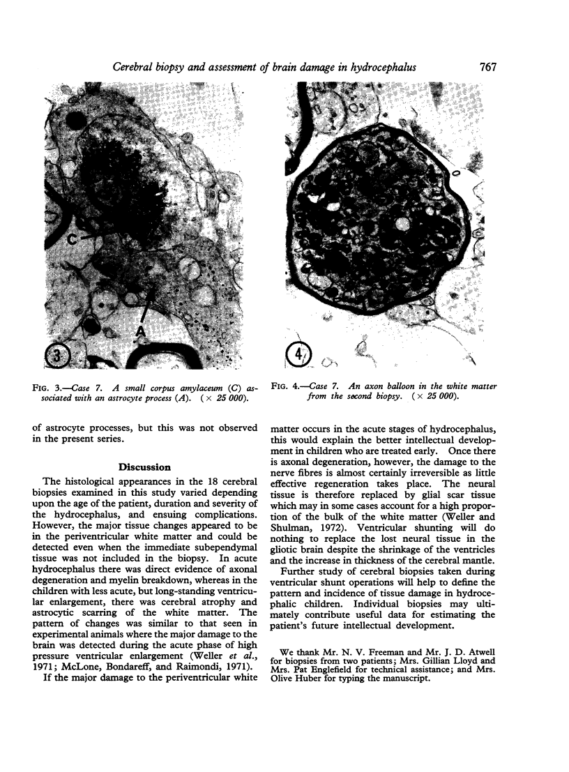
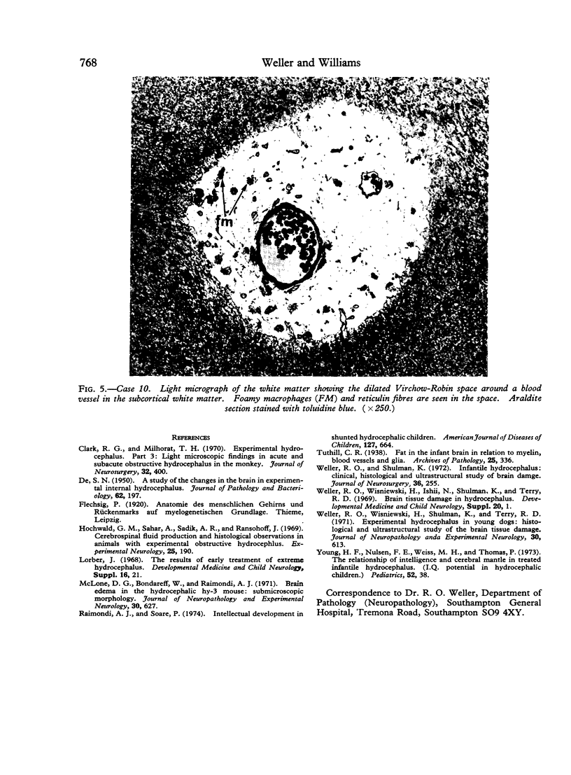
Images in this article
Selected References
These references are in PubMed. This may not be the complete list of references from this article.
- Clark R. G., Milhorat T. H. Experimental hydrocephalus. 3. Light microscopic findings in acute and subacute obstructive hydrocephalus in the monkey. J Neurosurg. 1970 Apr;32(4):400–413. doi: 10.3171/jns.1970.32.4.0400. [DOI] [PubMed] [Google Scholar]
- Hochwald G. M., Sahar A., Sadik A. R., Ransohoff J. Cerebrospinal fluid production and histological observations in animals with experimental obstructive hydrocephalus. Exp Neurol. 1969 Oct;25(2):190–199. doi: 10.1016/0014-4886(69)90043-0. [DOI] [PubMed] [Google Scholar]
- McLone D. G., Bondareff W., Raimondi A. J. Brain edema in the hydrocephalic hy-3 mouse: submicroscopic morphology. J Neuropathol Exp Neurol. 1971 Oct;30(4):627–637. doi: 10.1097/00005072-197110000-00007. [DOI] [PubMed] [Google Scholar]
- Raimondi A. J., Soare P. Intellectual development in shunted hydrocephalic children. Am J Dis Child. 1974 May;127(5):664–671. doi: 10.1001/archpedi.1974.02110240050005. [DOI] [PubMed] [Google Scholar]
- Weller R. O., Shulman K. Infantile hydrocephalus: clinical, histological, and ultrastructural study of brain damage. J Neurosurg. 1972 Mar;36(3):255–265. doi: 10.3171/jns.1972.36.3.0255. [DOI] [PubMed] [Google Scholar]
- Weller R. O., Wiśniewski H., Shulman K., Terry R. D. Experimental hydrocephalus in young dogs: histological and ultrastructural study of the brain tissue damage. J Neuropathol Exp Neurol. 1971 Oct;30(4):613–626. [PubMed] [Google Scholar]


