Abstract
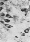
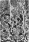
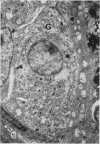
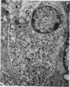
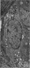
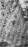
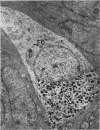
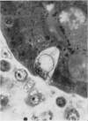
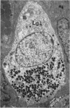
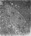
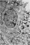
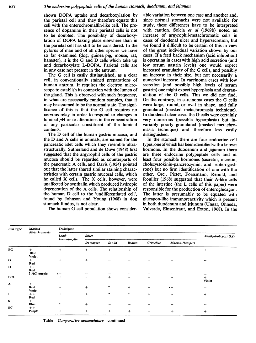
Thirty specimens of stomach, duodenum, and jejunum, removed at operation, were examined by optical microscopical, cytochemical, and electron microscopical techniques. The overall distribution of four types of endocrine polypeptide cell in the stomach, and three in the intestine, was determined. The seven cell types are described by names and letters belonging to a scheme for nomenclature agreed upon at the 1969 Wiesbaden conference on gastrointestinal hormones. The gastrin-secreting G cell was the only cell for which firm identification with a known hormone was possible. Although there was wide variation in the distribution of the various cells, from one case to another, striking differences were nevertheless observable, with respect to the G cell, between antra from carcinoma and from ulcer cases.
Full text
PDF
Images in this article
Selected References
These references are in PubMed. This may not be the complete list of references from this article.
- Bussolati G., Pearse A. G. Immunofluorescent localization of the gastrin-secreting G cells in the pyloric antrum of the pig. Histochemie. 1970;21(1):1–4. doi: 10.1007/BF00304798. [DOI] [PubMed] [Google Scholar]
- Capella C., Solcia E., Vassallo G. Identification of six types of endocrine cells in the gastrointestinal mucosa of the rabbit. Arch Histol Jpn. 1969 Aug;30(5):479–495. doi: 10.1679/aohc1950.30.479. [DOI] [PubMed] [Google Scholar]
- DAVIS J. C. The relation between the pancreatic alpha cells and certain cells in the gastric mucosa. J Pathol Bacteriol. 1954 Jan;67(1):237–240. doi: 10.1002/path.1700670128. [DOI] [PubMed] [Google Scholar]
- Forssmann W. G., Orci L., Pictet R., Renold A. E., Rouiller C. The endocrine cells in the epithelium of the gastrointestinal mucosa of the rat. An electron microscope study. J Cell Biol. 1969 Mar;40(3):692–715. doi: 10.1083/jcb.40.3.692. [DOI] [PMC free article] [PubMed] [Google Scholar]
- Grimelius L. A silver nitrate stain for alpha-2 cells in human pancreatic islets. Acta Soc Med Ups. 1968;73(5-6):243–270. [PubMed] [Google Scholar]
- HELLERSTROEM C., HELLMAN B. Some aspects of silver impregnation of the islets of Langerhans in the rat. Acta Endocrinol (Copenh) 1960 Dec;35:518–532. doi: 10.1530/acta.0.xxxv0518. [DOI] [PubMed] [Google Scholar]
- Håkanson R., Lilja B., Owman C. Cellular localization of histamine and monoamines in the gastric mucosa of man. Histochemie. 1969 Apr 17;18(1):74–86. doi: 10.1007/BF00309904. [DOI] [PubMed] [Google Scholar]
- Håkanson R., Owman C. Concomitant histochemical demonstration of histamine and catecholamines in enterochromaffin-like cells of gastric mucosa. Life Sci. 1967 Apr 1;6(7):759–766. doi: 10.1016/0024-3205(67)90133-6. [DOI] [PubMed] [Google Scholar]
- Johnson F. R., Young B. A. Undifferentiated cells in gastric mucosa. J Anat. 1968 Mar;102(Pt 3):541–551. [PMC free article] [PubMed] [Google Scholar]
- McGuigan J. E. Gastric mucosal intracellular localization of gastrin by immunofluorescence. Gastroenterology. 1968 Sep;55(3):315–327. [PubMed] [Google Scholar]
- Orci L., Pictet R., Forssmann W. G., Renold A. E., Rouiller C. Structural evidence for glucagon producing cells in the intestinal mucosa of the rat. Diabetologia. 1968 Jan;4(1):56–67. doi: 10.1007/BF01241034. [DOI] [PubMed] [Google Scholar]
- Pearse A. G., Bussolati G. Immunofluorescence studies on the distribution of gastrin cells in different clinical states. Gut. 1970 Aug;11(8):646–648. doi: 10.1136/gut.11.8.646. [DOI] [PMC free article] [PubMed] [Google Scholar]
- Penttilä A., Hirvonen J. Identity of the basally located parietal and enterochromaffin-like cells of the rat gastric mucosa. Scand J Gastroenterol. 1969;4(6):489–496. doi: 10.3109/00365526909180639. [DOI] [PubMed] [Google Scholar]
- Solcia E., Capella C., Vassallo G. Lead-haematoxylin as a stain for endocrine cells. Significance of staining and comparison with other selective methods. Histochemie. 1969;20(2):116–126. doi: 10.1007/BF00268705. [DOI] [PubMed] [Google Scholar]
- Solcia E., Vassallo G., Capella C. Selective staining of endocrine cells by basic dyes after acid hydrolysis. Stain Technol. 1968 Sep;43(5):257–263. doi: 10.3109/10520296809115078. [DOI] [PubMed] [Google Scholar]
- Solcia E., Vassallo G., Capella C. Studies on the G cells of the pyloric mucosa, the probable site of gastrin secretion. Gut. 1969 May;10(5):379–388. doi: 10.1136/gut.10.5.379. [DOI] [PMC free article] [PubMed] [Google Scholar]
- Solcia E., Vassallo G., Sampietro R. Endocrine cells in the antro-pyloric mucosa of the stomach. Z Zellforsch Mikrosk Anat. 1967;81(4):474–486. doi: 10.1007/BF00541009. [DOI] [PubMed] [Google Scholar]
- Unger R. H., Ohneda A., Valverde I., Eisentraut A. M., Exton J. Characterization of the responses of circulating glucagon-like immunoreactivity to intraduodenal and intravenous administration of glucose. J Clin Invest. 1968 Jan;47(1):48–65. doi: 10.1172/JCI105714. [DOI] [PMC free article] [PubMed] [Google Scholar]
- Vassallo G., Solcia E., Capella C. Light and electron microscopic identification of several types of endocrine cells in the gastrointestinal mucosa of the cat. Z Zellforsch Mikrosk Anat. 1969;98(3):333–356. doi: 10.1007/BF00346679. [DOI] [PubMed] [Google Scholar]


