Abstract
The use of automated biometrics-based personal identification systems is a ubiquitous procedure in present times. Biometrics has certain limitations, such as in cases when bodies are decomposed, burned, or only small fragments of calcified tissues remain. Dental enamel is the most mineralized tissue of organisms and resists post-mortem degradation. It is characterized by layers of prisms of regularly alternating directions, known as Hunter–Schreger bands (HSB). In this article, we show that the pattern variation of the HSB, referred here as toothprint, can be used as a biometric-based parameter for personal identification in automated systems.
Keywords: forensic science, Hunter–Schreger bands, dental enamel
1. Introduction
Human identification is becoming increasingly important in modern life. It may be required in simple procedures such as logging into a computer network or in more complex situations like post-mortem identification and criminal analysis. It is usually achieved by the use of passwords, physical tokens, photographs, iris and dental patterns, fingerprints and, more recently, DNA analysis (Weicheng & Tieniu 1999). These identification methods commonly fail or have certain limitations and may not be efficient when bodies are decomposed, burned, or in cases when only small fragments of calcified tissues are left (Perry et al. 1988).
The term ‘biometrics’ is used to refer to identification techniques based on physical characteristics. They are sometimes referred to as ‘positive identification’ because they are claimed to provide greater confidence that the identification is accurate. Biometric-based identification and verification methodologies such as fingerprint verification, iris scanning and facial recognition have been steadily improved and refined in automated systems and softwares, which have the capacity to distinguish individuals reliably.
To identify an individual uniquely based on biometric information, the following biometric data with the following characteristics are desirable: highly unique to each individual, easily transmittable, able to be acquired as unintrusively as possible and distinguishable by humans without much special training (Weicheng & Tieniu 1999). DNA testing is an example that has revolutionized human identification in forensic science. This method, however, is relatively expensive and time consuming, and the results take some time to generate. This is not ideal since the success of DNA analysis is also highly dependent on the preservation of the specimen.
Another well-known and widely used biometric is the fingerprint. Fingerprints are unique to each individual and can provide precise personal identification (Perry et al. 1988; Sweet & Sweet 1995). In the identification process, every fingerprint can be broken down into two basic features, called ridges and valleys. In a typical fingerprint, these features will appear as clear and dark lines. Minutiae are points at the ending lines and at the bifurcations when one line splits into two. Minutiae are the major parameters used for algorithm extraction in softwares for automated fingerprint identification systems (Nikodemuszszekely & Szekel 1993; Hiratsuka & Oshino 2002).
In most mammalian species, including humans, dental enamel is characterized by layers of prisms of regularly alternating directions known as Hunter–Schreger bands (HSB). HSB appear as dark and light bands under low-powered light microscopy (Koenigswald & Pfretzchner 1987; Koenigswald 1994). This phenomenon occurs because enamel prisms function like optic fibres when exposed to a directed source of light (Whittaker & Rothwell 1984). When observed from outside, the dark and light lines of HSB in dental enamel closely resemble a fingerprint as they form minutiae. Due to its similarity to a true fingerprint, the pattern of HSB will be referred to here as the toothprint. This similarity prompted us to use a fingerprint identification/verification software to evaluate the HSB singularity in human teeth as a biometric-based method for personal identification.
2. Material and methods
(a) Documentation and analysis of HSB
The sample was composed of 274 lower central incisors supplied by the museum of the Department of Morphology, Piracicaba Dental School, State University of Campinas, SP, Brazil. We have also performed in situ analysis of the HSB pattern in central inferior incisors directly from the mouths of 30 individuals.
This research project complies with resolution 196/96 from National Committee of Health Department (BR) and was approved by the ethical Committee in Research of the Piracicaba Dental School.
HSB were observed with a binocular microscope (magnification of 10–20×) and light shinning obliquely on the tooth (optic fibre–Fibre Light). HSB were clearly visible from outside in the cervical to the mid coronal regions of the teeth (average area of 6 mm2).
HSB were documented with a Pentax MZ-M photographic camera and ILFORD 50 ASA (Kodac). Digital images of the negatives were contrasted using the program Corel Photo Paint 9 (Corel Corporation). After contrast enhancement, the images were extracted on automated biometrics-based personal identification software (Verifinger Demo 4.2 SDK/Fingersec). The Verifinger recognition algorithm follows the commonly accepted fingerprint identification scheme, which uses a set of specific fingerprint points (minutiae). The software uses enhanced algorithmic solutions, which improve the system performance and reliability such as adaptive image filtration algorithm, which eliminates noises, ridge ruptures and stuck ridges. The Verifinger software is fully tolerant to fingerprint translation and rotation, and can recognize a fingerprint from any part of it since the software does not require the presence of the fingerprint core or delta points in the image. The software provides a numeral score of similarity (bits). Higher scores are given for more similar characters, and lower or negative scores for dissimilar characters. The similarity scores were plotted on similarity matrices, which express the similarity between two data points. The similarity scores given by the software were plotted on similarity matrices. The similarity matrix was numbered from 1 to 262 (according to the tooth number) in the ordinate and in the abscissa (N×N). The similarity scores were divided into four groups: group I, 2000–1001 bits (very high similarity); group II, 1000–101 bits (high similarity); group III, 100–10 bits (low similarity); group IV, 9–0 bits (very low similarity).
(b) Analysis of HSB thickness
The average thickness (micrometres) of HSB in the central portion of 70 human lower incisors was measured using the ImageMaster TotalLab Software v. 2.00 (Amersham Pharmacia Biotech).
(c) Effect of temperature
To test the effect of tooth burning on the visualization of HSB, teeth were incubated for 1 h in an oven at temperatures ranging from 100 to 500 °C.
3. Results
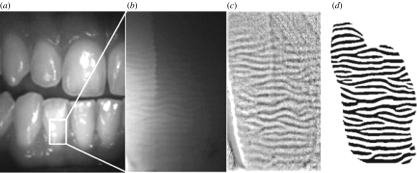
HSB could be examined and photographed without special preparation directly from the mouth of 30 individuals (figure 1) and from 262 isolated central incisors. Incisor teeth were chosen because they are frontal, and it is thereby easier to take measurements from them.
Figure 1.
HSB pattern documented in vivo with fibre light source. (a, b) Bands were visible from the cervical to the mid coronal region of the tooth. (c) Contrast enhancement by the software Corel Photo Paint 9 (Corel Corporation). (d) Extracted images on automated biometrics-based personal identification software (Verifinger Demo 4.2 SDK/Fingersec).
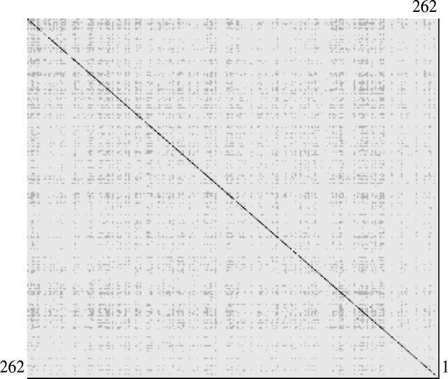
The similarity matrix shown in figure 2 represents the matching of each tooth with the teeth stored on database. The best match (i.e. the highest score) was always achieved when a tooth was compared with itself on the database. This indicates that toothprint analysis as fingerprint exhibits a high discrimination power.
Figure 2.

The similarity matrix: N×N (N: numbered teeth from 1 to 262). Toothprints were classified into four groups according to the similarity scores: group I, 2000–1001 bits (very high similarity) (black filled square); group II, 1000–101 bits (high similarity) (dark grey filled square); group III, 100–10 bits (low similarity) (light grey filled square); group IV, 9–0 bits (very low similarity) (open square). The dark line on the main diagonal shows that the highest scores (i.e. the best match) were always achieved when a tooth was compared with itself on the database.
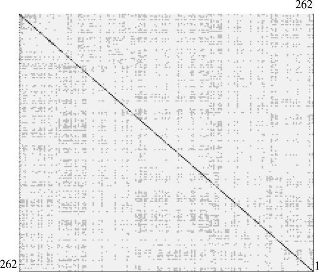
The light and dark bands of a toothprint can be reversed, creating a negative image, by changing the direction of the light source. In order to assess the reliability and robustness of these biometrics in identification/verification procedures, the toothprints of 100 teeth were measured with the light source directed to the mesial side of the teeth and stored in a new database. The toothprints of these 100 teeth were also measured on a separate occasion with the light directed to the distal side of the teeth. The toothprints of distally illuminated teeth were then compared with the database containing measurements from the mesially illuminated teeth. In this case, the best match was also always achieved when a tooth was compared with its template (i.e. negative image) on the database (figure 3).
Figure 3.

Similarity matrix comparing 100 toothprints. The images of each tooth were captured with light source directed to the mesial (abscissa) and distal (ordinate) sides. Toothprints were divided into four groups according to the similarity scores. Group I, 2000–1001 bits (very high similarity) (black filled square); group II, 1000–101 bits (high similarity) (dark grey filled square); group III, 100–10 bits (low similarity) (light grey filled square); group IV, 9–0 bits (very low similarity) (open square). The dark line on the main diagonal shows that the highest scores (i.e. the best match) were always achieved when a tooth was compared with its negative image on the database.
We have also performed an in vivo analysis directly from the mouths of 30 individuals. The comparison of right-left upper and lower central incisors showed that a tooth's print is distinct from that of its homologous tooth on the opposite side (not shown). This indicates that a toothprint pattern is highly specific, being unique for each teeth of an individual.
HSB could not be observed in 4.5% of the teeth examined. Teeth without HSB could not be included in the database. Scanning electron microscope analysis confirmed the absence of HSB in these teeth. Although most teeth presented HSB, 3.3% of the teeth analysed had only 0 or 1 minutia. These teeth gave low similarity scores, thereby increasing the false acceptance and rejection rates in the identification process.
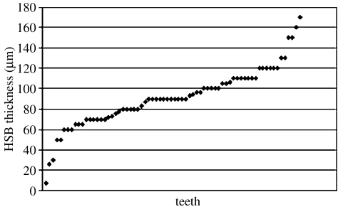
The toothprint differs from fingerprint in that the average thicknesses of HSB are variable, and this feature can be used as an additional parameter for automated identification (figure 4). After the use of two automated systems, the identification of the few remaining teeth with low similarity scores can be made by simple visual comparison. This work can be performed by individuals without much special training (see figure 5 of the electronic supplementary material).
Figure 4.
Average thicknesses (micrometres) of HSB in 70 human lower incisors.
HSB were clearly observed in the enamel of isolated teeth heated at temperatures of up to 300 °C for 1 h (see figure 6 of the electronic supplementary material). Above this temperature enamel becomes opaque, probably due to the incorporation of smoke residues resulting from the burning of organic matrix. This hinders the diffusion of the directed light through the HSB.
4. Discussion
Teeth have been extensively used as a source of information in human identification, especially when the soft tissues cannot provide reliable information (Stefen 2001; Holland et al. 2003). We show here that HSB can be observed and recorded directly from the mouth (figure 1) and from isolated teeth or even in tooth fragments collected in the field. Modern life is characterized by the concentration of large populations in urban areas, with the gathering of people in huge office buildings, schools, theatres and mass transportation systems. Unfortunately, with that comes an increase in the necessity for new and diverse methods of forensic investigation to identify the victims of mass incidents and perpetrators of terrorist acts. The attacks on the World Trade Centre towers on September 11, 2001 set a dramatic example. This event represented the single largest terrorist-related mass fatality incident in the history of the United States. More than 2700 individuals of varied racial and ethnic background lost their lives that day. Through the efforts of thousands of citizens, including recovery workers, medical examiners and forensic scientists, the identification of only approximately 1500 victims had been accomplished by June 2003 (Holland et al. 2003). Toothprint is a highly robust biometric-based procedure for personal identification that could be useful in mass disasters. The analysis of HSB pattern or toothprint may complement fingerprints and DNA, as well as others methods of identification. Accordingly, toothprint recording would be specially recommended for individuals working in dangerous occupations such as firefighters, soldiers, jet pilots, divers and people that live or travel to politically unstable areas.
HSB withstand extreme environmental conditions and are preserved after skin decomposition. HSB could be clearly observed and recorded in teeth burned at temperatures of up to 300 °C for 1 h. Experimental evidence indicates that teeth can withstand much higher temperatures in burned corpses. The preservation is achieved due to the protective effect of the tongue, mastication muscles and other soft tissues in the face and mouth (Valenzuela et al. 2000). Furthermore, well-preserved HSB have been observed from the outside in fossil teeth dating from several hundred thousand to over 60 million years ago (Koenigswald 1994; Ferretti 1999; Stefen 2001; Line & Bergqvist in press). Toothprint analysis is non-invasive, accurate and can be readily performed in automated systems. Toothprint is, therefore, a suitable physiological biometric trait for human personal identification and verification.
Acknowledgments
We gratefully acknowledge financial support from the FAPESP, proc 03/10073-3.
Supplementary Material
‘Find the match’ quiz, and the effect of incineration.
References
- Ferretti M.P. Tooth enamel structure in the hyaenid Chasmaporthetes lunensis lunensis from the late Pliocene of Italy, with implications for feeding behavior. J. Vertebr. Paleontol. 1999;19:767–768. [Google Scholar]
- Holland M.M, Cave C.A, Holland C.A, Bille T.W. Development of a quality, high throughput DNA analysis procedure for skeletal samples to assist with the identification of victims from the World Trade Center attacks. Croat. Med. J. 2003;44:264–265. [PubMed] [Google Scholar]
- Hiratsuka S, Oshino Y. The intelligent fingerprint authentication system SecureFinger. NEC Res. Dev. 2002;43:11–14. [Google Scholar]
- Line S. R. P.& Bergqvist L. P. In press. Enamel structure of Paleocene mammals of the São José de Itaboraí basin, Brazil. ‘Condylarthra’, Litopterna, Notoungulata, Xenungulata and Astrapotheria. J. Vert. Paleontol
- Nikodemuszszekely E, Szekel V. Immage recognition problems of fingerprint identification. Microprocess. Microsyst. 1993;17:215–218. [Google Scholar]
- Perry W.L, Bass W.M, Riggsby W.S, Sirotkin K. The autodegradation of deoxyribonucleic acid (DNA) in human rib bone and its relationship to the time interval since death. J. Forensic Sci. 1988;33:144–153. [PubMed] [Google Scholar]
- Koenigswald W. U-Shaed orientation of Hunter–Shereger bands in the enamel of moropus (mammalia Chalecotherudae) in comparison to some other perissodactyla. Annu. Carnegie Mus. 1994;63:49–64. [Google Scholar]
- Koenigswald W, Pfretzchner H. Changes in the tooth enamel of early Paleocene mammals allowing increased diet diversity. Nature. 1987;106:150–152. doi: 10.1038/328150a0. [DOI] [PubMed] [Google Scholar]
- Stefen C. Enamel structure of arctoid carnivora: Amphicyonidae, Ursidae, Procyonidae and Mustelidae. J. Mammal. 2001;82:450–452. [Google Scholar]
- Sweet D.J, Sweet C.H.W. DNA analyses of dental pulp to link incinerated remains of homicide victim to crime scene. J Forensic Sci. 1995;40:310–314. [PubMed] [Google Scholar]
- Valenzuela A, Martin-de las Heras S, Marques T, Exposito N, Bohoyo J.M. The application of dental methods of identification to human burn victims in mass disaster. Int. J. Legal Med. 2000;113:236–239. doi: 10.1007/s004149900099. [DOI] [PubMed] [Google Scholar]
- Weicheng S, Tieniu T. Automated biometrics-based personal identification. Proc. Natl Acad. Sci. USA. 1999;96:11065–11066. doi: 10.1073/pnas.96.20.11065. [DOI] [PMC free article] [PubMed] [Google Scholar]
- Whittaker D.K, Rothwell T.J. Phosphoglucomutase isoenzymes in human teeth. Forensic Sci. Int. 1984;24:219–222. doi: 10.1016/0379-0738(84)90187-7. [DOI] [PubMed] [Google Scholar]
Associated Data
This section collects any data citations, data availability statements, or supplementary materials included in this article.
Supplementary Materials
‘Find the match’ quiz, and the effect of incineration.


